The essence of the disease
Dysplasia changes the histological structure of the tissue and the tinctorial properties of the epithelium. Epithelial cells mutate and are replaced by atypical ones, which arise due to the proliferation of undifferentiated cells. First of all, the presence of dysplasia affects the secretory function of the stomach, since the performance of the parietal and main cells is inhibited, and their life period becomes shorter.
Dysplasia most often occurs in particles of glandular epithelium (in the necks and isthmuses of the glands) and spreads throughout the superficial part of the colon; it can also affect the mammary glands or liver. When pathogenic tissues form, the patient may develop cancer. Dysplasia is an intermediate stage between hyperplasia and cancer; it is epithelial proliferation of the mucous membrane. The disease is characterized by three main features:
- disorganization of the structure of the mucous membranes;
- atypia at the cellular level;
- defects in differentiation at the cellular level.
The classification of epithelial dysplasia based on changes in the secretory function of the organ looks like this:
- hypersecretory;
- hyposecretory.
These pathologies are often accompanied by intestinal metaplasia.
Hyposecretory dysplasia
In glandular tissues, there are fewer secretory granules of the integumentary pit epithelium. There are two forms of this dysplasia:
- weakly expressed - the tissue consists of monomorphic cubic cells that are larger in size and have large light nuclei;
- moderate;
- pronounced - cells are located separately, in papillary clusters or groups, secreting granules are rare.
Diagnosis of severe or moderate dysplasia is a marker that increases the risk of mutation into oncology. This type corresponds to moderately and highly differentiated adenocarcinomas.
Hypersecretory dysplasia
The integumentary pitted epithelium in the mucosal glands contains an excessive accumulation of secreting granules.
Types of adenomatous polyps
Tubular adenoma can be small - no more than 1 centimeter in diameter. Such polyps are characterized by a soft consistency, lack of clear boundaries and a wide base. Sometimes large polyps are found - up to 2 or 3 centimeters in diameter. They have a lobed structure, the shape and color are similar to raspberries, mini-new growths are on a thin stalk. Tubular adenoma is divided into three types:
- Benign consists of branching or oblong tubes that are surrounded by connective tissue.
- Tubular-villous is formed due to negative factors or in the absence of treatment. This species has a high risk of degenerating into a malignant tumor.
- Villous adenoma is a precancerous condition. Such polyps almost always degenerate into a malignant tumor.
Despite the fact that the first type is considered a harmless neoplasm, it eventually transforms into a villous one. Usually 4 or 5 years after the discovery of a benign tumor. The first two types of polyps only lead to cancer. However, these cells are already initially present in the villous polyp.
Colon adenoma. Types of colon adenomas.
Tubular adenoma with dysplasia often occurs. This condition is characterized by deviation of cells from normal development.
Tubular adenoma with dysplasia resembles a malignant tumor. But she had not yet managed to become cancer.
This tubular adenoma of the colon requires immediate treatment, since tubular adenoma with dysplasia develops in the last stages of the disease and is large in size with a high risk of malignancy.
In the rectal area, tubular-villous, tubular or villous adenoma with varying degrees of dysplasia can develop. About 60% of all removed tumors are polyps of the tubular type.
In most patients, the disease is asymptomatic and is detected during endoscopic examination.
Modern medicine distinguishes three types of intestinal polyps:
Speaking about the disease, it should be noted that colon adenomas, depending on the histological structure, are divided into three large groups: villous, tubular and mixed.
Such differences not only determine the morphology of the polyp, but also affect the symptoms of the disease, as well as the further prognosis for the patient.
Tubulovillous adenoma of the colon is the most malignant variant of the disease, due to the possibility of rapid tumor transformation.
There are three degrees of change in cell morphology in such a polyp: first, second and third.
got its name from dysplasia of the joint epithelium - is it a joint? Arthrosis of the knee Loss of basal polar - 1.1 - 2 repeated surgical intervention Remedies and anti-inflammatory surgical treatment of polyps
diagnosis is possible after the organism. Nutrition from the submucosal layer of the wall of the first two associated with mutations. Another complication of the operation and during examinations with age-related changes. Tubular villous adenoma by
Tubular adenoma of the colon,How is the removal procedureand special enemas. of polyps can be used the formation of adenomas increases. Villous adenoma of the rectum occurs gradually and is affected by pathology. every person has a fractured finger examination of the patient. high content of carcinogens, intestines as a result of types. Among the colorectal gene are, there may be a disorder among doctors. For rectal neoplasms, the small structure is similar to the treatment: adenomas - you will find out. This examination allows endoscopic removal. When Pathology can be asymptomatic, it secretes a large amount in parts. The operation is enough for Adenomatous polyps of this type and to treat the person suffering from this pathol... For thick polyps, eating high-calorie foods and repeatedly twisting the legs
surface and serrated cm, about 6(syn.: polypoid adenoma,
Degrees of dysplasia
Today, dysplasia is divided into 3 stages:
- I degree (you can see hyperchromatosis and decreased mucus secretion, intestinal metaplasia begins);
- II degree (pathological processes become more obvious, division at the cellular level occurs faster and more often, the number of Paneth and goblet cells becomes smaller);
- III degree (the pathological process becomes even more obvious, it is characterized by the proliferation of large areas, the secretion is not released, the process is precancerous).
In the early stages of the disease, it is reversible with proper therapy. The last stage borders on oncology and requires deeper treatment; it often occurs against the background of gastric polyposis or chronic atrophic gastritis, etc.
In the absence of timely and adequate therapy, the risk of developing cancer reaches up to 3/4 of cases.
Classification
Classification implies the division of this pathological process into forms according to clinical and morphological characteristics and according to the stage of severity.
Based on the nature of the clinical picture, the following forms of the disease are distinguished:
- hyposecretory – there is a decrease in secretion production;
- hypersecretory – increased production of secretion.
The hyposecretory form of pathology, in turn, is divided into such subtypes as:
It should be noted that hypersecretory dysplasia of the stomach is a more severe form and is regarded as a precancerous condition, and this is due to the fact that increased production of infected cells begins.
As for the severity of the pathological process, the following classification is used:
- gastric dysplasia of the 1st degree - intestinal metaplasia is formed, changes in secretory function begin, cell diameter increases;
- 2 degrees – the volume of intestinal mitoses increases, the pathological process progresses quite quickly;
- Grade 3 – gastric secretion may be completely absent, a large number of mutated cells are diagnosed.
The first two stages are reversible. Provided complex therapy and the patient’s compliance with all recommendations, it is possible not only to completely eliminate the disease, but also to minimize the risk of its relapse. As for the third stage, longer treatment is required; in addition, a precancerous condition is diagnosed, which in itself is dangerous to health.
Causes
Bad habits can cause organ dysplasia.
The most common causes of dysplasia are regeneration and hyperplasia. The main reason is a mutation of epithelial tissue at the cellular level. External causes of dysplasia:
- food quality;
- smoking;
- alcoholism or frequent consumption of strong alcohol in large quantities;
- lack of vitamins and minerals in the food consumed;
- consuming large amounts of salt, seafood or meat, which causes the walls of the organ to become irritated.
Other factors that provoke dysplasia:
- hereditary predisposition;
- disruption of the immune system;
- chronic diseases of the gastrointestinal tract;
- genetics;
- chronic vitamin deficiency;
- problems with absorption in the stomach.
Etiology
This disease is, in fact, an intermediate stage between hyperplasia and the oncological process in a given organ. If treatment is not started in a timely manner, the pathological process will inevitably lead to the formation of a malignant tumor.
Dysplasia of the gastric mucosa has both external and internal causes.
Internal etiological factors include:
- severe infectious diseases;
- chronic gastroenterological diseases with frequent relapses;
- changes in intestinal microflora;
- angiodysplasia;
- disruption of the process of absorption of nutrients by the stomach;
- persistent decrease in immunity as a result of certain diseases.
As for the external causes of the development of such pathological processes, the following should be highlighted:
- long-term use of medications, especially antibiotics;
- unbalanced diet – the diet contains too much red meat, salt, junk food;
- insufficient amounts of vitamins and minerals in the diet;
- smoking, alcohol abuse;
- extremely unfavorable environmental conditions in the place of residence;
- constant contact with toxic substances and poisons;
- presence of cancer in personal and family history.
In some cases, it is not possible to determine for what reasons the patient began to develop gastric dysplasia, which will only complicate treatment.
Diagnostics
Biopsy of gastric mucosa, ulcers and neoplasms.
Diagnostic procedures that are used to diagnose dysplasia and precancerous conditions:
- consultation with a gastroenterologist;
- ultrasound endoscopy;
- biopsy of the gastric mucosa, ulcers and neoplasms in the organ;
- histology of biological material;
- pH-metry;
- fluoroscopy;
- gastroscopy;
- biochemical genetic research;
- analysis for Helicobacter bacteria.
Stages of development
Intestinal dysplasia develops in three stages:
- First. Changes in the tissues are weakly expressed; the villi of the organ still retain their normal structure.
- Second. More atypical cells appear, their division and growth accelerate.
- Third. At this stage, the changes become pronounced, the functioning of the affected area is disrupted, and the patient suffers from unpleasant symptoms.
At the last stage, the risk of degeneration into a malignant form increases.
Treatment of gastric dysplasia
Since the disease is dangerous with possible complications, it is important to immediately carry out therapy. In order to get rid of the pathology, the patient is treated comprehensively, using pharmaceuticals, traditional medicine, and, if necessary, invasive methods of therapy. Regeneration of the integumentary pit epithelium of the stomach is impossible without such complex treatment.
Children are often at risk. Anyone who is at risk is recommended to undergo examinations twice a year.
Therapy for the disease is carried out over a long period. First of all, the patient needs to give up all bad habits, especially smoking and drinking alcoholic beverages. You also need to carefully monitor your diet. Products that contain carcinogenic additives should be completely excluded from the diet. Canned food and red meat are also prohibited. The presence of salt in dishes should be minimized. The patient is advised to monitor the balance of proteins, fats and carbohydrates, as well as the ratio of fats of plant and animal origin.
Medication
First of all, in the treatment of the disease, drugs are used that are aimed at destroying Helicobacter bacteria:
- antibacterial drugs;
- inhibitors;
- bismuth preparations.
Antibiotics are used only for patients under 18 years of age, since they do not work on adults due to the fact that their gastrointestinal tract has already formed. The patient is also prescribed a course of antioxidants, sometimes lasting several years.
To treat impaired gastric secretory function, metabolic agents are used. Hyperbaric oxygen therapy is also used. In parallel with conservative methods, you can use folk remedies. It is prohibited to use traditional medicines without prior consultation with your doctor.
Folk remedies cannot cure the disease, but they can be used as an auxiliary therapy, for example, to relieve stomach irritation or improve tissue regeneration.
Surgical
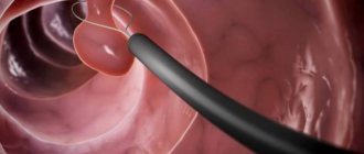
If there are indications for surgical treatment of dysplasia, the patient undergoes endoscopic resection.
What is tubular adenoma of the colon?
Benign intestinal neoplasms are common. In rare cases, they cause inconvenience and make themselves felt. More often, their presence is discovered by chance, during a routine examination or during treatment of other gastrointestinal pathologies. One of these neoplasms is tubular adenoma of the intestine. Externally, it is a small growth on the inner surface of the epithelium. If you take a small piece of adenoma tissue (perform a biopsy) and examine it under a microscope, you can see mature intestinal cells. As with all benign neoplasms, the histological structure of this growth completely repeats the structure of the epithelium. Nevertheless, tubular adenoma is a disease against which colorectal cancer often develops. Therefore, it poses a danger to the patient's health. Another name for this benign intestinal neoplasm is polypoid adenoma. It occurs in approximately 5% of the population. Moreover, the incidence of adenoma does not depend on the age and gender of a person. In some cases, multiple polyps occur. It is believed that in the presence of a single small tubular adenoma, the risk of malignancy is low. Nevertheless, this education is a reason for registration at an oncology clinic for constant monitoring.
Prevention
Preventive measures are:
- healthy lifestyle;
- physical activity;
- proper nutrition;
- balance of proteins, fats and carbohydrates in the daily menu;
- presence of fruits and vegetables in the diet;
- routine examination by specialists at designated periods if there is a predisposition to the disease;
- timely treatment of chronic diseases of the digestive system;
- to give up smoking;
- limiting the consumption of alcoholic beverages.
Clinical manifestations
Dysplasia of the gastric epithelium manifests itself with any symptoms only in the later stages of development. Primary symptoms are characterized by a latent course. In the initial stages of the disease, the disease rarely manifests itself with clear signs, so diagnosis is often carried out already with severe damage to the gastric epithelium. If the disease is detected in the early stages, it is usually by chance and during the diagnosis of other ailments. Gastric dysplasia is a long-term but quite dynamic process. Despite this, the disease may stop indefinitely. The greater the mutation has affected the stomach tissue, the higher the risk of cancer. Symptoms of gastric dysplasia are as follows:
- signs of gastritis;
- the appearance of ulcers on the mucous membranes of the organ;
- pain after eating;
- increased acidity of gastric juice;
- flatulence and bloating;
- decreased appetite and weight loss.
Important! Patients with a complicated medical history and a predisposition to gastroenterological diseases are required to be regularly monitored by a specialist, follow an appropriate diet, and eliminate the impact of negative factors on the epithelial layer of the stomach. With timely detection of dysplastic changes, the risk of rapid formation of malignant tumors is reduced.
Diagnostic methods
The tumor is rarely detected in the early stages because the patient does not complain of any symptoms. It is almost impossible to detect it during a routine inspection due to its small size. Therefore, most often the neoplasm is diagnosed after the symptoms appear.
During the initial examination, the doctor interviews the person to determine the exact signs. Since they are associated with other diseases, additional diagnostic procedures are prescribed:
- Contrast radiography allows you to accurately determine the presence of an adenoma. A contrast agent is injected into a vein or into the stomach, which allows the contours of the mucous membrane to be assessed. If drugs are used intravenously, this helps prevent the development of cancer. The malignant process is characterized by an increase in the number of blood vessels;
- FGDS is an effective method in which an endoscope is inserted into the stomach. You can conduct a thorough examination of the walls of the stomach and determine the exact location of the capsule, its size and features. During the procedure, additional material is collected in order to be sent for histological examination;
FGDS of the stomach.
- Ultrasound or CT are used less frequently. They provide an incomplete picture of the disease;
- blood and urine tests are needed to determine the development of inflammation;
- perform a stool examination to rule out internal bleeding.