Pemphigus is a fairly rare dermatological disease that affects people of different age categories. However, this disease is more often observed in adults 40-60 years old.
In this article we will introduce you to the causes, types, symptoms, methods of diagnosis and treatment of pemphigus in adults. This information will be helpful to you and your loved ones so you can take action to combat this difficult to treat disease.
Pemphigus is accompanied by the appearance of blisters filled with exudate on the body and mucous membranes. They are able to merge with each other and grow rapidly, causing the patient a lot of suffering. The disease is difficult to treat because it is of an autoimmune nature. As such, there is no specific therapy for this disease, and this fact often leads to the development of complications and severe consequences in the future.
Causes of pemphigus
The most likely cause of pemphigus is considered to be a disorder of the autoimmune system, which causes the body's cells to produce antibodies. Changes in the antigenic structure of the epidermis itself are observed due to the influence of external factors (for example, the effect of retroviruses or poor environmental conditions).
A negative effect on the epidermis and the production of antigens is caused by disruption of communication between cells, which is why blisters later appear. As for the risk factors for pemphigus, they have not yet been established, but it is known that people with a hereditary predisposition have a high incidence rate.
Causes of pathology
Viral pemphigus of the palms and soles is caused by a special group of viruses - enteroviruses, more specifically - Coxsackie viruses, subspecies A16 and the enteroviruses themselves, subspecies 71. The first type of microorganisms causes damage mainly only to the palms, which occurs easily and without complications. Enteroviruses of 71 subspecies cause a highly contagious, epidemic-causing pathology that can be complicated by enteroviral meningitis (inflammation of the membranes of the brain) and encephalitis (inflammation of the brain itself).
Viral pemphigus in adults occurs most often over the age of 40 years; Children under 10 years of age and those who have recently had another viral infection are more susceptible to it. They become infected in the following ways:
- airborne droplets when talking with a sick person;
- when eating from the same container with a sick person;
- after a handshake or other physical contact with a patient, when liquid from a bladder deprived of a tire gets on the skin of a healthy person;
- when kissing;
- when particles of the patient’s feces enter the digestive system of a healthy person - if hands were not washed after going to the toilet, and the patient took someone else’s dishes, towels, toys, shook hands, touched the handrails of public transport, and the healthy person did not wash his hands afterwards.
The danger of viral pemphigus is that you can become infected not only from the patient, but also from:
- a person who is in an incubation period lasting 3-6 days;
- a carrier of the virus - a person who, due to the activity of his immune system, is not sick and feels well, but excretes the virus in his feces;
- recovered from this infection, for about those months after the blisters in the mouth or on the extremities have disappeared.
People with reduced immunity and those who have a hereditary feature of the upper layer of skin (epidermis), which gives them a tendency to develop viral pemphigus, are more likely to get sick.
The disease is characterized by seasonality: the virus is usually activated in spring, summer and autumn, while high and low temperatures kill the microbe in the environment. People who have visited new places are more likely to become infected: the sea, nature, where other enteroviruses that differ from their own region “live” (the immune system of an adult usually develops protection against similar microorganisms in its own region).
Immunity is developed only to the strain of the virus that causes the disease. Other subspecies of enterovirus are still capable of causing viral pemphigus.
Video: Causes and symptoms of pemphigus in pregnant women and newborns
Pemphigus vulgaris
The first thing that occurs is pemphigus on the mucous membranes, so as a rule, the initial treatment will be incorrect - usually people are treated by a dentist or otolaryngologist. At this stage of the disease, patients complain of a sore throat when eating or even when talking. There is also hypersalivation and bad breath. The duration of such a symptomatic period ranges from three to four months to a year. After this, pemphigus is characterized by its spread over the surface of the body, involving an increasingly larger area of the skin.
Often, patients may not notice the presence of bubbles due to their small size, as well as a thin film on top. Typically, blisters can open quickly, so patient complaints are complaints about painful erosion. Long-term and almost always unsuccessful treatment of stomatitis is often carried out. Symptoms of pemphigus are primarily blisters localized on the skin. They are distinguished by the ability to open independently, exposing an eroded surface with the remains of a tire, which often dries out and takes on the appearance of a crust.
Diagnostics
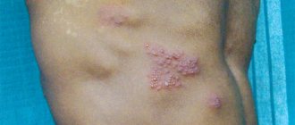
The active phase of pemphigus is easily identified by the appearance of characteristic blisters. Depending on the type of disease, formations can have different sizes, densities and localizations, but are always filled with liquid exudate during development.
Side signs of the disease are general weakness and fever.
In order to reliably identify the disease at an early stage, a dermatologist must conduct a series of immunological and cytological studies. Check the fingerprint smear for the presence of acantholytic cells, as well as histological analysis to identify the location among the layers of the epidermis of the vesicle. Often, before the blisters, a deep red rash appears on the patient’s skin.
It is very important that if you suspect pemphigus, you must undergo the “Nikolsky test”, which with one hundred percent probability will separate pemphigus from symptomatically similar diseases.
Pemphigus in adults
Pemphigus in adults may have a bright pink color and a smooth glossy surface. Pemphigus should not be confused with erosion. Pemphigus differs from the latter in its tendency to peripheral growth and the possibility of generalization with the further formation of extensive lesions. If pemphigus takes a similar course, the patient’s general condition almost always worsens, intoxication begins to develop, and a secondary infection may also occur. Without proper treatment, patients may die.
In the case of pemphigus vulgaris, Nikolsky syndrome will be positive in the lesion. On healthy skin, in the event of even a slight mechanical impact, detachment of the upper layer of the epithelium may be observed.
Symptoms
Regardless of the type and form, pemphigus has similar symptoms. A characteristic feature of the course of this disease is its undulation. In addition, in the absence of timely and adequate therapy, pemphigus rapidly progresses.
Acantholytic species
Pemphigus vulgaris (or pemphigus vulgaris)
With this type of acantholytic pemphigus, the blisters are localized throughout the body and have different sizes. They are filled with serous exudate, and their surface (tire) is thin and flaccid.
More often, the first blisters appear on the mucous membranes of the nose and mouth. This symptom leads patients to see a dentist or otolaryngologist for treatment, since the formations cause them:
- pain when talking, swallowing or chewing food;
- increased salivation;
- bad breath.
This period of illness lasts about 3 months or one year. After this, the pathological process spreads to the skin.
Blisters form on the skin with a flaccid and thin covering. Sometimes they burst, and the patient does not have time to notice the moment of their appearance. After opening the blisters, painful erosions and areas of tires that have shriveled into crusts remain on the body.
With pemphigus vulgaris, bright pink erosions with a glossy and smooth surface form on the body. Unlike other dermatological diseases, they grow from the center to the periphery and can form extensive lesions. The patient is diagnosed with a positive Nikolsky syndrome (or test, phenomenon) - with a slight mechanical effect on the skin in the affected area, and sometimes in a healthy area, the upper layer of the epithelium exfoliates.
During illness, the patient may feel general weakness, malaise and fever. Pemphigus vulgaris can last for years and lead to damage to the heart, liver and kidneys. Even with adequate treatment, the disease can cause severe disability or death.
Erythematous pemphigus
This type of acantholytic pemphigus differs from ordinary pemphigus in that at the beginning of the disease, blisters appear not on the mucous membranes, but on the skin of the neck, chest, face and scalp. They have signs similar to seborrhea - clear boundaries, the presence of yellowish or brown crusts of varying thickness. The covers of the blisters are sluggish and flabby and quickly open, revealing erosions.
With erythematous pemphigus, Nikolsky syndrome is localized for a long time, but after a few years it becomes widespread.
Pemphigus vegetans
This type of acantholytic pemphigus is benign, and many patients remain in satisfactory condition for many years. On the patient's body, blisters appear in the area of natural folds and holes. After opening, erosions appear in their place, at the bottom of which growths with a serous or serous-purulent fetid coating form.
Pustules appear along the edges of the formed erosions, and to make a correct diagnosis, the doctor has to differentiate the disease from vegetative chronic pyoderma. Nikolsky syndrome is positive only in the area where foci of skin changes appear and does not affect healthy skin.
Pemphigus foliaceus
This type of acantholytic pemphigus is accompanied by the appearance of blisters, which in most cases are located on the skin. Sometimes they can also be present on the mucous membranes.
A distinctive feature of this disease is the simultaneous appearance of both blisters and crusts. The blisters of the leaf-shaped type of pemphigus are flat in shape and only slightly raised above the skin.
Such lesions lead to the layering of similar elements of skin changes on top of each other. In severe cases, the patient may develop sepsis, leading to death.
Brazilian pemphigus
This variant of the disease is found only in Brazil (sometimes in Argentina, Bolivia, Peru, Paraguay and Venezuela) and has never been detected in other countries. The exact cause of its development has not yet been established, but most likely it is provoked by an infectious factor.
Brazilian pemphigus is most often observed in women under 30 years of age and affects only the skin. Flat blisters appear on the body, which, after opening, are covered with exfoliating scaly crusts. Beneath them there are erosions that have not healed for several years.
The lesions cause suffering to the patient - sensations of pain and burning. In the area of erosions, Nikolsky's syndrome is positive.
Non-acanotolytic species
Bullous pemphigus
This type of disease is benign and is not accompanied by signs of acantholysis (i.e. destruction). Blisters appear on the patient’s skin, which can disappear on their own, and no scar changes remain in their place.
Nonacantholytic pemphigus
This type of disease is benign and is accompanied by the appearance of blisters only in the oral cavity. The mucous membrane shows signs of an inflammatory reaction and ulceration.
Scarring non-acantholytic pemphigus
This type of disease is more often detected in women over 45-50 years of age. In the medical literature you can find another name for this form of pemphigus - “pemphigus of the eye.” The disease is accompanied by damage not only to the skin and oral mucosa, but also to the visual apparatus.
Erythematous type of pemphigus
The erythematous type of pemphigus will differ from the vulgar type in the localization of skin lesions, the presence of erythematous lesions on the chest, neck and face, as well as on the scalp. The latter is seborrheic in nature. Erythematous pemphigus is characterized by clear boundaries, its surface is covered with a yellow or brown crust. If such crusts are separated from the main surface, the entire eroded surface will be exposed.
In the case of erythematous pemphigus, the blisters are usually small in size, the crust is flabby and flaccid in appearance, they often open on their own, so it is very difficult to diagnose pemphigus. Nikolsky's symptom can be localized for a long time, and in case of generalization of the process it becomes similar to a vulgar appearance.
The erythematous type of pemphigus must be differentiated from the seborrheic type of dermatitis and lupus erythematosus.
Maintenance of the aquarium bladderwort plant
Most plants of the bladderwort family are common in tropical freshwater bodies, of which 2 - 3 species are firmly acclimatized in amateur aquariums. Most often in the aquarium they contain humpbacked bladderwort. It differs from common bladderwort by its thinner and more flexible stem and very narrow and short leaves. In winter, it feels good in an aquarium with sufficient lighting and a temperature of about 20 ° C.
Aquarium bladderwort can be preserved in the winter in the form of winter buds that form at the ends of the stems in the fall. These buds look like pale green, shaggy balls, consisting of a mass of crowded stems. They are stored in vessels with water, and in April they are placed on the window of a warm room, where they unfold and turn into long ribbon-like plants.
The aquarium plant bladderwort does well in the summer months and will sometimes bloom in bright sunlight. It is difficult to preserve bladderwort in an aquarium in winter. But if there are buds left, taken in the fall from a natural reservoir, they are kept in a vessel with water on the window at a temperature 2 - 3 ° C below room temperature. With the onset of spring, with additional lighting, the buds begin to grow and young plants are transferred to the aquarium.
Leaf type pemphigus
The leaf-shaped type of pemphigus, in its symptoms, is a rash of the erythematous-squamous type; the blisters have thin walls and often appear on areas that were already affected. After the bubbles open, the eroded surface becomes bright red. When the surface dries out, lamellar crusts appear. Since with this form of pemphigus, blisters also form on the crusts, the affected area of the skin can become covered with a massive layer of crust due to the abundant exudate.
The leafy form of pemphigus involves the entire skin, but rarely also the mucous membranes are involved. Pemphigus leaf quickly covers the entire skin, on which blisters, erosions and fresh crusts immediately appear. Uniting with each other, the damaged areas form a large wound surface. Nikolsky syndrome will be positive even on a healthy area of skin. If pathogenic microflora attaches, sepsis will begin to develop, which is why a person’s death occurs.
Complications
Caused by enteroviruses or Coxsackie viruses, viral pemphigus can be complicated by such severe conditions as:
- pneumonia;
- encephalitis;
- meningitis;
- myocarditis – inflammation of the heart muscle tissue. It occurs mainly with Coxsackie lesions. This is due to the fact that there is a similar area in the structure of the virus and in the myocardium. The immune system, starting to attack the microbe, discovers that a similar locus exists in the structure of cardiac muscle cells. Mistaking it for a virus, the immune system attacks the heart;
- viral pemphigus during pregnancy, developing in the first trimester, can cause spontaneous abortion or fetal malformations that may be incompatible with life.
Enterovirus infection is usually mild, but in some cases its complications can cause death.
Vegetating type of pemphigus
The vegetative type of pemphigus is benign. Patients can feel well for many years. Bubbles are located around the holes, as well as in the area of skin folds. When the bubbles open, they reveal erosions, at the bottom of which soft vegetations appear that have a fetid odor. Such vegetations are covered with serous-purulent or simply serous fluid. Pustules can be seen along the contour of the neoplasms, so pemphigus vegetans must be distinguished from the chronic form of pyoderma. Nikolsky syndrome will be positive only near the affected areas of the skin, however, in the terminal stages, pemphigus vegetans is similar to pemphigus vulgaris in its clinical manifestations.
Prevention
To protect yourself as much as possible from viral pemphigus, you should not go to foreign regions or to the sea after an illness or if a person is constantly taking hormonal drugs such as Prednisolone or Dexamethasone. In these situations, weakened immunity is easily exposed to the enterovirus and may even lead to a more severe course of viral pemphigus.
There is no vaccine against a huge number of enteroviruses, only some of which cause pemphigus. If you come into contact with a sick person, you need to try to provide yourself and your child with adequate nutrition, start taking calcium supplements in age-appropriate dosages: “Calcium gluconate”, “Calcium-D3” or others. In addition, it is important to wash your hands after transport, outside, going to the toilet and before eating.
If you need to care for a patient with viral pemphigus, you should only touch skin with a rash while wearing gloves.
Treatment of pemphigus
Treatment of pemphigus, first of all, consists of eliminating roughage, simple carbohydrates, canned food, salted and salty foods from the normal diet. In case of damage to the oral cavity, you need to add soups and rare cereals to the diet, so as not to completely exclude food from the diet. It is known that products rich in protein content accelerate the process of cell regeneration and influence the process of epithelization of open erosions.
All patients suffering from pemphigus are monitored by a dermatologist. For such people, a lighter work schedule, cessation of physical activity, and avoidance of insolation are indicated. Frequently changing bedding and underwear will help prevent secondary infections.
Treatment of pemphigus also involves the use of glucocorticosteroids, using them in large doses. Otherwise, positive dynamics in treatment will not be achieved. After relief of acute symptoms of pemphigus, the dosage of the drugs used is reduced to the minimum level. For treatment, the method of extracorporeal hemocorrection is also used, which includes cryoapheresis, membrane plasmapheresis and hemosorption.
For local treatment of pemphigus, non-aggressive antiseptic solutions and aniline dyes are used. The prognosis of pemphigus is almost always unfavorable, since in the absence of adequate treatment, a person’s death can occur quite quickly due to complications that arise. Long-term hormone therapy significantly increases the risk of side effects, but if you stop taking glucocorticosteroids, pemphigus will recur.
Diagnosis of pemphigus
To make a diagnosis, the following studies are necessary:
- clinical examination of the patient;
- definition of Nikolsky's symptom;
- cytological examination for acantholytic cells in fingerprint smears from the bottom of fresh erosions of the mucous membranes and/or skin (the presence of acantholytic cells is not pathognomonic, but a very important diagnostic sign; at the onset of the disease, especially with seborrheic pemphigus, acantholytic cells may be absent);
- histological examination (allows you to detect the intraepidermal location of cracks and/or blisters);
- indirect immunofluorescence method (allows you to detect circulating IgG autoantibodies against intercellular binding substance antigens), the patient’s blood serum is used for analysis;
- direct immunofluorescence method (allows you to detect class G immunoglobulins in the intercellular adhesive substance of the epidermis in a biopsy of apparently healthy skin obtained near the lesion);
- determination of antinuclear antibodies (for differential diagnosis of erythematous pemphigus).
To determine the patient’s condition, identify possible complications of previous therapy with glucocorticosteroids and other immunosuppressive drugs and prescribe concomitant therapy, the following studies are necessary:
- clinical blood test (with mandatory determination of platelet levels);
- biochemical blood test (determining the level of bilirubin, transaminases, glucose, creatinine, protein, potassium, sodium, calcium);
- clinical urine analysis;
- determination of bone tissue density in accordance with clinical guidelines for the diagnosis, prevention and treatment of osteoporosis [11];
- chest x-ray;
- ultrasound examination of internal organs.
In case of damage to the mucous membranes, consultations with an otorhinolaryngologist, ophthalmologist, gynecologist, or urologist are recommended (if there are appropriate indications). If side effects from treatment occur, consultations may be required with: a therapist, cardiologist, gastroenterologist, endocrinologist, psychiatrist, surgeon, traumatologist, phthisiatrician.
Differential diagnosis
Differential diagnosis is carried out with bullous pemphigoid, Dühring's dermatitis herpetiformis, chronic benign familial pemphigus Gougereau-Hailey-Hailey, cicatricial pemphigoid, discoid lupus erythematosus, seborrheic dermatitis, Lyell's syndrome, erythema multiforme, chronic pyoderma vegetans, etc.
Lever's bullous pemphigoid differs from pemphigus by the presence of tense blisters with a dense covering, fairly rapidly epithelializing erosions (in the absence of secondary infection), the absence of Nikolsky's symptom, the subepidermal location of the blisters, the absence of acantholytic cells and the location of class G immunoglobulins along the basement membrane of the epidermis.
Lever's bullous pemphigoid
Dühring's dermatitis herpetiformis is characterized by a polymorphic itchy rash, dense, tense grouped blisters on an edematous hyperemic base, rapid epithelization of erosions, absence of Nikolsky's sign and acantholytic cells in the impression smear from the bottom of erosions, subepidermal location of blisters, deposition of immunoglobulins A in the area of the dermal papillae, high content eosinophils in cystic fluid and/or peripheral blood.
Dühring's dermatitis herpetiformis
In chronic benign familial pemphigus Gougerot-Hailey-Hailey, the distinctive features are the familial nature of the lesion, a benign course, deterioration of the skin process in the summer, localization of lesions (lateral surface of the neck, axillary, inguinal folds, navel area), the presence of maceration of the skin with the formation tortuous fissures like “brain convolutions”, pathognomonic for this disease. Nikolsky's symptom is not always positive and only in lesions. Acantholytic cells are found, but without signs of degeneration, and immunoglobulin deposition is unusual. The disease occurs with periods of remission and exacerbation, mainly in the summer. Rashes often regress when only external therapy is prescribed (without the use of systemic drugs).
pemphigus Gougereau-Hailey-Hailey
Cicatricial pemphigoid differs from pemphigus in the absence of acantholytic cells, a negative Nikolsky symptom, the development of cicatricial changes on the oral mucosa, skin and conjunctiva, the subepidermal location of the blisters, as well as the absence of IgG in the intercellular substance of the epidermis in an immunomorphological study.
Scarring pemphigoid
Discoid lupus erythematosus is distinguished by a characteristic triad of symptoms in the form of erythema, hyperkeratosis and atrophy. Acantholytic cells and intraepidermal blisters are not detected. Nikolsky's symptom is negative.
Discoid lupus erythematosus
With seborrheic dermatitis, there is no acantholysis, damage to the mucous membranes, or histological and immunofluorescent signs characteristic of pemphigus.
seborrheic dermatitis
Lyell's syndrome (toxic epidermal necrolysis) is an acute disease accompanied by fever, polymorphism of rashes, extremely severe general condition and usually associated with taking medications. The disease is characterized by detachment of the epidermis with the formation of extensive painful erosions. Nikolsky's symptom is sharply positive. Possible damage to mucous membranes.
Lyell's syndrome
With exudative erythema multiforme, along with spots and papules, vesicles, blisters, and blisters may appear. Blisters form on the mucous membranes, which open to form painful erosions. An edematous ridge forms along the periphery of the spots and/or edematous papules, and the center of the element, gradually sinking, acquires a cyanotic tint (symptom of “target”, or “iris”, or “bull’s eye”). Subjectively, the rash is accompanied by itching. The rashes tend to merge to form garlands and arcs. The rash appears within 10–15 days and may be accompanied by a deterioration in the general condition: malaise, headache, fever. Then, over the course of 2–3 weeks, they gradually regress without leaving scars; in their place, pigmentation may be observed.
erythema multiforme exudative
Chronic vegetative pyoderma, in addition to signs reminiscent of pemphigus vegetans, has symptoms of deep pyoderma: erosions, ulcers, deep folliculitis. Nikolsky's symptom is negative, and there are no paraclinical signs of pemphigus.
Chronic vegetative pyoderma
Sneddon-Wilkinson disease (subcorneal pustular dermatosis) is characterized by the development of superficial conflict pustules with a diameter of up to 1.0–1.5 cm with a flabby tire, located on a hyperemic background, a slightly edematous base, prone to grouping and herpetiform arrangement. Due to the fusion of elements, scalloped lesions are formed, along the periphery of which fresh elements appear, and in the central part of the lesion the rash is in the stage of resolution. The pathological process is localized mostly on the skin in the abdomen and limbs (flexor surfaces), in the armpits and under the mammary glands. Among the subjective sensations, in rare cases, mild itching is noted. The general condition of patients is usually satisfactory. The disease occurs in paroxysms with incomplete remissions. Circulating and fixed IgA in the intercellular spaces of the multilayer epithelium are not detected.
Sneddon-Wilkinson disease
In some cases, it is necessary to carry out differential diagnosis between different forms of pemphigus.
Treatment
Treatment for viral pemphigus is purely symptomatic and is aimed at improving the patient’s well-being. Therapy consists of following semi-bed rest, drinking plenty of fluids, taking vitamins and increasing immunity. The disease most often ends on its own after 10–14 days.
To eliminate the discomfort and symptoms of cystic lesions in adults, the following groups of drugs are used:
- antiallergic - Diazolin, Fenistil, Citrine, Erius, Suprastin;
- antipyretics - Aspirin, Paracetamol, Nimesil, Tylenol, Ibuprofen;
- corticosteroids - Dexamethasone, Prednisolone;
- cytostatics - Methotrexate, Zeksat, Sandimmune, Cytarabine, Azathioprine.
Among the antiviral drugs for viral pemphigus, Laferon, Cycloferon and Viferon are most often taken.
Local treatment involves the prescription of antiseptics (Miramistin, Chlorhexidine) and combined drugs with an analgesic and disinfecting effect (Oflocain ointment). To heal the skin, Bepanten, Solcoseryl, Vishnevsky liniment, Levomekol are used.
Treatment of viral pemphigus in the oral cavity is carried out by rinsing with antimicrobial and anesthetic solutions - Orasept, Forteza. You can use infusions of anti-inflammatory herbs: chamomile, calendula flowers, oak bark, cornflower. Lotions with agave juice or fresh nettle and sea buckthorn oil help well.
Maintenance treatment is based on taking vitamin preparations, in particular, ergocalciferol, since it is it that is involved in the formation of skin peptides necessary for the development of local immunity. You should also consume more microelements - magnesium, calcium, potassium, selenium and zinc.
Diet
When vesicles appear in the oral cavity, an adult patient is recommended to exclude hot, sour and spicy foods from the diet, and limit the consumption of foods that can provoke an allergic reaction. You should give up smoking and alcohol.
Treatment of viral pemphigus will be more effective if you give preference to fresh vegetables, fruits, liquid porridges and puree soups. Such a diet will speed up the restoration of the mucous membrane and will not cause additional harm.
Features of cultivation
Despite the exoticism that bladderwort has acquired due to its predatory behavior, the plant is not particularly demanding in terms of growing conditions. Pemphigus vulgaris is especially unpretentious and can live in almost any water, except very hard water. Other varieties will develop better in the shallow waters of small bodies of water in soft water with an acidic reaction - infused with peat or rain. Bladderwort is a plant that easily reproduces on its own. If you need to move it to another body of water, in the summer the shoot is separated from it and simply thrown into the water.
Pond use
The culture is excellent for mini-ponds, where it is easier to provide it with water of the required quality. It also grows well in the shallow waters of large bodies of water, decorated in a natural style. Some even specifically keep it in small ponds and aquariums in order to observe the original “hunt” of this predator.
When thinking through the design of a pond, it is important to take into account that during flowering the bright flowers of bladderwrack are noticeable from a distance, but the rest of the time it is completely immersed in water and is completely invisible.
You should also understand that thickets of bladderwort are a rather dangerous place for fish eggs and fry. Even if it is possible to hide from the enemy in the thick thread-like shoots, the bubble valve may slam shut on the head or tail, and the still weak fish, not having the strength to get out, dies.
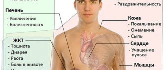
Clinical manifestations
Pemphigus is a disease whose symptoms, regardless of its type, have certain similarities. This skin disease has a wavy course. In the absence of adequate therapy, the patient's general health is impaired.
Symptoms common to all types of pemphigus:
- weakness;
- temperature increase;
- slowing down the epithelization of erosions;
- increase in cachexia;
- loss of appetite.
There are certain differences in the symptoms of different types of this disease:
- In the vulgar form, the blisters have different sizes. The membrane of the blisters is thin and flaccid. Most often, a sick person, having discovered these blisters in his mouth, goes to the dentist for treatment, although this disease should be dealt with by a dermatologist. The patient complains of pain while eating and talking, and bad breath. This period lasts 3-12 months and, in the absence of appropriate treatment, becomes widespread. Next, the inflammatory process moves to the skin. An advanced disease leads to a deterioration in the patient’s condition, and he develops intoxication. Pemphigus is accompanied by a secondary infection. Without proper treatment, the patient faces death.
- In the erymatous form, the blisters are small in size. Their tire is sluggish and flabby. Very often they spontaneously burst. This form is difficult to diagnose.
- In the leaf form, all skin is affected, but sometimes the mucous membranes also become inflamed. It differs from other types in that it can simultaneously cause blisters, erosions and crusts, which, merging with each other, form a large wound surface. With the leaf form, sepsis often occurs, which leads to the death of the patient.
- In the vegetative form, pustules appear in erosions around the formations, so it is differentiated from chronic pyoderma. In terms of its clinical manifestations, this pemphigus is similar to vulgaris.
How to treat pemphigus?
The main treatment pemphigus is the use of glucocorticosteroid hormones; all other medications are of auxiliary value.
The general principles for the use of these hormones are:
- initial loading doses for stabilization and regression of the rash;
- gradual dose reduction;
- individual maintenance doses, in most cases throughout life.
There is no consensus on initial loading doses. Some experts believe that in the case of active generalization of the process, 150-180 to 360 mg of prednisolone per day should be prescribed, while others recommend 60-80-100 mg/day and only if this dose does not produce an effect for 6-7 days, it should be doubled. There are methods according to which 150-200 mg of prednisolone per day is prescribed for 4-6 days, then the dose is reduced to 60 mg or half, and this dose is again used for a week, followed by a reduction by 50%, and then the dose is reduced gradually.
The administration of 1 g of methylprednisolone sodium succinate for 3 days (pulse therapy) was effective when this dose was administered over 15 minutes and in subsequent days was reduced to 150 mg per day.
The question of the duration of use of maximum (loading) doses of corticosteroids and tactics for reducing them is important. Most authors are of the opinion that the maximum daily dose should be maintained until a pronounced therapeutic effect and epithelization of erosions occur.
One of the options for reducing the maximum dose is as follows: during the first week, the dose is reduced by 40 mg, the second - by 30 mg, the third - by 25 mg to a daily dose of 40 mg, the dose reduction is carried out against the background of the use of cytostatics: methotrexate (20 mg per week ), cyclophosphamide (100 mg per day) or azathioprine (150 mg per day). Against this background, the daily dose of prednisolone is reduced by 5 mg monthly, and with a dose of 15 mg per day - by 5 mg every 2 months. Please note that these are only general recommendations because each patient responds differently to corticosteroids and the rate at which they are tapered.
It should be noted that erosions on the oral mucosa epithelialize very slowly and therefore it is not worth continuing treatment with high doses of corticosteroids.
The form of administration of steroids is also of practical importance. One of the options is this: with an active disseminated process, 60 mg of prednisolone (12 tablets) is prescribed orally, taking into account the daily biorhythm of the release of steroids into the blood, and 60 mg of prednisolone (2 ampoules of 30 mg each) is prescribed intramuscularly. In the process of reducing the daily dose, first of all, the injection form is discontinued (30 mg - 1 ml per week).
It should be noted that in some cases the process is resistant to steroids and, in general, to individual drugs. In this case, prednisolone can be replaced with triamcinolone, methylprednisolone, dexamethasone, betamethasone in equivalent doses.
It should be noted that in the treatment of pemphigus there are practically no contraindications for the use of corticosteroids, since without their use the disease is fatal.
In order to reduce the dose of corticosteroids, in addition to their combination with cytostatics, heparin, plasmapheresis, hemosorption, and proteinase inhibitors (contrical) are used simultaneously. Injections of gammaglobulin, interferon, riboxin, vitamins, blood transfusion, plasma, diphenyl sulfone are indicated.
Sometimes, in addition to steroids, riboflavin or benzaflavin is recommended for the treatment of erythematous pemphigus.
Maintenance therapy, selected individually for each patient, must be carried out permanently for years. Apart from clinical ones, there are no other objective criteria for monitoring steroid dose reduction.
If pemphigus recurs, the maintenance dose is doubled and, if necessary, increased further. When erosions are localized on the oral mucosa, doxycycline, methotrexate, nizoral, dipheny are periodically indicated; in case of complications with candidiasis - nizoral and fluconazole, pyoderma - antibiotics, steroid diabetes - antidiabetic drugs after consultation with an endocrinologist.
External therapy for pemphigus is of secondary importance. Aerosols with corticosteroids and antibiotics (oxycyclosol, oxycort, polcortolone), corticosteroid creams, fucorcin, xeroform, syntomycin liniment are used. When the process is localized in the mouth, frequent rinsing with a solution of soda, boric acid with the addition of a 0.5% novocaine solution is recommended. Insolation is strictly contraindicated for patients with pemphigus.
The prognosis is difficult both for life and for recovery. Only in a few patients after long-term therapy is it possible to completely discontinue GCS. Life is threatened by the disease itself and its complications, as well as long-term exposure to corticosteroids. Depending on the condition, such patients are transferred to the appropriate disability group. Patients die from complications: pneumonia, sepsis, cardiovascular failure, cachexia, etc.
Prevention of pemphigus has not been developed.
Forecast
The prognosis of this disease is always unfavorable, since in case of inadequate treatment, the death of patients from additional complications and secondary infection occurs quite quickly.
Pemphigus requires the use of potent hormonal drugs, which in high doses can lead to various side effects, which also adversely affects the patient's health. Refusal of corticosteroid treatment leads to relapse of this skin disease. This “vicious circle” causes unfavorable forecasts from experts.
Therapy for pediatric pemphigus
Pemphigus is diagnosed after a visual examination. It is differentiated from syphilitic, which is congenital and in which inflammation of the skin is localized on the palms. The basis of treatment for this disease is antibiotic therapy, which can significantly reduce the percentage of deaths. Antistaphylococcal gamma globulin is most often used. Locally, the use of aniline dyes and non-aggressive antiseptics is prescribed. The doctor prescribes maintenance and detoxification therapy and plasma transfusion.
With timely and adequate treatment, pediatric pemphigus gives a favorable prognosis. As preventive measures, frequent changes of underwear and bed linen are required, removal from newborns of people with pustular inflammation of the skin, and timely treatment of such rashes on the epidermis of a pregnant woman.
What is pemphigus
Viral pemphigus in children tends to spread quickly, especially in groups of children.
The disease is contagious: the pathogen from saliva and secretions from wounds is transmitted by coughing, sneezing, or through objects.
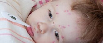
Blisters filled with clear liquid contents appear on the patient’s skin and mucous membranes. The blisters cover the mouth, feet, hands, buttocks, and genital areas, cause itching, and can merge into larger formations.
A type of pemphigus called pemphigus occurs in newborns and is bacterial in nature. The cause of skin lesions is a staphylococcal infection in a child.
Causes of the disease
Viral pemphigus is caused by Kosaka ecterovirus. You can get it in the following cases:
- with a weakened immune system after an illness;
- if personal hygiene is not observed;
- when eating from common dishes;
- during exacerbation of a chronic disease;
- when communicating with a carrier of this infection;
- with bad heredity.
Being in the same conditions, the child’s body reacts differently to viral pemphigus. Doctors explain that the reason for this is the formation of auto-aggressive bodies, which, at the slightest disruption in the body, appear in the form of watery blisters on the skin or mucous membrane (as with enteroviral vesicular stomatitis).
What kind of disease is viral pemphigus and how is it contracted?
Pemphigus is a viral disease in which blisters filled with clear fluid form on the skin. Their number is rapidly increasing, small neoplasms can merge and, together with other blisters, cover large areas of the body. The disease is accompanied by itching and burning of the skin, and patients suffer severe pain.
The disease is considered seasonal; the virus is most active in autumn and spring. Blisters can appear not only on the skin, but also on the oral mucosa, sometimes they spread to the buttocks and genitals.
You can get infected with the virus anywhere. There are two methods of infection:
- visiting public places - kindergarten, school, beach, transport, etc.;
- contact with objects touched by a sick person.
At risk are children with reduced immunity and hereditary predisposition, as well as those children who do not adhere to the rules of personal hygiene. Infection is caused by insufficient food processing and lack of the habit of washing hands after going outside.
It is worth remembering that lasting immunity to the disease is not developed. There are many strains of pemphigus, and the baby receives immune protection only from the one that he has had. For this reason, parents of children who have already encountered this disease should remain vigilant.
Types of pemphigus
With this disease, transformation from one form to another is often observed.
There are several types of pemphigus:
- Vulgar, which occurs most often. Its main symptoms are blisters on the mucous membrane of the gums, cheeks, and palate. They quickly burst, and in their place painful red erosions are formed, which are bordered by the remains of a blister. Sometimes these wounds become covered with a whitish coating. Over time, blisters appear on the skin of the chest and back of a sick person. Moreover, they can be of different sizes. The blisters contain clear serous fluid. After a few days they dry out and become crusty. In some cases, the blisters burst and red erosions appear in their place. Medical history is important in treating this disease. Pemphigus vulgaris often appears in those whose parents suffered from this disease. Once a hereditary connection is established, it will be easier for the doctor to prescribe the most effective type of therapy.
- Erymatous, in which blisters first appear on the skin. They form on the face, chest, neck, and scalp. At the beginning of the disease they are seborrheic in nature. The bubbles have clear boundaries, and their surface is covered with yellow-brown crusts. When they are separated, the eroded surface of the skin is revealed. Erymatous pemphigus is a disease that experts differentiate from seborrheic dermatitis or lupus erythematosus.
- Leaf-shaped, which manifests itself as erythema-squamatous rashes. With it, thin-walled blisters appear on previously affected areas of the skin. Once opened, a red, eroded surface is exposed. When it dries, lamellar crusts form. With this form, blisters may reappear directly on them. Because of this, a thick layered crust forms on the skin. There is a constant separation of exudate.
- Vegetative, which is characterized by a sluggish course. With it, blisters most often affect the skin around openings on the body and in the area of skin folds. After opening them, erosions with a foul odor remain. Vegetations (pathological growths of tissue) appear on them, which are covered with serous-purulent plaque.
Which doctors should you contact if you have pemphigus?
- Dermatologist
Diagnosis of pemphigus is based on the following signs:
- resistance to any local therapy;
- frequent damage to the mucous membranes of the mouth;
- positive Nikolsky sign;
- identification of acantholytic cells using the Tzanck method - this study is carried out to confirm the diagnosis by identifying the so-called acantholytic cells, which are formed as a result of acantholysis (breaking the connections between cells).
The Tzanck method involves placing a glass slide on fresh erosions and acantholytic cells (imprint smear) sticking to it. A sterile gum is applied to mucous membranes with erosions, and then this surface of the gum is applied to a glass slide, thus transferring acantholytic cells onto it. Staining using the Romanovsky-Giemsa method is used.
Morphological features of acantholytic cells:
- they are smaller in size than normal epidermocytes, but their nuclei are larger than those of normal cells;
- the nuclei of acantholytic cells are stained more intensely;
- there are always 2-3 nucleoli in the nucleus;
- the cytoplasm of the cells is sharply basophilic, stained unevenly, a blue zone is observed around the nucleus, and an intense blue border is observed along the periphery.
Acantholytic cells in pemphigus often have several nuclei. However, acantholytic cells can be found in Lyell's syndrome, Darier's disease, and transient acantholytic dermatosis. These cells must be differentiated from cancer cells.
As part of the diagnosis of pemphigus, immunomorphological studies using direct immunofluorescence are used - in 100% of cases, IgO class antibodies are detected in skin sections, which are localized in the intercellular spaces of the epidermis. The method of indirect immunofluorescence detects circulating antibodies of the IgO class against antigenic complexes of the intercellular substance of the epidermis.
Histological examination reveals intraepidermal (suprabasal) bullous elements and fissures.
Differential diagnosis of pemphigus herpetiformis is carried out with bullous pemphigoid, Lyell's syndrome, dermatitis herpetiformis and other bullous dermatoses.
Differential diagnosis of pemphigus vegetans is carried out with syphilitic condylomas lata, chronic familial benign pemphigus, and pyoderma vegetans.
Differential diagnosis of pemphigus foliaceus is carried out with erythroderma, Lyell's syndrome, subcorneal Sneddon-Wilkinson pustulosis, erythematous (seborrheic) pemphigus.