Endometrium during pregnancy - the role of the endometrium
Of course, for every mother, pregnancy is a very exciting period.
This is the blossoming of every woman and at the same time the expectation of a baby, which cannot but please and excite at the same time. In order not to worry once again, you need to know everything about your body during such a crucial period as pregnancy. After all, anxiety can harm your unborn baby. Endometrium - what role does it play during pregnancy? The endometrium is a layer inside the uterus that creates a kind of lining for the inner surface of the uterus. It is into the endometrium that part of the placenta (scientifically chorion) grows into the endometrium in early pregnancy.
- Thus, fetal nutrition and breathing occur through the placenta. The peculiarity is that the placenta is a “coming and going” thing, and the endometrium is always in a woman’s body. It is by the endometrium that one can assess the condition of a woman’s body when it comes to reproductive functions. For example, during menstruation, the endometrium grows so much that it forms glandular structures and folds.
- The thickness of the endometrium also changes during pregnancy. Structurally, the endometrium is a tissue very susceptible to hormonal changes. In the third phase of the cycle, the endometriotic tissue begins to be abundantly supplied with blood and filled with glands. In the early stages, this process is the body’s preparation for placing an embryo.
- If fertilization does not occur during ovulation or another period, then the overgrown endometrium is excreted by the body. The reasons for the inability to get pregnant can be very different; perhaps the thickness of the endometrium in a particular case will have nothing to do with it. In another case, if conception has occurred, the hormonal background changes, preventing the rejection of the endometrium. It is hormones that prevent the endometrium from being released. Thus, it remains and serves as a “seating” place for the soft landing of the unborn child.
Anatomy and physiology of the reproductive system
The uterus is a hollow muscular organ that is a kind of container for the developing fetus. The endometrium is the inner mucous layer of the uterine cavity. It is represented by two layers:
- Functional - changes with each menstrual cycle in which fertilization does not occur
- Basal - contains secretory glands and a rich network of blood vessels that supply blood to the entire organ.
The structure of the mucous membrane is very complex. It consists of epithelial, mucous and stromal elements.
The main function of the endometrium is to create favorable conditions for the implantation of a fertilized egg. For conception, the minimum thickness of the endometrium of the uterus must be at least 7 mm. When pregnancy occurs, the structure of the internal layer changes significantly.
In the early stages of pregnancy, the number of secretory glands increases, the circulatory network becomes more branched, which increases blood flow into the uterine blood supply. These changes are the basis for the placenta, which will form soon.
Regular cyclic transformations of the uterine mucosa
In women of reproductive age, cyclical fluctuations in the level of sex hormones occur in the body every month, and the mucous layer is sensitive to these fluctuations. Under the influence of the hormone progesterone, the mucous membrane increases in size, blood circulation is more intense in it, and the number of glandular cells increases. All these changes reach their maximum by the middle of the cycle, at which time an endometrial thickness of 11 mm is a good indicator for pregnancy and implantation of a fertilized egg into the uterine cavity. But it is important to know that the thickness of the mucous membrane for pregnancy to occur must be at least 7 mm. So, with an endometrial thickness of 5 mm, even if the egg is fertilized, its implantation will be impossible.
If pregnancy does not occur, the functional layer peels off and menstruation occurs. Menstrual discharge consists of cells of the functional layer of the endometrium, blood, which is released from damaged vessels in the uterine cavity, and a mucous component, which is the secretion of the gonads. Immediately after the end of menstruation, the mucous membrane begins to grow again and change its structure, thus preparing fertile ground for conception in a new cycle.
Now it becomes clear why the thickness of the endometrium plays an important role for conception. One of its main functions is to create conditions in the uterine cavity that will be conducive to implantation and further development of the fertilized egg until the placenta is formed and begins to function. Such changes in the reproductive system are normal during pregnancy.
Normal state of the endometrium by day of the cycle:
Norms for endometrial thickness:
- 1-2: the first two days of the cycle 0.5-0.9 cm
- 3-4: third, fourth day 0.3-0.5 cm
- 5-7: from the fifth to the seventh day of the cycle 0.6-0.9 cm
- 8-10: from the eighth to the tenth day 0.8-1 cm
- 11-14: from the eleventh to the fourteenth 0.9-1.3 cm
- 15-18: from the fifteenth to the eighteenth 1-1.3 cm
- 19-23: from the nineteenth to the twenty-third day of the cycle 1-1.4 cm
- 24-27 days of the cycle 1.0-1.3 cm
Follicle diameter depending on the day of the cycle:
- 10th day - 10 mm
- 11th day - 13.5 mm
- 12th day - 16.6 mm
- 13th day - 19.9 mm
- Day 14 - 21 mm, at the time of peak ovulation
What happens during fertilization?
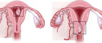
After fertilization of the egg occurs, the latter transforms into an embryo. It grows, moving parallel to the uterus through the fallopian tube over several days. Under normal conditions, the fertilized egg penetrates the uterus around the sixth day after conception, then it attaches to the walls of the organ.
But sometimes the normal activity of the fallopian tube is disrupted, and the patency of the tube is reduced. In this case, the fertilized egg remains in the tube, implanting into its wall (less often it ends up in the abdominal cavity or ovary). This is how an ectopic pregnancy forms. This condition is dangerous for a woman’s health, since after a few weeks such an abnormal change can cause the fallopian tube to rupture.
What should the endometrium look like during pregnancy?
The size of the endometrium is always dynamic, therefore it changes during pregnancy. In the first week of conception, the thickness of the endometrium should ideally be between eight and fifteen millimeters. Later, when it is necessary to undergo the first ultrasound examination, when the fertilized egg is already visible, the endometrium should be about two centimeters. The size of the endometrium during pregnancy should initially be at least seven millimeters. If the thickness of the endometrium is significantly less, then the likelihood of pregnancy is significantly reduced. Of course, this does not completely exclude the possibility, but it is still more difficult to get pregnant with an endometrial layer of less than seven millimeters. This may be one of the reasons for infertility in women. A very important point is that a non-developing endometrium during pregnancy is dangerous for the fetus. Depending on the abnormal thickness of the endometrium, there may be two diagnoses:
- Hypoplasia (thin endometrium)
- Hyperplasia (thick endometrium)
Both hypoplasia and hyperplasia can be a significant obstacle to pregnancy. If disturbances occur during the growth process, this can lead to endometriosis, which can lead a woman to infertility. Therefore, it is very important to undergo an examination by a gynecologist whom you trust in order to identify the problem in time.
In general, the endometrium has a very important function during pregnancy. This is a very complex system that consists of various components:
- Glandular epithelium
- Covering epithelium
- Blood vessels
- Stromas (histiocytes, mast cells and fibroblast-like) and much more
In turn, the epithelium consists of:
- Secretory cells
- Ciliated
- Argyrophilic cells
When are disturbances in endometrial thickness observed?
- Disruptions in the hormonal system. If the hormone prolactin increases, it is produced in large quantities during breastfeeding. If it rises when a woman is not yet breastfeeding, serious problems arise with the endometrial layer.
- Uterine hypoplasia - a woman has congenital disorders in the production of sex hormones. In this situation, many organs are not fully developed.
- Blood flow in the pelvic organs is disrupted. The pathology may be congenital or a consequence of an abortion or injury.
- Serious endometrioid injury. Previously, uterine curettage was performed poorly. This negatively affected the development of the mucous membrane.
Thin endometrium and pregnancy
The endometrium during pregnancy should be within the normal range described above, but it happens that deviations lead to dire consequences, so it is necessary to diagnose the problem in time and consult a doctor. So: why is thin endometrium dangerous during pregnancy?
Therefore, with a pathology of this kind, pregnancy is difficult. Even before pregnancy occurs, the chance of conceiving a child decreases. Even if fertilization and development of the embryo occurs normally at the initial stage, then there is a very high probability that the body will terminate the pregnancy, in other words, there may be a miscarriage. If you have a similar problem, then it’s too early to worry. The problem can be solved, but only if you consult a doctor in a timely manner and have the correct treatment algorithm.
Treatment and diagnosis
Methods for diagnosing endometrial pathologies include:
- Ultrasound. It is used to determine the size of the inner layer of the uterus. In order to detect deviations, the available volume is compared with that which should correspond to the phase of the cycle.
- Echosalpingoscopy. Examination of the uterus can detect pathologies and neoplasms.
- Scraping. When removing the abnormally overgrown inner layer of the reproductive organ and polyps, particles of biomaterial are taken for examination to exclude the presence of malignant formations.
- Biopsy. A piece is plucked from the affected area to be sent for analysis. By studying it under a microscope, you can determine the nature of the tissue damage.
- Hysteroscopy. The technique identifies and eliminates uterine pathologies.
Treatment of endometrial pathologies must be carried out comprehensively:
- Such diseases are caused by a violation of hormone secretion. Therefore, first of all, they take medications aimed at stabilizing hormonal levels.
- For hyperplasia, surgical techniques such as curettage and ablation are used. The latter includes therapy with nitrogen, radio waves, laser, and chemical solutions.
- Combined method. It combines surgery and hormones.
Each treatment method chosen by the doctor depends on the condition of the individual woman. Only a qualified gynecologist should make a diagnosis and prescribe adequate therapy. Self-medication can only aggravate the situation and lead to sad consequences associated not only with pregnancy, but also with life.
The uterine mucosa is responsible for attaching the fertilized egg and nourishing the embryo until the placenta forms. The normal course of these processes is impossible if the thickness of the endometrium is too small or, conversely, large. How thick should it be for a woman to be able to bear a child? Is it possible to cure the pathology, and what methods of therapy are used?
Symptoms
Before talking about how compatible endometriosis and a condition such as pregnancy are, it is necessary to point out the symptoms of the disease.
A woman should pay special attention to them, and if they are present, she should consult a doctor. If the size has increased by 10 mm or more, the symptoms are as follows:
- mild but debilitating pain inside the abdomen, occurring periodically;
- prolonged pain in the lower abdomen, perineum and lower back;
- discomfort during and after sex;
- periodic bleeding or bloody discharge from the rectum, coinciding with the onset of menstruation (if there are polyps in the intestines);
- pain when urinating, urine discoloration in an uncharacteristic color;
- painful menstruation;
- the appearance of blood when sputum or coughing.
An accurate diagnosis is made after examination, questioning and hysteroscopy.
Hyperplasia or hypoplasia of the endometrium can manifest itself stronger or weaker depending on the age and characteristics of the woman’s body, her general condition. The location of the polyps also plays a role - on the ovaries, in the uterus, cervix or intestines.
During pregnancy, there is relief in the manifestations of endometriosis. Just like during menopause. But it is very difficult to get pregnant if you have this disease, since pregnancy and endometrial hyperplasia should not be combined.
How to bring the endometrium back to normal
In order to establish all the processes occurring in the thickness of the endometrial layer, an accurate diagnosis of its condition is necessary. Diagnosis is carried out using ultrasound and other techniques that allow one to assess the thickness of the functional layer by week, and sometimes by day of the cycle. Tests are required to determine hormone levels and determine the presence or absence of concomitant pathologies.
In order for a woman to become pregnant, it is necessary to completely cure all pathologies that impede the process of conception. Hormone replacement therapy helps to cope with the condition of hypoplasia. It helps to “grow” the endometrial layer (sometimes by 6 mm), which increases the chances of the egg to firmly penetrate the uterine layer. Additionally, to stimulate the growth of thin endometrium, physiotherapy is used, mainly aimed at improving blood microcirculation.
The progression of hyperplastic processes is stopped by medication or surgical methods (hormone therapy, curettage of the uterine cavity). How many days, what courses and medications are needed for hormone therapy is determined for each patient individually. It is necessary to monitor the thickness of the endometrial layer over time in order to assess the effectiveness of treatment and timely prevent relapses of the disease.
The successful development of the fetus largely depends on the condition of the endometrium, so every woman should know as much as possible about the state of her female health. And this is possible with regular visits to the gynecologist and following his recommendations.
The uterine mucosa is responsible for attaching the fertilized egg and nourishing the embryo until the placenta forms. The normal course of these processes is impossible if the thickness of the endometrium is too small or, conversely, large. How thick should it be for a woman to be able to bear a child? Is it possible to cure the pathology, and what methods of therapy are used?
Main signs of ectopic pregnancy
When an ectopic pregnancy occurs, women often experience bleeding. They can be both scanty and abundant, very painful. They are not the onset of menstruation, they are a reaction of the endometrium to pathology in the fallopian tube.
In this case, the woman complains of pain in the lower back and pelvic area. Painful sensations of a pulsating nature may be observed, which radiate to the legs and stomach.
Learn more about the symptoms and signs of ectopic burden week by week.
If you have any suspicions, you should not delay your visit to the doctor, since an ectopic pregnancy is fraught with rupture of the fallopian tube and various complications. Modern diagnostic methods make it possible to detect pathological changes even in the early stages.