The article is under development.
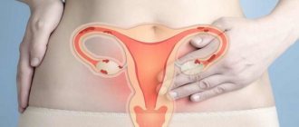
Endometriosis is functioning endometrium outside of its normal location. Internal endometriosis (adenomyosis) includes fragments of the endometrium in the thickness of the myometrium, and external endometriosis includes lesions in the ovaries, utero-rectal space, uterosacral ligaments, rectum, bladder, ureters, vagina, etc.
Click on pictures to enlarge.
Endometriomas can be nodules, infiltrates and cysts, ranging in size from 1 to 40 mm. Under the influence of hormones, cyclic changes occur in them, just like in the uterus. Perifocal inflammation is a constant companion of all types of endometriosis, which leads to the formation of small adhesions around. Often the adhesive component predominates over the endometriotic component. Over time, this leads to the formation of an endometroid-scar nodule, which, having reached a certain size (3-5 mm), becomes visible on ultrasound. Visualization of “fresh” and very small formations is not possible.
Drawing. Pathomorphology of adenomyosis: in the thickness of the myometrium, endometrial glands are visible surrounded by stroma with a scar-lymphoplasmacytic reaction.
With endometriosis, the main complaint is painful, heavy and prolonged periods. Retrocervical endometriosis has the most aggressive course. Characterized by severe pain during sexual intercourse and, to a lesser extent, during bowel movements; constant aching, and during menstruation, sharp shooting pains in the lower abdomen, radiating to the sacrum, rectum, vagina, and thigh.
Diffuse form of endometriosis of the uterine body (adenomyosis) on ultrasound
A 3.5-7 MHz convex sensor is used. Position the patient lying on his back. Bladder of varying degrees of filling. Smoothly reduce the intensity of the echo-positive component of the image: many elements of the picture disappear, but high-density pathological details of the image are highlighted against a general dark background. Repeated performance of this technique from different angles ensures reliable visualization of heterotopias whose dimensions exceed 3-4 mm.
On ultrasound, the uterus is diffusely enlarged, spherical in shape, with a clear and even contour. Compared to the cervix, the echogenicity of the uterine body is increased, the myometrium is heterogeneous due to many hyperechoic point and linear inclusions, and blood flow is often diffusely increased. With TV-ultrasound, tortuous dilated vessels are often visible in the peripheral parts of the uterine wall. In half of the cases, the endometrium is thicker than it should be. In young patients, the echogenicity and echostructure of the uterus is often normal, but the uterus is always spherical in shape.
"God is in the details"
The size of the uterus can be increased in tall women, in those who have given birth many times, before menstruation, and in the presence of an intrauterine contraceptive. In contrast to endometriosis, the uterus retains an oval or pear-shaped shape, and the density of the myometrium is regarded as low.
With a pronounced bend, the size of the uterus may be larger than normal, and the shape may approach spherical. In such cases, the absence of a diffuse increase in the echogenicity of the myometrium, endometrial hyperplasia and complaints is important.
Before menstruation, the echogenicity of the uterus may decrease due to vasodilation and edema.
Diffuse fibrous changes in the myometrium during adenomyosis are often mistakenly regarded as diffuse fibromatosis of the uterus.
Table. Difference between adenomyosis and diffuse form of uterine fibroid.
| Adenomyosis | Diffuse uterine fibroma | |
| Complaints | Algomenorrhea | More often, asymptomatic |
| Uterus size | Increased | Increased |
| Nodes | No | No |
| Form | Regular spherical | Irregular oval or pear-shaped |
| Circuit | Smooth | Wavy or finely lumpy |
| Myometrium | Diffusely heterogeneous due to point and linear hyperechoic inclusions | Multiple hypoechoic zones with unclear contour |
| Echogenicity | Diffusely increased | Hypoechoic areas |
| Endometrium | Often hyperplasia | Usually not changed |
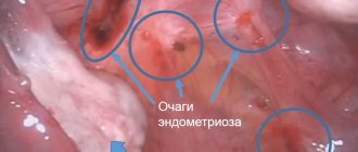
Adhesions in the pelvis with endometriosis
The disease is often complicated by the formation of adhesions, which on MRI may appear as spicule-shaped cords, characterized by a low-to-moderate signal intensity on T1 and T2 weighted tomograms.
Adhesions can fix the pelvic organs, leading to posterior dislocation of the uterus and ovaries, superior displacement of the posterior vaginal vault, and angulation of intestinal loops, which can lead to hydronephrosis, although in most cases hydronephrosis is caused by secondary fibrosis.
On T1 and T2 weighted tomograms in a patient with endometriosis, “kissing” ovaries are visualized, located close to each other due to multiple adhesions. Also visible is a small hemorrhagic cyst in the left ovary and a superficial hemorrhagic plaque, giving a hyperintense signal (red arrows)
Endometriosis complicated by hydronephrosis. T2-weighted tomograms reveal dilatation of the left ureter in its distal parts, caused by pronounced deep endometriotic infiltration with damage to the left uterosacral ligament and extension to the sigmoid colon
Local form of endometriosis of the uterine body on ultrasound
In the myometrium, individual bright hyperechoic inclusions without an acoustic shadow, irregular round, oval or blocky shape, size 2-6 mm, are found. These are areas of fibrosis around one or more endometriomas in the thickness of the myometrium. While cyclic processes occur in the foci, they can increase in size and take on the appearance of small, clearly defined nodes of irregular shape. In the local form of endometriosis, the uterus is of normal size and typical shape, the endometrium is not changed.
In almost all such cases, there is a habitual overdiagnosis of intramural fibromatous nodes with a predominance of fibrosis and calcification. Please note that the clear dependence of the lesion on the phase of the cycle indicates local fibronodular endometriosis.
What does the doctor see during an ultrasound examination?
Thanks to ultrasound, doctors will be able to recognize the following problems:
- uneven and unclear border of the endometrium;
- presence of a nodal structure;
- violation of the symmetry of the walls of the uterus.
If endometriosis affects the ovaries, then the following symptoms are characteristic of this pathology:
- the presence of a round-shaped formation that is located on the side or in the back of the uterus;
- heterogeneous and small-point structure of soft tissues;
- focal formations have different shapes, sizes and appearances.
Endometriosis of the cervix on ultrasound
Endometriosis of the cervix is rare and does not cause pronounced manifestations. The only complaints may be bleeding before and after menstruation.
On ultrasound, cysts are detected in the myometrium of the cervix or the area of the cervix is thickened compared to the intact sections. The outer contour in this place is clear, smooth or wavy. The echogenicity of the cyst-free myometrium is not changed. The configuration of the neck is club-shaped, pear-shaped or fusiform. Cysts are round in shape, the wall is hyperechoic, thin, the effect of enhancement is behind, the contents are homogeneous or finely dispersed, size 4-15 mm. It is especially visible with a TV sensor.
Nabothian cysts are found in the cervix much more often than endometroid cysts. With long-term glandular pseudo-erosion, the stratified squamous epithelium of the vaginal part of the cervix covers the mouths of the glands, which leads to the formation of thin-walled cavities. Nabothian cysts are asymptomatic, very slowly increase in size to 15-20 mm, and then empty; contents are colorless, sterile, cell-free liquid. On ultrasound, Nabothian cysts are located superficially, without wall thickening and contour deformation; long-existing cysts are immersed in the myometrium.
Types of studies
There are three methods for detecting endometriosis on ultrasound:
- transvaginal - the most reliable method, involves inserting a sensor into the vagina followed by scanning the pelvic organs and uterine cavity;
- transabdominal - a less revealing diagnostic method, involving scanning through the abdominal wall;
- transrectal - the sensor is inserted through the anus and shows a picture similar in information content to transvaginal examination (for young girls who are not sexually active, it is better to do an ultrasound in this way).
There are more reliable methods for identifying endometriosis.
Diagnosis of a hormone-dependent disease involves the following manipulations:
- colposcopy – determines cervical endometriosis;
- hysteroscopy – identifies lesions in the cavity of the reproductive organ, in the muscle layer and the mouth of the tubes;
- laparoscopy – detects endometriosis of the abdominal cavity, ovaries, intestines and other organs;
- MRI, CT - makes it possible to determine the location of lesions, cysts and the stage of development of the disease.
It is not immediately possible to see endometriosis on an ultrasound. If the disease is at the initial stage of development, repeated examinations are prescribed - once a month on the 5th–7th day of the menstrual cycle.
Additionally, the doctor can determine by ultrasound:
- changes in the pipes and neck;
- condition of appendages;
- structure of internal tissues - nodular inclusions, heterogeneity, thickening;
- echogenicity of the myometrium and uterine body, its size;
- any neoplasms and nodular seals.
The presence of pathological processes is indicated primarily by changes in the size of the uterus. Normally, its length is 7 cm, width – 6 cm, and thickness – up to 42 mm. The contours must be clear, uniform, without foreign inclusions. If the thickness of the endometrium is higher than normal, they speak of the beginning of the development of endometriosis.
The detection of hyperechoic inclusions indicates pathological tissue growth. This is also indicated by a violation of the structure of the cervix and cervical canal.
Additionally, the internal view of the uterus and the localization of all pathological foci are visualized. Today, women are most often diagnosed with ovarian endometriosis. When carrying out differential diagnosis, cysts and polyps are detected.
Ultrasound examination is also used to monitor the effectiveness of therapy, especially when carrying out conservative treatment. In this case, it is better to do an examination every three months to assess the degree of tissue proliferation.
DETAILS: Biowave and permanent coloring alternative to eyelash extensions
Ovarian endometriosis on ultrasound
Ovarian endometriosis comes in two forms: endometrioid cysts and superficial endometriosis.
Endometrioid cysts can reach large sizes (up to 10-15 cm in diameter). On the smooth inner surface, compactions are found, which, upon microscopic examination, turn out to be areas of the endometrium; chocolate-colored contents. Ultrasound reveals a round formation with a double contour, the capsule in 30% of cases contains hyperechoic foci; there are no dense inclusions in the lumen, the contents are hypoechoic, homogeneous, and there is no internal blood flow. The echo structure does not change at different periods of the menstrual cycle.
Ultrasound for superficial endometriosis reveals a small (2-9 mm) hyperechoic formation of a round, oval or lumpy shape on the ovarian capsule; the contour is clear, smooth or spicule-shaped due to single short fibrous strands. The structure is homogeneous, the echogenicity is high or very high. In the affected area there is some retraction of the ovarian contour; the endometrioma is partially immersed in the ovarian tissue, but is always clearly limited from it by a thickened and compacted capsule. With purely adhesive changes paraovarian, the most typical are multiple linear hyperechoic inclusions along the edge of the ovary without retraction of the contour.
Most of these patients are observed and treated for adnexitis, and the possibility of endometrioid damage to the ovarian capsule is not taken into account. Long-term, untreated ovarian endometriosis often leads to adhesions in the pelvis, creating conditions for chronic salpingitis. It is necessary to look for hydrosalpinx/hematosalpinx and peritoneal cysts - indirect signs of adhesions in the pelvis.
Drawing. Diffuse paraovarian fibrosis as a consequence of external endometriosis.
Drawing. Under the influence of hormone therapy, the lesions shrink and can even resolve.
Endometriosis of the fallopian tubes, outer wall, round and broad ligaments of the uterus is not visible on ultrasound.
Why is endometriosis dangerous?
It cannot be said that the disease can provoke infertility, because many women live with this diagnosis for many years and successfully give birth to children.
Before answering the question about why endometriosis is dangerous, it is necessary to identify symptoms that will help build an accurate picture of health risks:
- painful and profuse critical days (in some cases, the degree of the painful syndrome is so unbearable that the patient needs medical help);
- clots in menstrual blood;
- severe dizziness;
- pain in the abdomen and lower back;
- pain during sexual intercourse, urination;
- loss of consciousness that requires emergency medical attention;
- anemia;
- psychoemotional disorder;
- changes in relation to hormonal levels: hair loss, obesity, the appearance of vegetation (mustaches, hair around the nipples, as well as on the chest), dermatological changes.
In addition, endometriosis is classified depending on the severity of the disease and the location of the lesion. Having considered the entire characteristics of the disease, an answer will automatically form to the question of how dangerous this disease is if it is not treated in time. Classification of endometriosis:
- The first stage – the microflora of the myometrium is affected.
- The second stage is damage to the middle region of the myometrium.
- The third stage - the endometrium is affected to the serous layer.
- Stage four – the disease progresses to the area of the parietal peritoneum.
If endometriosis is not cured, the consequences will become radical, affecting the entire diseased organ, as well as affecting neighboring areas.
Endometriosis of the ovarian ligaments on ultrasound
TA-ultrasound is optimal with a full bladder, then the ovaries are pushed upward, the ligaments are stretched and fully included in the image. During TV-ultrasound on an empty bladder, the ovaries descend, the ligaments hang and occupy an almost vertical position in relation to the vaginal vaults, the image includes transverse and oblique sections of the ligaments, which merge with the surrounding tissues.
On ultrasound, endometriosis of the ovarian ligaments is a hyperechoic nodule or a large linear adhesion up to 30-32 mm that covers the ligament in a muff-like manner.
Preparation for the procedure
There are several ways to conduct research:
- Transabdominal (through the anterior abdominal wall);
- Transvaginal (through the vagina);
- Transrectal (through the rectum);
- Obstetrics (for pregnant women).
Each type of study has its own separate preparation, which should be explained in detail by the attending physician. However, there are general rules:
- 1-2 days before the procedure, do not eat foods that can cause gas (high-fiber vegetables, peas, beans and other legumes, yeast bread, dairy products, alcohol).
- If a few days before the ultrasound the patient underwent a contrast X-ray examination using barium preparations, it is necessary to postpone the procedure, since this substance may blur the results of the examination.
- On the day of the ultrasound, you must have a bowel movement.
This study does not have any contraindications.
Deep infiltrating endometriosis on ultrasound
TV-ultrasound has a clear advantage over TA-ultrasound. During examination, the bladder is slightly full. It is necessary to determine the number, position, size (in three planes) of endometriomas, and echostructure.
Four stages of TV-ultrasound for suspected deep infiltrating endometriosis:
- Examination of the uterus and ovaries. Assess the mobility of the uterus - normal, decreased, fixed (“question mark”);
- Indirect signs of endometriosis: local pain and fixed ovaries increase the likelihood of endometriosis and adhesions. By applying pressure between the uterus and ovary, it can be assessed if the ovary is attached to the uterus medially, to the side wall of the pelvis laterally, or to ligaments.
- Assess the pouch of Douglas using the “sliding sign” during dynamic TV-ultrasound. When the uterus is in anteversion, gentle pressure on the cervix using a transvaginal sensor is established as the rectum slides freely along the posterior surface of the cervix (retrocervical region) and the posterior wall of the vagina. One hand is then placed on the anterior abdominal wall to move the uterus between the palpating hand and the transvaginal probe to assess how the anterior bowel wall slides over the posterior surface of the upper uterus and fundus. When the sliding sign is considered positive in both of these anatomical regions (retrocervix and posterior wall of the uterus), it is recorded that the pouch of Douglas is not obliterated.
- Assess the anterior and posterior cervical space.
The nodular form is a hyperechoic, compactly located heterotopia fused to each other in the space between the posterior surface of the cervix (or isthmus) and the anterior wall of the rectum. The shape of the lesion is irregular oval, less often irregular round or blocky. The contours are uneven (lumpy) and heavy. The heaviness of the contours is a consequence of adhesions and local infiltrative spread of endometriosis. The size of the lesion is from 3 to 30 mm. Retrocervical endometriosis is characterized by very high density, often with an acoustic shadow.
Drawing. Heterotopia group
The scar-infiltrative form is characterized by a significant predominance of the connective tissue component. In other words, a minor endometrioid lesion initiates the development of a pronounced adhesive process. The distribution of changes occurs along the posterior wall of the cervix: the vaginal vault, the uterosacral ligaments, the peritoneum covering the body of the uterus, the broad uterine ligament and the wall of the uterus, the anterior wall of the rectum, bladder and ureters. Ultrasound shows a hyperechoic, heterogeneous compaction of an elongated shape - a scar cord - spreading along the posterior wall of the cervix, the anatomical and topographical features of which determine the position and shape of the changed area. The pathological focus forms a flat platform - straightening of the cervix at the level of the retrocervical lesion. The contours are heavy. Heaviness (spiculosity) is a reliable indicator of locally invasive growth.
Drawing. Perifocal inflammation appears before menstruation or immediately after its end - a hyperechoic focus is outlined by a hypoechoic rim. Perifocal inflammation is a constant companion of all variants of endometriosis, but only with retrointestinal localization can it be seen with TV-ultrasound.
One of the objects of spread of retrocervical endometriosis is the uterosacral ligaments - from the posterolateral surfaces of the cervix and isthmus, cover the rectum in an arcuate manner, and are attached to the pelvic fascia of the sacrum. Isolated lesions are rare, more often secondary lesions due to ingrowth from the retroisthmic-uterorectal recess. On ultrasound, the uterosacral ligaments are not visible. A survey ultrasound is used with a weakly filled bladder, vigorous compression of the anterior abdominal wall, the beam is directed towards the intended focus - a round hyperechoic formation in one of the parametric areas at the level of the isthmus. In such patients, scar-infiltrative changes often spread to the posterior wall of the bladder, sometimes to one of the ureters - narrowing, ureterectosis, hydronephrosis.
Indirect signs of endometriosis invasion into the rectum are large size of the node, pronounced heaviness of the lower edge + pain during defecation, intensifying during menstruation, admixture of blood in the stool during menstruation.
The “kissing” sign of the ovaries indicates the presence of serious pelvic adhesions. Endometriosis of the intestine and fallopian tube is significantly more common in women with kissing ovaries versus those without kissing ovaries.
Anterior cervical space on ultrasound
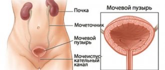
Assess the anterior cervical space, where the bladder, anterior wall of the uterus and ureters are located.
We must not forget that TA-ultrasound and TV-ultrasound are complementary techniques; in the form of a two-stage study, they are a powerful diagnostic tool for diagnosing endometriosis.
It is best to scan the bladder if it contains a small amount of urine. Four zones of the bladder on ultrasound:
- (I) in the trigonal zone, which is within 3 cm of the urethral opening, the smooth triangular area is divided into two ureteral openings and the internal urethral opening;
- (ii) at the base of the bladder, which faces back and down and lies next to both the vagina and the supravaginal uterus;
- (III) the bladder dome, which lies superior to the base and is intra-abdominal;
- (IV) extra-abdominal bladder.
Bladder endometriosis is more common in the base and dome of the bladder than on the peritoneal surface of the bladder. On ultrasound, endometriosis in the anterior compartment can be variable, including hypoechoic linear or spherical lesions, with or without clear contours involving muscle (most often) or (sub)mucosa of the bladder. Bladder endometriosis is diagnosed only when the muscles of the bladder wall are affected; lesions involving only the serosa represent superficial disease.
Drawing. The four zones of the bladder are: the trigone, the base of the bladder, the dome of the bladder, and the extra-abdominal bladder. The point of demarcation between the base and the dome of the bladder is the uterine bursa.
Obliteration of the uterovesical region can be assessed using the “sliding” sign, i.e., a transvaginal probe is placed in the anterior fornix and the uterus moves between the probe and one hand of the operator placed in the suprapubic region. If the posterior wall of the bladder slides freely on the anterior wall of the uterus, then the uterine region is not obliterated. If the bladder does not slide freely along the anterior wall of the uterus, one can think about obliteration of the uterovesical area with adhesions. Adhesions in the anterior pelvis are present in almost a third of women following a cesarean section and are not necessarily a sign of endometriosis.
The distal ureters should be examined. The ureter can be found by identifying the urethra in the sagittal plane and moving the probe to the lateral wall of the pelvis. The intravesical segment of the ureter is identified and follows its course to where it exits the bladder and further to the lateral wall of the pelvis and to the level of the bifurcation of the common iliac artery. This is useful to see how peristalsis occurs and confirms the patency of the ureters.
On ultrasound, the ureters usually appear as long tubular hypoechoic structures, with a thick hyperechoic wall extending from the lateral surface of the bladder, from the base to the common iliac vessels. Ureteral dilation due to endometriosis is caused by a stricture (either external compression or internal penetration) and the distance from the distal ureteral opening to the stricture should be measured. All women with deep endometriosis have their kidneys examined to rule out hydronephrosis as a consequence of obstruction by endometriosis.
Indications
When to do an ultrasound for endometriosis? The answer would seem obvious: as soon as endometriosis is suspected, there are symptoms. These include:
- painful menstruation,
- spotting before and after menstruation,
- problems with conception, infertility,
- less often – pain during intimacy.
It is worth giving some explanations on the symptoms. Women who are concerned about endometriosis also suffer from infertility - up to 40% of patients.
Pain in the pelvic area occurs in almost 25% of patients, and the pain can be localized either in a specific area or throughout the entire pelvic area.
During sexual intercourse there may be not so much pain as a feeling of discomfort. Women do not always even pay attention to this, considering it a kind of norm or attributing it to the cause of fatigue or reluctance to initially have sex.
You need to additionally pay attention to other symptoms that should concern you. Reasons to think about performing an ultrasound:
- long, heavy menstruation,
- the usual discharge is darker, even brown,
- painful urination,
- pain during menstruation (also called dysmenorrhea) – in the first three days of a new cycle, 40-60% experience this,
- cycle failures,
- posthemorrhagic anemia due to large loss of blood during periodic discharge - weakness, pallor or yellowing of the skin, severe fatigue and attacks of dizziness are noted,
- excessive emotionality,
- increase in body temperature.
The earlier the diagnosis is made and endometriosis is detected at the initial stage, the better for the patient. But the “insidiousness” of the pathology lies in the fact that sometimes its course is asymptomatic, or it can be difficult to identify the signs, since they are weak and not so disturbing. Because of this, it turns out that the disease is diagnosed at a late stage.
In order to detect a pathological condition in time, doctors recommend coming for an ultrasound once or twice a year.
Time spending
Since the endometrium is hormone-dependent, its thickness and features change throughout the menstrual cycle. Therefore, it is very important to conduct gynecological ultrasound examinations on the very day of the cycle that the specialist prescribes. In the case of endometriosis, the most favorable days of the cycle are considered to be from the fifth to the seventh, if you count from the first day of menstruation.
It is very important to carry out diagnostics at this time. Therefore, it is necessary to do it, even if residual menstrual bleeding is still present. It should not be carried out during this period only if the bleeding is still severe.
However, sometimes doctors recommend doing several repeated studies in a row in the following cycles. Only in this case will it be possible to accurately confirm or refute the pathology. There is no need to be afraid of this, since ultrasound is a relatively safe procedure that does not have a negative effect on the body. If any treatment was prescribed, then at certain stages of it, studies are also necessary to assess the effectiveness of therapy.
Determining the optimal day of the cycle for ultrasound
To obtain the most accurate result when diagnosing endometriosis, it is necessary to do an ultrasound on days 5-7 of the menstrual cycle.
An ultrasound examination is performed even if there is residual spotting. To track the dynamics of the uterus and get more accurate results, it is recommended to perform ultrasound in subsequent cycles.
This diagnostic technique will help determine the effectiveness of the prescribed treatment and therapy. When women notice the first signs of endometriosis, they must immediately make an appointment with a gynecologist and an ultrasound examination.
Ultrasound is most effective for endometriosis of the uterus and ovaries. Diagnostics is carried out in several ways:
- inserting a sensor into the vagina;
- external examination through the abdominal cavity.
Gynecologists note that in some cases, using ultrasound, they are unable to accurately determine the presence of the disease. In this case, they recommend laparoscopy in combination with a biopsy. In 20% of patients, during an ultrasound examination with the progression of endometriosis, bending of the uterus, dilatation of blood vessels, and deformation of the bladder due to excessive pressure and the presence of tumors are noted. The most important and serious manifestation of endometriosis is endometrial nodes on the walls of the uterus, which are small in size. They come in regular and irregular shapes.
In the cavity of endometrial nodes there is often a cystic cavity, which on ultrasound looks like an inclusion with a minimal level of echogenicity.
Refers to severe gynecological diseases that can significantly reduce a woman’s quality of life and hinder her ability to become a mother. It is thanks to ultrasound that this disease can be diagnosed at an early stage and treatment can be started in a timely manner, avoiding fatal complications. This article is devoted to the features of ultrasound for endometriosis, as the most effective diagnostic method.
Endometriosis in gynecology is a chronic disease that causes benign growth of the intrauterine membrane in various atypical places in the female body.
This disease ranks second among the causes of female infertility.
This disease is insidious in that it does not manifest itself in any way for many years. And the many forms of endometriosis and the varieties of its manifestations seriously complicate the diagnosis of this pathology.
What exactly may be the cause of endometriosis is still not fully understood. It is believed that the disease is based on immune and endocrine disorders of the body. However, the main impetus for its development are the various consequences of endometrial trauma for the following reasons:
- scraping;
- abortions;
- use of intrauterine contraceptives.
All of the above factors seriously affect female hormonal levels and the cyclical nature of its development.
With endometriosis, foci of endometrial growth are periodically rejected (in accordance with the phases of a woman’s menstrual cycle). At the beginning of the cycle, the endometrium grows, reaching its maximum thickness towards the end of the cycle and being shed during menstruation.
It is with the cyclical behavior of endometriotic lesions that the timing of ultrasound examination in this disease is associated.
Main forms and symptoms of endometriosis
Endometriosis usually affects women of reproductive age. Often this disease is discovered unexpectedly, for example, during a preventive examination.
It is transvaginal ultrasound that is considered the main method for identifying this pathology.
The types of this disease are distinguished from the location of the foci of tissue proliferation:
- perineal, cervical and vaginal;
- pipe;
- ovarian.
Women suffering from endometriosis may most often experience the following unpleasant symptoms:
- duration of menstruation and their pain;
- the appearance of clots during menstruation;
- chronic pain in the lower abdomen, starting in the middle of the cycle;
- spotting brown discharge outside the menstrual cycle (in the middle, a few days before and after the cycle);
- pain during sexual intercourse, urination, defecation, exertion;
- infertility for unknown reasons.