Is it possible to do an ultrasound during menstrual bleeding? In the shortest formulation, the answer to this question will be positive. Yes, you can, but the question is not entirely correct. What organ or system do you plan to examine? Ultrasound studies have found application in the diagnosis of a huge number of pathological processes affecting most existing organs and systems of the human body, and not only in the practice of gynecology.
If you are worried about pain in the heart area, you suspect a pathology of the thyroid gland, lymph nodes or urinary system, you want to examine the abdominal organs, you can go to any medical institution equipped with ultrasound diagnostic equipment and calmly do an ultrasound of the abdominal cavity or heart, no matter what It's your cycle day. But if you want to examine the pelvic organs, it is not advisable to perform an ultrasound during menstruation. It is possible, of course, but it is better to postpone the study to a more suitable date. The mammary glands are part of the reproductive system of the female body, and they are also not examined during menstruation.
Unlike other types of ultrasound, it is not advisable to conduct an examination of the pelvic organs during menstruation - this is done only in extreme cases
Main aspects of ultrasound examination of the reproductive system organs
For diagnosing pathological processes of the pelvic organs, there are deadlines based on the characteristics of the functioning of the female body. Violating them will result in an uninformative answer. This is why it is not advisable to do an ultrasound during menstruation. Assessing the condition of the uterine wall, when they look like a wound and the uterus itself is filled with blood clots, is quite problematic. A blurred ultrasound picture during menstruation can lead to the fact that the doctor at the functional diagnostics office misses polyps, or worse, a tumor.
In addition, to diagnose the condition of the pelvic organs, in contrast to ultrasound of the abdominal organs, when examining adults, not pregnant women, vaginal sensors are most often used. The examination procedure may simply not be pleasant or hygienic for the person being examined.
Indications and contraindications
Ultrasound diagnostics is carried out as a preventive annual measure or if the patient has specific complaints. Indications for ultrasound are as follows:
- presence of a lump in the thyroid gland;
- visible enlargement of the left or right lobe of the organ;
- swelling of tissues;
- baldness;
- complaints of tenderness of the gland on palpation;
- tremor;
- heart diseases;
- increased fatigue;
- hoarseness of voice;
- labored breathing.
Ultrasound of the thyroid gland is justified in cases of postoperative monitoring of the condition of the organ and during pregnancy to prevent complications during childbirth. Using this type of diagnosis, you can draw conclusions about the health of the fetus and avoid the development of hypothyroidism.
Ultrasound examination is harmless, the procedure has no contraindications, and it is allowed to be performed even on children under 1 year of age. But the doctor does not prescribe an ultrasound for a cold, because the thyroid gland becomes inflamed due to infection, and the diagnostic results may be unreliable.
Diagnosis of the gland can be performed during menstruation and pregnancy. Most often, the examination is carried out on the last day of the cycle to reduce stress levels. Such a delay is especially advisable when performing Doppler ultrasound, since pathological processes in the body can affect blood flow.
Indications for ultrasound during menstrual bleeding
Menstruation is not an absolute contraindication to ultrasound examination of the pelvic organs. And it is not a contraindication to the diagnosis of abdominal organs. There are a number of pathologies that require urgent examination. Then it ceases to matter whether the procedure is hygienic or not.
If, during the examination, the gynecologist reveals signs of pathology of the mucous membrane and uterine cavity, an ultrasound examination is carried out on the 1st to 3rd day of the cycle due to the fact that the endometrium thickens with each day of the cycle. And performing an ultrasound after this period becomes less informative.
In this case, the following pathological phenomena are revealed:
- myoma;
- polyp(s);
- hyperplasia.
Determining the type of cystic formations of the ovaries is another indication for ultrasound during menstruation, since small (no more than 1 centimeter) cysts are visualized up to 5 days from the beginning of the cycle. With ultrasound diagnostics after the specified period, it is problematic to determine the type of cyst (pathological/physiological).
In gynecology, for screening examination of follicles, ultrasound is prescribed at the beginning of menstrual bleeding and again before the 15th day of the cycle. This study is part of a set of measures to restore a woman’s reproductive function.
In gynecological practice, menorrhagia (excessive bleeding) also requires an ultrasound examination to assess the condition of the woman’s pelvic organs. The causes of excessive blood discharge by the female body can be:
- endometriosis;
- myoma.
The cause of menorrhagia may be endometriosis - inflammation of the inner layer of uterine tissue.
Using ultrasound diagnostics, a doctor at a functional diagnostics office can assess the thickness of the outer layer of the endometrium. If during the examination polypous growths or thickening of the endometrium are detected, a procedure is prescribed to remove the pathological area of the mucous membrane of the cervical canal and the body of the uterus.
There are situations when ultrasound diagnostics are not only permitted, but also necessary. In all other cases, it is better to postpone diagnosis of the genital organs until after the 5th day of the cycle.
Decoding the results
In adults, an endocrinologist deciphers the results. If pediatric ultrasound data are analyzed, among other things, consultation with a pediatrician is required.
It is necessary to pay attention to the size, volume, structure, density, presence of pathological elements (nodules, pseudonodules, cysts, fluid) and other parameters. Also, in the process of analyzing the ultrasound report, it is worth taking into account age and gender characteristics. The normal size of the gland in women and men varies significantly at different ages.
Normally, on ultrasound, the thyroid gland appears as two round lobes connected by an isthmus. The structure of the fabric should be uniform and granular. This is due to the fact that the entire gland consists of small follicles in which thyroid hormones are produced.
The most important parameters are the size of the shares. For an adult they are as follows:
- length – 25-40 mm;
- width – 15-20 mm;
- thickness – 10-15 mm.
It is worth remembering that for most people the sizes of the right and left lobes may differ slightly. Such variations are not pathology. In addition to the above parameters, the volume of the gland is important. It is calculated based on the size of the shares.
Formula for calculating volume:
Volume = [(ShP x DP x TP)] + [(ShL x DL x TL)] x 0.479
- ШП – width of the right lobe;
- DP – length of the right lobe;
- TP – thickness of the right lobe;
- ШЛ – width of the left lobe;
- DL – length of the left lobe;
- TL – thickness of the left lobe;
- 0.479 – ellipsoidal correction factor.
The maximum volumes of the gland, taking into account the weight of the subject, are shown in the table:
| Weight, kg | 50 | 60 | 70 | 80 | 90 | 100 |
| Volume, cubic cm | 15,5 | 18,5 | 22 | 25 | 28,5 | 32 |
If the value exceeds the normal volume, one should speak of hyperplasia or the presence of a tumor.
In addition to the thyroid gland, the condition of the parathyroid glands is analyzed. They are involved in the regulation of calcium and phosphorus metabolism in the body. Enlargement of the parathyroid glands indirectly indicates the presence of hyperparathyroidism. Also during the study it is necessary to evaluate the condition of the lymph nodes in the neck. A change in their structure or density indicates the presence of an inflammatory process.
Pathologies identified during the study:
- Diffuse toxic goiter.
- Nodular goiter.
- Thyroiditis.
- Hypothyroidism.
- Autoimmune thyroiditis.
- Thyroid adenoma.
- Cancer.
- Cyst.
- Calcifications.
- Diffuse toxic goiter is characterized by a significant increase in the size of the gland, while the structure remains homogeneous. The diagnosis is made based on complaints (fatigue, weight loss, fever) and ultrasound images.
- The main symptom of nodular goiter is the heterogeneous structure of the gland. Nodes are located in the thickness of the tissue. They are foci of increased density with clear, even boundaries.
- Thyroiditis is an inflammatory process in the thyroid gland. The volume of the organ increases due to edema. The thyroid gland becomes loose and loses its elasticity and density. With a long course, foci of purulent destruction appear. They are visualized as hypoechoic formations with clear contours.
- When most people talk about hypothyroidism, they mean a decrease in thyroid hormone levels. However, in addition to changes in thyroid status, there is a decrease in the size and density of the thyroid gland.
- The main symptom of autoimmune thyroiditis (AIT) is a change in the structure of the thyroid gland. Most often, a large number of small nodules with clear boundaries are found. They are denser than the main tissue and appear lighter on ultrasound.
- During ultrasound examination, tumors of various natures can be detected. Benign adenomas have the appearance of focal shadows, clearly demarcated from healthy tissue. In addition to adenomas, cysts are occasional findings. They have a hypoechoic color and clear boundaries.
- Markers of malignant neoplasms are invasive growth, lack of clear boundaries, and a large number of feeding vessels. This is what thyroid cancer most often looks like.
- During the study, structural changes that are not pathological may be detected. Their occurrence is often associated with age-related involution of the thyroid gland. These include calcifications and diffuse structural changes. The deposition of insoluble calcium salts is the result of past inflammatory diseases or degenerative changes in the thyroid gland.
Diffuse changes in the structure of the thyroid gland more often occur in postmenopausal women. This is due to changes in hormonal levels or aging of the body. Such findings, in the absence of complaints or hormonal imbalances, are not considered gross pathologies.
Summarizing all of the above, when making a diagnosis, it is important to be guided not only by the results of ultrasound, but also by laboratory and clinical data.
Additionally, see ultrasound of the thyroid goiter:
Preparatory procedures before ultrasound during the menstrual period
Minimal preparation is required before the test. You need to have a disposable oilcloth diaper with you. Before visiting a doctor, it is recommended to toilet the external genitalia.
There is no need to douche after hygiene procedures. This will not bring any benefit, but it can cause significant harm to health. The cervix opens slightly during bleeding, which facilitates access for infectious agents. Before the examination, you should visit the toilet. A full bladder causes discomfort during examination.
To diagnose the organs of the abdominal region, you should exclude foods that cause increased gas formation 2-3 days before the procedure. Regardless of the time of the diagnostic procedure, ultrasound diagnostics of the pelvic organs will not entail consequences, either immediate or long-term.
Since menstrual flow is an integral and completely natural process for every woman of reproductive age, and ultrasound is one of the most popular modern methods for diagnosing almost all existing gynecological diseases, you should know whether it is possible to do an ultrasound during menstruation.
If a woman is worried about heart pain, or there is a suspicion of diseases of the thyroid gland, lymph nodes, urinary system or abdominal organs, an ultrasound examination can be performed on any day of the cycle, including during menstruation. If an ultrasound of the mammary glands or organs of the reproductive system is necessary, you should consult your doctor in advance about the timing of the procedure. In the vast majority of cases, routine ultrasound is not recommended at this time, but there are exceptions, which will be discussed in this article.
Is preparation necessary?
Ultrasound requires little preparation. No significant lifestyle changes are required. If it is necessary to study the reproductive organs, the doctor recommends the transvaginal method.
The study is carried out on an empty bladder. Before diagnosis, you need to visit the toilet. The intestinal tract should not be filled either. If you are prone to bloating, reconsider your diet. Excluded from the menu:
- cabbage;
- beans;
- flour products;
- carbonated and alcohol-containing drinks;
- fat;
- fried;
- spicy;
You should not eat cabbage before the examination.
- pickled;
- canned.
If you are prone to constipation, you should do an enema first. Douching is prohibited.
In the morning the girl takes a shower and puts on clean underwear. The ultrasound is painless, so there is no need to worry before the examination. Take with you:
- absorbent diaper;
- medical card;
- referral for diagnostics.
What can an ultrasound detect?
Ultrasound of the pelvic organs is a fairly common diagnostic method. Based on the results of this study, the echogenicity and size of organs can be assessed. The gynecologist determines the position, structure and size of the uterus, its cervix, the size and location of the ovaries. Ultrasound also determines the presence of follicles and neoplasms in the uterus and its appendages. Additionally, the condition of the bladder, kidneys and intestines can be assessed for the presence of stones, sand and tumors. After diagnosis, a conclusion is made orally or in writing, where the doctor describes all the indicators and their compliance with the norm, and a preliminary conclusion is made about the woman’s health.
If there are any deviations from the norm in the examination results, this is a clear sign of the presence of disorders or diseases. In some cases, to clarify the diagnosis, the examination will need to be repeated on different days of the cycle.
Hardening of the wall of the uterus and fallopian tubes may indicate the possible development of a cancerous tumor. If oval or round segments are visible on ultrasound, it may be a cyst or fibroma. If the uterus is reduced in size, and the ovaries, on the contrary, are enlarged, this is a sign of polycystic disease. If the echogenicity of the entire organ is changed, this may indicate the development of endometriosis or the presence of a benign tumor. Depending on the expected diagnosis, the examination is recommended to be done on different days of the cycle; in some cases, an ultrasound may be required during menstruation. The examination results are provided in the form of screenshots and a diagnostic report.
Experts consider ultrasound one of the safest and most informative diagnostic methods. Thanks to ultrasound, it is possible to determine the presence of ovarian fibroids in 90% of cases, and a cyst in 98%. But there are a number of factors that can significantly affect the reliability of the result, including the presence of a woman’s critical days at the time of the procedure. Based on this, a qualified specialist should make a decision on the need for intravaginal ultrasound during this period.
If a woman’s menstrual cycle is characterized by irregularity, then it is quite natural that on the pre-planned date of an ultrasound of the pelvic organs, menstruation may begin unscheduled. In this case, the study is not urgent, and therefore it is recommended to postpone it to another day of the cycle. There are several reasons why the procedure is not recommended during menstrual periods:
- during a routine ultrasound it is impossible to assess the condition of the endometrium, since it looks like a heterogeneous structure;
- minor pathologies in the endometrium and uterine walls may be hidden and not visible to the doctor’s eye;
- the tissue of the mammary glands is modified during regulation, and such an examination will be uninformative;
- conducting an ultrasound with a vaginal sensor will cause psychological discomfort to the woman;
- It is unhygienic to perform a transvaginal examination during menstruation; this is done only when absolutely necessary.
When is the best time to perform an ultrasound?
Ultrasound is usually prescribed when other diagnostic methods have not made it possible to establish a final diagnosis. The best time for testing depends on the suspected diagnosis.
If the mammary gland is affected, the examination is carried out approximately 6-15 days of the menstrual cycle. If it is necessary to study the reproductive organs, the study is scheduled for 8-10 days.
Immediately after menstruation, an examination is carried out if necessary to assess the condition of the endometrium. You should not go to the doctor even if you have a bloody smear. You need to wait until the blood stops coming out completely.
Blood may make it difficult for your doctor to examine the uterine cavity.
Blood and clots make it difficult to see the uterine cavity. You should inform your doctor about the progress of your menstruation. Emergency diagnosis is carried out only if there are serious reasons.
An abdominal examination can be performed on any day of the cycle. This is convenient, for example, if you need to confirm or deny pregnancy.
When menstruation is not a hindrance and an ultrasound is worth doing
The date of the pelvic ultrasound is determined by the attending physician; there are situations when it is impossible to wait for the end of the regulation and an emergency examination is required; there are also a number of pathologies that can only be identified with the onset of critical days. We list all the situations in which an ultrasound examination is necessary during the regulatory period:
- too much discharge during regulation is a direct indication for immediate ultrasound diagnosis, since many pathologies can only be distinguished during this period;
- if there is a suspicion of a short-term pregnancy, any spotting, even the most scanty, may be a sign of an ongoing spontaneous abortion;
- ectopic development of the fetus is suspected;
- if menstruation turns into uterine bleeding or bleeding between menstruation;
- if tumors of various types, polyps, endometrial hyperplasia are suspected, gynecological ultrasound is best done in the first days of the cycle;
- in the first days of the cycle, you need to do an ultrasound diagnosis, the main purpose of which is to recognize a specific type of cyst with a size of up to 1 cm. If you miss time, the formations become denser and diagnosis becomes difficult;
- Follicle screening is carried out throughout the entire menstrual cycle, including during menstruation, in order to determine the period of ovulation, the woman is examined from the 1st to the 15th day of the cycle. A diagnostic procedure may be prescribed again for the treatment of infertility or in case of irregularities in the menstrual cycle;
- acute inflammatory processes in the organs of the reproductive system;
- sharp and unbearable pain in the lower abdomen.
How is an ultrasound performed before menstruation?
Ultrasound of the abdominal cavity before menstruation is most often done in two common ways:
- Transabdominal. This is an external study. The sensor moves along the surface of the lower abdomen, onto which a special gel is previously applied. Most often, such a study is carried out on girls who have not had sexual intercourse, as well as women who are pregnant. Using this ultrasound, the presence or absence of uterine fibroids is often determined;
- Transvaginal. This is an internal test where an ultrasound probe is inserted into the vagina. This method is used most often, as it provides the most accurate data during the examination of the abdominal organs. The sensor follows the shape of the vagina and is small in size.
Doctors call the first days of the cycle the best time to perform an ultrasound. But quite often there are situations when a specialist prescribes a study before menstruation. What is the reason for this?
What kind of ultrasound can be done
During an ultrasound examination, the patient does not feel any discomfort; this is an absolutely safe diagnostic procedure that is not capable of affecting the patient’s well-being. If a specialist has prescribed an ultrasound of organs that are not related to the reproductive system, then it can be performed on any day of the menstrual cycle. During menstruation, you can do an ultrasound of the kidneys, abdominal cavity, bladder and other internal organs without any fear.
But there are a number of emergency situations in which a gynecological ultrasound can be performed during menstrual periods:
- heavy bleeding;
- suspicion of an ectopic fetus or miscarriage in the first weeks of pregnancy, especially with abnormally scanty periods;
- if you suspect the presence of cysts, hyperplasia, benign and malignant neoplasms and other pathologies of the reproductive system;
- severe pain syndrome of unknown etymology;
- when studying folliculogenesis, for such studies it is necessary to do ultrasound at all stages of the menstrual cycle.
Only a specialist can prescribe an ultrasound examination during the procedure, based on the existing symptoms; self-examination during this period is not recommended.
Thyroid hormones and cycle length
A noticeable change that an unhealthy thyroid gland brings to the reproductive system is a delay in menstruation. Violations of its operation can be of different nature.
If the organ is not able to produce the required amount of active substances, this inhibits the functioning of the ovaries. The isolation of the follicle becomes difficult, then the maturation of the female reproductive cell slows down, and in some cycles it does not occur at all.
Hormonal imbalance caused by insufficient thyroid function is aggravated by the fact that the ovaries produce active components in smaller quantities. All this prevents the development of the endometrium of the uterus. As a result, menstrual mucus becomes noticeably thinner.
Hormonal disorders also affect how you feel before your period, as well as how you feel during it. Menstruation becomes painful, and many other symptoms of PMS intensify.
We recommend reading the article about the peculiarities of menstruation when taking birth control pills. You will learn about the principle of action of contraceptives, the nature of menstruation when using them, as well as the medications prescribed by doctors.
Which diagnosis is most likely to delay?
What thyroid diseases cause a delay in menstruation:
- Delayed menstruation due to hypothyroidism
Hypothyroidism. Actually, this is its insufficient function. In addition to the long wait for menstruation, a woman finds a decrease in the volume of discharge and its duration, but an increase in the time intervals between critical days. The development of the disease can lead to the cessation of menstruation. And if they do occur, then rarely, with variable (“yesterday a lot, today little”) discharge, pain, but they are not preceded by ovulation;
- Thyroiditis. These are inflammations of the thyroid gland, becoming frequent precursors of hypothyroidism. Delays are one of the characteristic signs of malaise. Another symptom is bleeding outside of menstruation;
- Tumors. Atypical cells formed in the tissues of the gland spread, interfering with its work. The volume of hormones decreases, which leads to disruption of ovarian activity. The thyroid gland with a neoplasm makes menstruation rare. The interval between them can be up to 90 days;
- Diffuse toxic goiter, in which the body experiences intoxication from excess thyroid hormones. The first signs are a violation of nervous reactions, and as a result, delays;
- Hyperthyroidism. This is an excess production of thyroid hormones by the gland, usually leading to a shortening of the cycle. But in some cases, the violation may be in the nature of a delay. Despite excessive amounts of thyroxine and triiodothyronine, an overactive thyroid gland and scanty periods are a common combination.
What day is the best day to get tested?
In very rare cases, women plan in advance to have an ultrasound scan during the procedure; usually during this period they have to do an emergency examination. In most cases, patients are advised to schedule an ultrasound after their period. But what to do in cases where your period unexpectedly arrived on the date planned for the examination, or it was delayed a little, and at the time of the ultrasound there is still spotting? The answer to this question can only be obtained from your attending physician.
There are pathologies for the detection of which it is necessary to strictly adhere to the timing of the ultrasound. For example, in order to detect inflammatory processes in the uterus and appendages, an ultrasound examination is carried out in the first phase of the cycle, between its fifth and tenth day. It is important to monitor on what day of the cycle a gynecological ultrasound is performed if endometriosis is suspected, because more reliable information will be provided by the study in the second phase of the cycle, when the thickness of the endometrium is maximum.
If the gynecologist suspects the presence of inflammatory processes in the uterus and appendages, and also suspects the presence of oncological tumors, he recommends ultrasound diagnostics immediately after menstruation. In case of postoperative complications, ultrasound of the pelvic organs is performed as prescribed on any day of the cycle.
Only a doctor can determine the exact date when to perform an ultrasound of the pelvic organs, judging by the symptoms, expected diagnosis and general health of the patient.
Research of the uterus and ovaries
The examination of these pelvic organs is carried out during the second period of the menstrual cycle when a woman is undergoing treatment for infertility. At this time, an ultrasound will show the condition of the follicle and confirm the onset of ovulation. In addition, it is possible to monitor the condition of the uterus itself. It should be located in the center of the pelvis, have a homogeneous structure without any formations. An ultrasound before menstruation will show its size - they differ depending on what day of the cycle occurs.
Before menstruation, the uterus should be at its largest size. Using an ultrasound machine, you can obtain specific data. What diseases of the uterus can an ultrasound scan performed before menstruation show:
- Uterine fibroids are a benign tumor. A woman can pay attention to various symptoms herself and consult a doctor. But an ultrasound examination will help to accurately determine the presence of this problem. Thanks to timely detection, fibroids can be effectively treated;
- Endometriosis. An ultrasound before menstruation does not make it possible to diagnose this disease with 100% accuracy. But, thanks to it, you can detect characteristic symptoms - small bubbles in the muscular cavity of the uterus and carry out all the necessary additional research;
- Cervical cancer is a malignant tumor. An ultrasound before menstruation will be able to show the size that the tumor has reached, the location of its location and spread, as well as its effect on nearby organs.
Examinations of the uterus and ovaries may be prescribed simultaneously. This is quite convenient and understandable, since their work depends on each other. The follicle, which is monitored by the doctor, develops in the ovary in the second phase of the cycle. It is often called a corpus luteum cyst. This is a completely normal phenomenon that is characteristic of the follicle preparing for ovulation.
In order to exclude the presence of a real cyst that develops in the ovaries, the doctor may order an ultrasound scan several times throughout the menstrual cycle. The size of the ovaries is also measured. There are normal indicators that doctors use as a guide. But if the ovary on a certain day of the cycle has a discrepancy with standard indicators, this does not indicate the presence of any disease. The doctor makes a diagnosis individually for each specific situation. But, nevertheless, by performing an ultrasound of the uterus and appendages before menstruation, the following ovarian diseases can be diagnosed:
- Cyst. This is the formation of a small space in the ovary that fills with fluid. Their size can be completely different depending on what stage of development the cyst is at. Thanks to ultrasound, you can clearly determine the size of the tumor, as well as its location. Most often, a cyst can only be gotten rid of through surgery. But timely detection makes it possible to monitor the progression of the disease;
- Polycystic disease is a disease caused by hormonal imbalance. An examination of the uterus and appendages will show that the ovaries have increased significantly in size, the walls have become thicker, and there is also the formation of a large number of cysts.
Thanks to the examination of the uterus and ovaries, it is possible to detect possible problems at the earliest stages and effectively deal with them.
Preparation for the procedure
Preliminary preparation for the examination is required on any day of the menstrual cycle, but ultrasound of the pelvic organs during menstruation requires the woman to do this more carefully. A transvaginal examination is usually performed, so following general recommendations will help make the result as objective as possible.
Before the ultrasound, you must empty your bladder and bowels. If a woman’s digestive system is prone to gas formation and bloating, then on the eve of a visit to the doctor, foods that cause fermentation and accumulation of gases should be excluded from the diet. A few days before the ultrasound, you should not eat cabbage, legumes, pastries, brown bread and soda.
If a woman is prone to constipation, then to achieve a more reliable examination result, it is best to do an enema first, but douching before the procedure is strictly prohibited, because this can cause infection in the uterine cavity.
Before undergoing the procedure, it is recommended to take a shower and also take moisture-absorbing diapers and wet wipes with you. In private clinics, the medical staff usually takes care of such hygiene products, but to be completely sure, you should still have them with you.
Ultrasound examination is a universal method for diagnosing gynecological diseases and pathologies. It is believed that critical days are an obstacle to examination. Women who need high-quality diagnostics should know whether it is possible to do an ultrasound during menstruation.
This is not to say that ultrasound is prohibited during menstruation. Doctors do it in situations where a diagnostic examination is needed immediately.
If the need for an ultrasound of the abdominal cavity is due to suspicions of uterine cancer, then even the secreted menstrual blood will not become an obstacle.
There are the following indications for an ultrasound procedure:
- Painful menstrual flow. During menstruation, you can do a pelvic ultrasound if bleeding occurs due to gynecological diseases or uterine pathology.
- Spotting, indicating pregnancy.
- Suspicion of ectopic pregnancy.
- Frequent bleeding of unknown etymology, occurring at different stages of the cycle.
- Suspicion of endometrial hyperplasia. In this case, an ultrasound examination will help confirm or refute the presence of the disease. Timely diagnosis is necessary for quality treatment, which will help avoid the development of complications.
- Determining the type of cyst. Such an examination should be carried out at different times, including the period of menstruation.
- Ovarian follicle screening.
- Diseases of the pelvic organs accompanied by inflammation.
Also an indication for this diagnostic procedure is acute pain in the lower abdomen that occurs during menstruation.
When is an ultrasound scan required?
Ultrasound of the thyroid gland makes it possible to promptly detect and begin to treat such serious ailments as adenoma, hypothyroidism, cysts, goiter and oncology.
Symptoms that indicate the need to immediately consult a doctor are:
- unexplained sudden weight loss or, conversely, obesity;
- chills, feeling of constant cold or, on the contrary, increased sweating;
- unmotivated loss of strength;
- indifference to external factors, depression;
- feeling of constant sleepiness;
- causeless mood swings (not only during menstruation);
- unexplained rise in temperature (37-38oC);
- swelling in the neck area;
- sensation of a “lump” in the throat;
- tachycardia;
- peeling of the skin, brittle nail plates, hair loss;
- tremor;
- sleep disturbance;
- swelling of the limbs;
- increased irritability.
In addition to the listed symptoms, ultrasound of the thyroid gland without preparation for the procedure is prescribed in the following cases:
- growth of goiter or lymph nodes,
- irregular menstrual cycle, pregnancy planning, causeless infertility,
- deviations of hormone levels in the blood from the norm,
- monitoring the functioning of the thyroid gland after surgery,
- frequent changes in climate zones,
- age over 40 years for men, over 35 for women,
- constant use of hormonal medications,
- prolonged exposure to ultraviolet radiation,
- genetic predisposition to pathologies of this organ.
We also recommend viewing: How to determine pain from the thyroid gland
The doctor can also order an ultrasound of the thyroid gland if nodules are palpated on the organ, the patient lives in environmentally hazardous areas, or works in hazardous industries. Those at risk should have an ultrasound scan every 6 months.
When is the best time to do an ultrasound of the pelvic organs?
If performing an ultrasound of the pelvic organs during menstruation is justified, the doctor must comply with the timing of the examination. We are talking about the specifics of some ailments, which need to be diagnosed within a specified time frame, for example, on the 5th and 15th day of the cycle.
The ultrasound diagnostic procedure these days is performed for a specific purpose.
To evaluate the endometrium
If there is a suspicion of endometrial pathology, when the inner mucous membrane of the uterus becomes inflamed, an ultrasound examination is necessary. In this case, it is recommended to carry it out from the 21st to the 24th day of the cycle.
If you suspect it, it is best to do it after the end of your critical days. This diagnostic procedure will help detect the inflammatory process in the appendages.
Also, using ultrasound, you can determine the thickness of the endometrium, which will help judge women's health. Knowing the value of this indicator, the doctor will be able to determine the etymology of the development of a gynecological disease, as well as the optimal strategy for its treatment.
Check ovulation
To accurately identify the pelvic organs, it is necessary to carry out on the 7th, 10th, 14th and 17th day. Women who cannot get pregnant need this examination. In this case, checking ovulation is needed to determine the optimal day of the cycle for conception. It is recommended to do ultrasound 3-4 times a month.
Also, using this diagnostic measure, ovulation is checked during ovarian follicle screening. It is necessary to determine the cause of the cycle shift, due to which the follicle malfunctioned.
Identify pathology
Diagnosis of the pelvic organs, which is carried out in the presence of gynecological pathologies of the ovaries and uterus, must necessarily include ultrasound. There are a large number of diseases that cause the condition of the genital organs to deteriorate. To determine the cause of dysfunction of the reproductive system, this examination is performed on any day of the cycle, including menstruation.
For example, if symptoms of pathology of the uterine mucosa appear, an ultrasound examination is performed on the first and third days of menstruation. This is due to rapid thickening of the endometrium.
Pathologies that ultrasound examination can help identify:
- Uterine fibroids.
- Hyperplasia.
- Uterine ectropion.
- Ovarian cyst.
- Endometriosis.
- Polyps.
When to go for an ultrasound
If a pregnant woman chooses a medical center and a specialist with a good reputation, then already on the 5th or 6th day of the delay she can do an ultrasound scan for pregnancy.
A sonologist will be able to determine pregnancy, which will already be approximately three weeks old. This means that conception occurred three weeks ago, although the delay was only the 5th day. Don't expect to get too much information about the embryo in the uterus.
This is due to the gestation period being too short. You can get more data on the development of the embryo in another five days, that is, on the 10th day of the delay. It is also worth knowing that at this time an ultrasound will not show whether the pregnancy is uterine or ectopic. If the specialist conducting the ultrasound examination suspects an abnormal development, he will indicate this in the examination report and recommend that the procedure be repeated.
Why do they try not to do an ultrasound during menstruation?
Some doctors refuse to do an ultrasound during menstruation if the results of other diagnostic methods are disappointing. There are reasons for this.
Firstly, it is not easy to conduct an ultrasound examination of the pelvis during menstruation due to bleeding. There is a risk of receiving information that does not objectively reflect reality. Blood and clots prevent the doctor from seeing tumors on the organs of the reproductive system
. Hence the risk of inaccurate diagnosis. Trying to examine pathologies through clots in the uterine cavity is useless.
Secondly, bleeding prevents the doctor from conducting an examination, which can lead to incorrect treatment prescription. The main difficulty in performing such an examination during menstrual flow is the inability to assess the thickness and condition of the endometrium.
There are other reasons why it is not recommended to do an abdominal ultrasound during menstruation. This procedure involves the use of instruments that may cause discomfort.
The vaginal sensor will cause discomfort in the woman when it comes into contact with the walls of the vagina. Moreover, using this device for diagnostic purposes during menstruation is unhygienic.
Also, do not forget that during menstruation, the cervix is slightly open, which means it is vulnerable to infections.
You are scheduled for an ultrasound examination of the pelvis. The day and time have been set, and you are already looking forward to finally receiving information about the state of your health. However, literally before the procedure, a monthly female illness suddenly begins... What to do: postpone the ultrasound or come to it as if nothing had happened?
Specialist position
According to the official position, it is not advisable to undergo ultrasound diagnostics during menstruation. This is due to a number of reasons.
Firstly, the presence of liquid blood and clots in the uterine cavity makes it difficult to examine its lumen and see some pathologies, for example, constrictions between the walls of the organ. Secondly, the doctor cannot assess the thickness and condition of the endometrium - the inner mucous membrane of the uterus. Meanwhile, this is very important, because based on what changes are present in the endometrium, conclusions are often drawn about the nature of the existing disease. If you undergo the procedure during menstruation, all that the specialist will see is an uneven, “torn” mucous membrane, by the appearance of which it is impossible to determine whether there are abnormalities in the endometrium or not. Thirdly, while bleeding continues and the mucous membrane is unevenly torn away from the underlying layers of the wall, it is impossible to notice many small formations, for example, polyps in the uterus. As a result, the diagnosis may remain uncertain.
Another inconvenience is associated with the actual conduct of the study. When dealing with gynecological problems, specialists often use vaginal sensors, and their use during menstruation is inconvenient, unhygienic and unpleasant for the patient herself. Thus, it is advisable to abandon the idea of carrying out an ultrasound during menstruation, and postpone it until a more appropriate time.
When can you undergo a pelvic ultrasound?
The optimal period for performing an ultrasound examination is the period from 8 to 11 days of the menstrual cycle (remember, the beginning of the cycle occurs on the first day of menstruation). At this point, a new mucous membrane is forming and developing; it is of moderate thickness and is not yet hypertrophied (too thickened), so all changes in the endometrium will be very visible to the doctor.
Exceptions
Sometimes menstruation is not a reason to refuse an ultrasound. Firstly, the study can be done in an emergency situation, when specialists need at least some information about the condition of the pelvic organs. In this case, you can neglect the subtleties of diagnosis and find out what can be established at a given point in time. Second, an ultrasound can be performed to determine the condition of the muscular lining of the uterus (myometrium) or ovaries, for example, when looking for cysts. The ovaries during menstruation have a peculiar structure, but some information about them can still be obtained.
Finally, ongoing bleeding does not serve as an obstacle to performing an ultrasound if we are talking about diagnosing a threatened pregnancy. In this case, of course, we are not talking about real menstruation, but simply about uterine bleeding associated with the onset of spontaneous abortion, but the meaning is the same. The presence of blood in the uterus does not prevent the visualization of the fetal egg and the determination of the strength of its attachment to the wall.
Menstruation and other types of ultrasound
We hope you understand in absentia that a possible ban on performing an ultrasound during menstruation exists only when it comes to examining the uterus and appendages. However, the word “only” is unnecessary here: after all, those that are part of the reproductive system must also be done at a certain time, on days 5-14 of the cycle. But the abdominal cavity, kidneys, joints and everything else can be examined at any time.
I would like to believe that now the question of whether it is possible to do an ultrasound during menstruation is closed for you. The procedure does not increase bleeding or worsen the woman’s health, but, unfortunately, in many cases its results are not credible. If you still have doubts, tell your doctor about the timing of the examination: he will explain to you what he expects to find from the ultrasound data and tell you when it is best to conduct it.
PS:
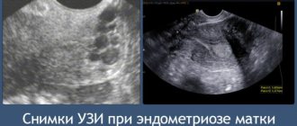
Endometriosis
Or pathological growth of the endometrium. A common disease that occurs in almost a third of all women, according to statistics from gynecologists. On what day of the cycle is it better to do a pelvic ultrasound to make such a diagnosis?
But if we are talking about endometrial hyperplasia, studies are carried out several times, the first time before menstruation, the second time immediately after it. During the first examination, the thickness and structure of the endometrium is determined, and during the second examination, the effectiveness of its detachment is determined.
Where can I get an ultrasound of the thyroid gland?
Free ultrasound examinations of the organ are practically not performed in public clinics. The reason for this is the unsuitability of the equipment they have for a detailed examination or the lack of software required for ultrasound. In extreme cases, the patient may receive a referral to a private clinic from his or her physician. It is possible to pay through an insurance company; in this case, it is enough to present your compulsory medical insurance policy.
Ultrasound of the thyroid gland is most often paid for and is performed in commercial clinics and diagnostic centers. A patient who wants to have an ultrasound scan can always sign up at a branch located closer to home, or get a recommendation from the attending physician at his clinic. The average price for the procedure is from 1,500 rubles for adults and from 1,000 rubles for children.
What does an ultrasound of the thyroid gland show?
The diagnosis depends on the results of the ultrasound examination. The procedure allows you to assess the size, structure and volume of the organ, as well as determine the presence of neoplasms. In combination with Doppler ultrasound - a color mapping method - you can see the condition of the walls of blood vessels and assess the speed of blood flow. The color flow diagram shows the movement of blood in a visually understandable form with color highlighting of the flows.
During pregnancy, a thyroid examination is carried out to prevent diseases caused by iodine deficiency and subsequent complications. Preventive ultrasound allows you to prevent abnormalities in the development of the unborn child before alarming symptoms are detected.
Using ultrasound diagnostics in women and men it is possible to identify:
- thyroid cyst;
- tumor formations;
- inflammatory processes;
- increased blood flow, indicating pathological changes in the organ;
- diffuse goiter;
- pathologies associated with hormonal imbalance;
- thyroid nodule.
Errors during ultrasound occur due to the poor sensitivity of the device and lack of experience on the part of the doctor. All data is recorded by a specialist, who may make errors in measurements and assessment of the boundaries of the organ. If the examination results appear unreliable, the patient can always request additional verification through tests.
Features of gynecological ultrasound examination
Normal study indicators deserve special attention:
- the reproductive organ is located in the central part of the pelvis, closer to the front wall;
- with longitudinal scanning, the uterus is pear-shaped, and with transverse scanning, it is ovoid;
- The echogenicity of the reproductive organ is average. Its internal structure is homogeneous;
- the ovary has an oval shape. The presence of echogenic inclusions inside the organ is noted;
- the condition of the endometrium directly depends on the phase of the cycle. Therefore, when drawing up a protocol, it is imperative to indicate in what period the examination was carried out.
Thanks to ultrasound, it is possible to identify various disorders in the female reproductive system. The main thing is to choose the right date to perform these manipulations. Only if this condition is met will accurate information be obtained.
The main advantage of ultrasound diagnostics is its safety and painlessness, which allows for an effective examination even during gestation. In some cases, pregnancy is one of the indications for scanning. Other common indications for screening include:
- violation of the frequency of menstruation;
- inflammatory processes;
- tumor formations;
- suspicion of infertility;
- monitoring complications when taking oral contraceptives or having an intrauterine device;
- pathologies of the urinary system.
Before the examination, it is necessary to calculate on what day of the cycle an ultrasound of the pelvic organs will provide objective results. If screening is planned in advance, then it is scheduled for 5-7 days from the start of menstruation. In the following days, the reproductive organs receive intensive blood supply, which changes the picture of their correct size and condition.
What is the essence of the procedure
An ultrasound of the pelvic organs is performed while lying on your back on a couch. The light in the office is dim for better visualization of the picture on the screen. The procedure varies slightly depending on the examination method chosen. Currently, there are 4 types of ultrasound examination of female reproductive organs:
- transabdominal,
- transvaginal,
- transrectal,
- intrauterine.
When performing transabdominal ultrasound, the ultrasound sensor is placed on the anterior abdominal wall. The study is carried out using a special gel that improves ultrasound performance. This method is actively used in the second and third trimesters of pregnancy, in women who are not sexually active, and also to identify gross gynecological pathology.
Transvaginal ultrasound
Transvaginal ultrasound obtains the necessary information using a sensor inserted into the vagina. This procedure is painless, but a woman may feel discomfort during it. To reduce discomfort, the patient is advised to relax. Transvaginal ultrasound is used to diagnose ectopic pregnancy, during short gestation, for endometrial polyps, endometriosis and other gynecological pathologies. There is no need to be afraid of transmission of infection, since a condom is used during the examination.
In difficult diagnostic situations, transrectal and intrauterine ultrasound are used.
Optimal time for the procedure
Gynecologists recommend that women regularly undergo pelvic ultrasound. This procedure will allow us to identify many diseases in the early stages, at a time when they can be successfully treated with conservative treatment.
It is necessary to carefully plan your visit to an ultrasound specialist. It is not recommended to carry out the procedure on any day of the cycle. Gynecologists advise doing an ultrasound 3 days after the end of menstrual flow. Already on days 7–10 the data will not be as reliable as possible.
Such a narrow gap is associated with changes in the uterus, ovaries and other pelvic organs during the female cycle. All these processes are controlled at the hormonal level by brain structures - the hypothalamus and pituitary gland. They influence the duration of the menstrual cycle and the duration of its individual parts.
It makes sense to perform an ultrasound of the pelvic organs only in the first half of the menstrual cycle, in its follicular phase. At this time, the following pathologies can be detected:
- hyperplasia;
- polyps;
- structural anomalies;
- myoma;
- cervical cancer
These conditions can only be diagnosed in the first days after the end of menstruation. During menstruation, the loose endometrium lining the inside of the uterus is shed. This layer grew and thickened throughout the previous cycle in order to serve as an attachment site for the fertilized egg. If a mature oocyte does not meet a viable sperm on its way, conception does not occur, and the egg leaves the uterine cavity along with menstrual blood. In parallel with this, unnecessary endometrium is eliminated.
Immediately after menstruation, the inner layer of the uterus has a minimum thickness. Therefore, even small deviations from the norm can be seen on ultrasound. It is for this reason that gynecologists advise carrying out diagnostics from 3 to 5 days after the end of menstruation.
There is another reason why it is worth checking your pelvic organs a few days after your period. Towards the middle of the cycle, follicles begin to grow in the ovaries. One will release a mature egg at the time of ovulation. At this time, ultrasound can detect compactions in the ovaries, the size of which reaches 3 centimeters in diameter.
Normally, this may be a corpus luteum cyst or a protruding part of the follicle itself. However, starting from the 10th day after menstruation, it is difficult to distinguish a typical lump from a pathological one.
At first after menstruation, normal formations will have a small diameter. Typically it ranges from 7 to 12 millimeters. These cysts are normal. They should not arouse suspicion.
The first half of the cycle is optimal for ultrasound
Ultrasound during menstruation
Can an ultrasound be done during menstruation or is it better to postpone it? Often women are faced with this choice, especially if the examination is prescribed by the attending physician, as planned, under a compulsory health insurance policy. In this case, you usually have to wait more than one day, or even a month, and it is not possible to calculate in advance what day the examination will fall on, especially if the cycle is irregular. And yet - what could this threaten?
The correct answer to the question will be - depending on which organs are being examined. When asked whether it is possible to do an abdominal ultrasound during menstruation, the doctor will definitely answer yes. Since menstrual bleeding will not interfere with the doctor. It’s another matter if you need to check the pelvic organs - the ovaries and uterus. In this case, the examination is prescribed in the first half of the menstrual cycle, but not earlier than the 5th day, since menstruation usually lasts 5-7 days, that is, during this period the uterus has already been completely cleared of the remnants of the old endometrium, and vaginal discharge is minimal, which does not create psychological discomfort for a woman if an ultrasound is performed using a vaginal sensor. It is during this period that it is recommended to conduct examinations for infertility (monitoring the maturation of follicles and the formation of a dominant one), uterine fibroids, etc.
However, there are many reasons when testing should be done immediately, no matter how severe the bleeding.
This need may arise in the following situations:
- suspected ectopic or intrauterine pregnancy (threat of miscarriage);
- severe pain;
- suspected of a large cyst in the ovary, etc.
There is no need to be ashamed of the discharge, even if you undergo a vaginal examination. Preparation for the examination is minimal. You need to wash yourself (for your own comfort) and grab a waterproof diaper (you can buy it at the pharmacy if the discharge is heavy. Your vagina doesn’t need a lot of “cleaning.” Do not douche under any circumstances, it is useless to do this, and moreover, it is dangerous if there is bleeding, since the cervix is slightly open at this time, the uterus is not protected from the penetration of infectious pathogens into it.
That is, the correct answer to the question - is it possible to do an ultrasound of the uterus during menstruation is “yes”, but only for emergency indications. If it concerns ordinary menstruation and there is no urgency in the study, it is better to wait at least until the 5th day of the cycle, and not go to the doctor on days 1-3 of monthly bleeding.
| 15.10.2019 09:11:00Lose Weight Easily with Low Carb Eating! Why reduce the proportion of carbohydrates in the diet, because this is a very important element for the body? To lose weight! A low-carbohydrate diet can help you lose weight significantly without requiring you to give up many foods like other diets. Let's find out what you need to do to lose weight without harm to your health! |
| 14.10.2019 18:43:00What can fat deposits tell you about your health? Almost every woman has a place on her body that she would like to change - to make it thin and toned. Usually these are problem areas - places of active fat deposition. But what do they mean for health? |
| 12.10.2019 09:45:008 answers to questions about proper weight loss What to do when severe hunger appears? Why do men lose weight easier than women? And for what reasons does weight come off quickly at the beginning of a diet, and then stop? Read answers to frequently asked questions about weight loss! |
| 11.10.2019 20:03:008 common causes of excess weight Lack of sports, vegetables and protein in the diet, love for fast food is guaranteed to lead to excess weight. However, the causes of extra pounds are not limited to gluttony and lack of physical activity. Let's find out what else we can get better from. |
| 11.10.2019 06:40:00How much fat should a woman have in her body? Women who want to lose weight without harming their health cannot ignore the question of their body fat percentage. Contrary to popular belief, fat deposits are not bad - they are vital for the body. |
| 10.10.2019 21:33:0038 healthy snack ideas Who doesn't love snacking between meals? But some foods then settle on the thighs as fat. It's better to replace them with healthy snacks. They will give you energy, but won't make you fat! |
All news