How to donate blood for a coagulogram
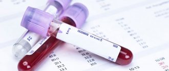
Blood is drawn from a vein in the elbow area. To avoid distortions and misinterpretation of the results, you need to prepare accordingly for the analysis.
Basic rules that are important for the patient to follow:
- 8-12 hours before the test you should not eat;
- the day before you should not overeat at night;
- alcohol, tea, juices and other drinks are excluded - you can only drink clean water;
- people with nicotine addiction should not smoke at least an hour before the test;
- It is important to exclude physical and mental stress 15 minutes before the analysis.
Important: if the patient is taking pharmacological anticoagulants, he must inform the doctor about this! If, during the process of collecting material for a blood coagulogram study, dizziness appears or a fainting state begins to develop, you should immediately notify health care workers about a change in your state of health.
What is a hemostasiogram
A hemostasiogram, or coagulogram, is a study of venous blood that helps to study the state of its coagulation system, or hemostasis. The fact is that in a woman’s body, thanks to special mechanisms, a certain blood consistency is always maintained. They are called that - the coagulation system (it is responsible for the absence of severe bleeding) and the anticoagulant system (it is responsible for the absence of blood clots). If there are no heart diseases, they work harmoniously, although sometimes only for the time being.
Hormonal imbalances, the appearance of an additional circulation (uteroplacental) and the preparation of the body for childbirth can lead to failures, the consequences of which will be disastrous. A hemostasiogram during pregnancy helps to identify them in a timely manner.
Depending on the number of indicators, it can be:
- basic;
- extended.
When is a blood coagulation test necessary?
Indications for analysis:
increased susceptibility to thrombosis;
- previous heart attacks and strokes;
- vascular pathologies;
- liver pathologies;
- pregnancy;
- preparation for surgery.
Blood is drawn with a sterile syringe or using a special vacuum system. The tourniquet is not applied to the arm. The puncture must be atraumatic to avoid data distortion due to large amounts of tissue thromboplastin entering the material. Two tubes are filled with blood, but only the second is used for research. The sterile tube contains an anticoagulant - sodium citrate.
What kind of analysis is a coagulogram?
A hemostasiogram, the same as a coagulogram, is a special comprehensive analysis that can be used to determine the level of blood clotting. Translated from Greek “coagulatio” means condensation and “gramma” means image.
The process of blood clotting is a protective mechanism that prevents blood loss. Bleeding is stopped by platelets - blood cells that are in a fluid state under normal conditions and form into plugs during various injuries: cuts, scratches, etc.
This comprehensive analysis is mandatory for patients in the following cases:
- before performing a planned or emergency operation - to determine the degree of risk of possible bleeding or blockage of blood vessels;
- for cardiac diseases - to control the degree of blood thinning, to prevent the rupture of a blood clot in the heart;
- in post-stroke condition - to reduce the risk of vascular accidents;
- during pregnancy - to determine the degree of safety of childbirth;
- for varicose veins to prevent thrombosis;
- when taking blood thinning medications (for example, Aspirin tablets), etc.
A coagulogram is also performed for a number of other diseases and conditions in which there is a possibility of complications in the form of severe bleeding.
Coagulogram indicators: interpretation
In a standard coagulogram analysis, a number of indicators are studied and evaluated together.
Clotting time is the time interval between the onset of bleeding and its cessation when a fibrin clot forms. Capillary blood clots in 0.5-5 minutes, and venous blood in 5-10. The duration of bleeding increases against the background of thrombocytopenia, hypovitaminosis C, hemophilia, liver pathologies and taking drugs from the group of indirect anticoagulants (including acetylsalicylic acid, Trental and Warfarin). The duration of coagulation is reduced after massive bleeding, and in women, even with the use of oral contraceptives.
PTI (prothrombin index) reflects the ratio of the duration of blood clotting in normal conditions to the clotting time of the subject. Reference values (variants of the norm) – from 97 to 100%. In pregnant women, the rate increases (up to 150% and above), which is not a pathology. PTI numbers make it possible to identify the presence or absence of liver pathologies. The index increases when taking hormonal contraceptives. An increase in values relative to the norm indicates the risk of developing thrombosis, and a decrease indicates the likelihood of bleeding.
Important: for the prothrombin index to be normal, the body requires a constant nutritional supply of vitamin K.
Thrombin time reflects the rate of conversion of fibrinogen to fibrin. The normal interval is 15-18 seconds. A shortening of the time interval most likely indicates an excess of fibrinogen, and its lengthening indicates a low concentration of this protein compound in the serum or severe functional liver failure due to hepatitis or cirrhosis.
Please note: regular monitoring of this blood coagulogram indicator is very important during heparin therapy!
APTT (activated partial thromboplastin time) is an indicator that reflects the duration of clot formation after CaCl2 (calcium chloride) is added to plasma. Normal values are within 30-40 seconds. Changes are noted when the remaining blood coagulogram parameters deviate within 30%. An extension of this time interval may indicate liver pathology or hypovitaminosis K.
AVR (activated recalcification time) in a healthy person ranges from 50 to 70 seconds. The indicator allows you to evaluate the course of one of the stages of coagulation. A decrease in AVR is a sign of thrombophilia, and prolongation is observed with thrombocytopenia, taking anticoagulants (heparin), serious injuries, extensive burns and the development of a state of shock. A low AVR indicates an increased risk of massive and life-threatening bleeding.
PRP (plasma recalcification time) is a coagulogram indicator that correlates with AVR and reflects the coagulation time of citrate serum after the addition of calcium salts. Normal time is 1 to 2 minutes. Its reduction indicates increased activity of hemostasis.
The fibrinogen content in the absence of pathologies varies from 2 to 4 g/l. This protein compound is synthesized in the liver and, under the influence of coagulation factors, is transformed into fibrin, the threads of which are the structural basis of blood clots.
If a blood coagulogram shows a significant decrease in the indicator, this may be a sign of the following pathologies:
- violation of hemostasis;
- severe liver damage;
- toxicosis during pregnancy;
- hypovitaminosis of group B and ascorbic acid deficiency.
The level falls during therapy with anticoagulants and anabolic steroids, as well as against the background of consumption of fish oil.
An increase in fibrinogen content is recorded in cases of hypothyroidism, large-area burns, acute circulatory disorders (strokes and heart attacks), acute infections, after operations, during hormone therapy, and in women - during pregnancy.
Fibrinogen B is not normally detected.
The fibrinogen concentration in a healthy person is 5.9-11.7 µmol/l. Its decrease is observed with liver problems, and its increase is observed with malignant neoplasms and hypofunction of the thyroid gland.
The RFMC indicator (soluble fibrin-monomer complexes) characterizes changes in the structure of the fibrin protein at the molecular level under the influence of coagulation factor II (thrombin) and plasmin. A value not exceeding 4 mg/100 ml is considered normal. The variability of the indicator is due to the same reasons as changes in fibrinogen concentration.
Please note: RFMK is a marker that allows you to take timely measures to prevent the development of DIC syndrome.
Fibrinolytic activity is an indicator of a coagulogram that reflects the ability of the patient’s blood to dissolve formed blood clots. A component of the body's anticoagulant system, fibrinolysin, is responsible for this function. With its high concentration, the rate of dissolution of blood clots increases, and accordingly, bleeding increases.
Thrombotest allows you to visually determine the volume of fibrinogen in the test material. The norm is thrombotest grade 4-5.
Plasma tolerance to heparin is a characteristic that reflects the time of formation of a fibrin clot after adding heparin to the test material. Reference value – from 7 to 15 minutes. The analysis reveals the level of thrombin in the blood. A decrease in the indicator most likely indicates liver damage. If the interval is less than 7 minutes, cardiovascular pathologies or the presence of malignant neoplasms can be suspected. Hypercoagulation is characteristic of late pregnancy (III trimester) and conditions after surgical interventions.
Blood clot retraction describes a decrease in the volume of the blood clot with complete separation from the plasma. Reference values are from 44 to 65%. An increase in values is observed in various forms of anemia (anemia), and a decrease is a consequence of thrombocytopenia and erythrocytosis.
Duke bleeding time is a separate test that examines capillary rather than venous blood. The fingertip is deeply pierced (4 mm) using a special lancet. The blood that comes out of the puncture is removed with special paper every 15-30 seconds (without contact with the skin). After each blotting, the time until the next drop appears is measured. The normal time for bleeding from small blood vessels to stop is one and a half to two minutes. This indicator is influenced, in particular, by the level of the mediator serotonin.
Indications for analysis in pregnant women, types of hemostasiograms
During pregnancy, a coagulogram is taken 3 times: after registration, at 22-24 weeks, and at 30-35 weeks. Indications for additional testing:
- infertility before real pregnancy;
- history of miscarriages;
- delayed fetal development;
- autoimmune diseases;
- gestosis;
- Rh conflict between mother and fetus;
- multiple pregnancy;
- long-term use of anticoagulant drugs;
- bleeding gums;
- frequent nosebleeds;
- liver diseases;
- varicose veins;
- fetoplacental insufficiency;
- smoking, drinking alcohol;
- the appearance of hematomas, bruises on the body for no apparent reason or after minor blows;
- deviation from the norm of coagulogram parameters during a scheduled test.
There are 2 types of hemostasiogram: simple, detailed. In the first, platelets, prothrombin, MHO, thrombin time, fibrinogen are determined. With a detailed coagulogram, antithrombin, D-dimer, and lupus anticoagulant are examined.
Important! A detailed analysis is prescribed when violations of indicators are detected in a simple coagulogram.
Blood coagulogram in children
Normal blood coagulogram values in children differ significantly from normal values in adult patients. Thus, in newborn babies, the normal level of fibrinogen ranges from 1.25 to 3.0 g/l.
Indications for studying a child’s coagulogram are:
- suspicion of hemophilia;
- diagnosis of pathologies of the hematopoietic system;
- upcoming surgery.
Normal values
The question of what indicators can be called adequate is quite complex. Because clinics use different counting methods. Even doctors are confused. When deciphering, they take into account the research technique that the laboratory uses.
In adults and children
The normal APTT in adults is within 25-35 seconds. Then everything depends on the specific medical center.
Deviations up or down by 1-2 seconds are possible. In women, the cause is most often due to altered hormonal levels. Deviations depend on the menstrual cycle and its phase.
Anything that goes beyond this permissible error almost completely indicates pathology. You need to understand it in detail.
There is also no significant difference in APTT rates in children. The rate of coagulation with chemical provocation will be approximately the same. All other rules, such as deviations and the need for further diagnostics, are also valid.
During pregnancy
Gestation is a serious test for the circulatory system. Changes begin in the second trimester. At first, as a rule, there are no deviations. Approximate APTT values can be presented as follows:
| Trimester | APTT rate in seconds |
| I | 25-35 |
| II | 30-33 |
| III | 31-36 |
An increase in the indicator indicates that the blood is clotting more slowly than expected. It's too runny.
During gestation, other tissue properties and laboratory levels also change. Including fibrin, platelet concentration. This is fine.
However, too strong changes in the coagulogram, even if there is no connection with pathologies, do not bode well. Need treatment.
Attention:
When the APTT is at the border of 35-36, and even more so moves beyond these limits, the expectant mother needs to be helped. Because with natural resolution of childbirth, there is a high probability of fatal uterine bleeding.
Of course, hematologists are limited in their choice of drugs. Pregnant women should not take many medications. The problem is solved by a group of specialists.
Blood coagulogram during pregnancy
Important: during pregnancy, a blood coagulation test is carried out at least three times (in each trimester).
During pregnancy, hemostasis parameters normally change, which is caused by significant hormonal changes in the female body, an increase in the total volume of circulating blood and the formation of an additional (uteroplacental) circulation.
In the first trimester, the clotting time, as a rule, increases significantly, and in the third, it is significantly shortened, thereby providing the woman with protection from possible blood loss during delivery. A blood coagulogram allows you to identify the threat of spontaneous abortion or premature birth due to the formation of blood clots. Disturbances in the coagulation system of a pregnant woman negatively affect the central nervous system of the unborn child.
Important: having blood coagulogram data and comparing them with the norm allows obstetricians to take adequate measures to prevent serious bleeding during delivery.
A mandatory blood coagulogram study is necessary if a woman has vascular diseases (in particular, varicose veins) or is diagnosed with liver failure. A blood coagulogram is also examined in cases of decreased immunity and negative Rh factor.
Reference values for individual blood coagulation parameters in pregnant women:
- thrombin time – 11-18 seconds;
- APTT – 17-20 sec.;
- fibrinogen – 6 g/l;
- prothrombin – 78-142%.
Important: deviation of the prothrombin level from normal values may indicate placental abruption!
Lotin Alexander, medical columnist
69, total, today
( 36 votes, average: 4.64 out of 5)
X-ray and fluorography of the lungs: indications, rules of conduct and interpretation of results
Fluorescein angiography of the retina: indications, procedure, study results
Related Posts
How to suspect deterioration of coagulation hemostasis
The first signs indicating poor blood clotting
Prolonged bleeding occurs with minor skin injuries or after injections. Normally, cuts or injections should not bleed for more than 3-5 minutes, but if there is pathology, this time can increase significantly. Sometimes such people have hemorrhages under the skin.
Another symptom that indicates this condition is prolonged nosebleeds that are difficult to stop. Women with hemocoagulation disorders may experience menorrhagia and metrorrhagia. Sometimes traces of blood may even be present in urine and feces.
If these symptoms appear, it is recommended to donate blood for a coagulogram. Research conducted by our specialists will help identify disorders of coagulation hemostasis. All analyzes are carried out using modern equipment and reagents.
Content:
- Features of hemostasis
- Importance of analysis to determine the level of coagulation
- Who is recommended to undergo testing?
- Blood clotting during pregnancy
- Features of the analysis
- Causes of development of blood clotting abnormalities
- Causes and consequences of increased blood clotting
- Main manifestations of poor clotting
Analysis for homocysteine: reasons for the increase in men and women →
Who is recommended to undergo testing?
A blood clotting test is recommended during pregnancy.
A test for blood protein coagulation should be carried out to prevent possible disruptions in the hemostasis biosystem in the following patients:
- Persons who have reached the age of forty.
- Pregnant women, since hemostasis during pregnancy can change significantly.
- During menopause.
- Anyone preparing for surgery.
- Patients who have been taking medications and products that thin the blood for a long time.
We have previously written about platelet levels during pregnancy and recommended bookmarking this article.
In children, the need to undergo these tests arises only in preparation for operations and if the physiology of the hemostatic system is impaired.
Basic tests, their normal values
The clotting test ( coagulogram ) is performed using three methods.
During the study, the period of time during which the clotting protein fibrin is produced is determined.
Sukharev's method
This method measures two parameters : the start time of coagulation and the end time. For research, capillary blood is taken from a finger and a test tube is filled with 30 ml of biomaterial. The sample begins to be shaken until a clot begins to appear.
Watch a video on this topic
Morawitz method
Drops of biomaterial are placed on special glass. Every 30 seconds the drop is checked with a glass rod.
The time of appearance of the fibrin thread is observed.
According to the Duque method
The simplest research method.
Ask your question to clinical laboratory diagnostics doctor Anna Poniaeva. Graduated from the Nizhny Novgorod Medical Academy (2007-2014) and Residency in Clinical Laboratory Diagnostics (2014-2016).Ask a question>>
The earlobe is pierced with a special needle. After 15 seconds, the puncture is blotted with paper.
The time the spot disappears is the time of coagulation.
According to Lee-White
1 ml of venous blood is placed in a test tube and kept at a temperature of 37 degrees. The formation of a clot is monitored by periodically tilting the test tube.
What can a hemostasiogram tell you?
We list the main parameters of the study that enable the doctor to determine the state of the coagulation and anticoagulation systems of the blood:
APTT is activated partial thromboplastic time or, in other words, an indicator of blood clotting time. The norm for this indicator is considered to be 24-35 seconds. A reduction in time indicates accelerated blood clotting, which in turn is an indicator of DIC syndrome. And, conversely, if the APTT is more than 35 seconds, the blood does not clot well and there is a risk of postpartum hemorrhage.
USEFUL INFORMATION: PGD file extension
Prothrombin or prothrombin index is an indicator that also reflects the quality of blood clotting. Values from 78% to 142% are considered normal. A decrease or increase in the percentage indicates slowed or accelerated blood clotting.
Antithrombin III is a blood protein that inhibits clotting processes. Its norm is considered to be values from 71% to 115%. A decrease in the level of antithrombin III entails the possibility of blood clots, and an increase leads to the risk of postpartum hemorrhage.
Thrombin time (TT) is the time of the last stage of blood clotting. Normally, this process takes from 11 to 18 seconds. A shorter time (up to 11 seconds) indicates signs of disseminated intravascular coagulation syndrome, a longer time warns of the possibility of postpartum hemorrhage.
D-dimeter is the most important indicator of a hemostasiogram, which allows you to find out about increased blood clotting. Normally, the D-dimeter value should be less than 248 ng/ml. A higher value indicates blood that is too thick, viscous, and prone to forming blood clots.
RCM is a marker of intravascular coagulation, the excess of which (more than 5.1 mg per 100 ml) indicates disseminated intravascular coagulation.
The parameters listed above are basic and can be supplemented by other indicators. It should also be noted that the result of a hemostasiogram can be affected not only by pregnancy, but also by other factors, for example: diseases of internal organs, lack of vitamins and microelements, taking certain medications, injuries and bruises. All this is taken into account by the doctor when deciphering the analysis results.
Normal in adults
It is worth recalling that in medical centers, test results and methods used may vary. Therefore, the verdict on the tests is made directly by the doctor.
| Name | Norm |
| GRP | 1-2 min. |
| TPH (Plasma Tolerance to Heparin) | 3-11 min. |
| Blood clot retraction | 40-65%. |
| D-dimer | up to 250 ng/ml |
| Lupus anticoagulant | No |
| Protein C | 70–130% |
Deviations from the norm
Accelerated clotting
The reason is blood thickening . Circulation through the vessels is disrupted, less oxygen is transferred, and the risk of thrombosis increases.
Accelerated clotting is called thrombophilia.
Prerequisites for thrombophilia:
- Infectious diseases.
- Diseases of the heart and blood vessels.
- Taking medications to increase coagulation.
- Dehydration.
- Unstable hormonal levels.
- Low physical activity.
- Risk of genetic pathologies.
- Metabolic disease.
- Autoimmune disorders.
- Pregnancy.
Thrombophilia affects overall health.
Symptoms are easy to notice:
- Spider veins, spider veins on the legs.
- Bruises from light blows.
- Headache.
- There is swelling and pain at the site of the blood clot.
The greatest danger is heart attack and stroke. An area of tissue dies due to blockage of blood vessels by blood clots. During a stroke, damage occurs to the brain and its important centers.
The result can be paralysis .
Delayed clotting
Slow clotting time causes bleeding that is difficult to stop . Any injury or bleeding wound causes anemia and large blood loss. The indicator may indicate oncology. The reasons are:
- liver diseases;
- anemia;
- allergies (the release of histamine increases the permeability of vascular walls);
- leukemia;
- tumors;
- heparin overdose;
- abuse of acetylsalicylic acid.
Rules for taking the analysis
An in vitro hemostasiogram is performed - the patient’s blood must be taken for analysis. Blood for a hemostasiogram is taken from a vein in the morning on an empty stomach. For women, it is advisable to take this test not during menstruation, since blood loss will lead to a physiological increase in blood clotting.
The results of a hemostasiogram are usually ready 1.5 - 2 hours after the test. If you have been prescribed a hemostasiogram, deciphering the analysis by parameters may be difficult for a non-specialist.
USEFUL INFORMATION: The most interesting things about a woman’s life. How to take plantain seeds to get pregnant. Sage, hogweed or plantain, what folk remedies to take to get pregnant Contraindications for treatment with plantain
You can roughly determine your performance. Find the form with the heading coagulogram or hemostasiogram. The norm or reference values are usually indicated in brackets or in a separate column. Compare your performance with the norm. However, only a doctor can make the final interpretation of the analysis.
Why is coagulation disrupted and what are its consequences?
The norm of indicators of the hemostasis system can be exceeded for the following pathological reasons:
- An increase in the level of platelets due to their excessive production by the bone marrow;
- Infectious-toxic and septic diseases;
- Any intoxication that occurs against the background of severe pathology of internal organs;
- Widespread atherosclerotic vascular lesions;
- Congenital and genetic abnormalities of anticoagulant system factors;
- Artificial heart valves and vascular prostheses;
- Autoimmune diseases;
- Endocrine pathology with metabolic disorders in the body;
- Blood stagnation due to heart failure and physical inactivity;
- The first phase of DIC syndrome.
Indicators of clotting tests may be lower than the generally accepted norm. The following reasons will lead to this:
- Thrombocytopenia;
- Hemophilia and other hereditary defects of clotting factors;
- Hemolytic anemia;
- Leukemia;
- Decompensated liver failure with cirrhosis;
- Insufficient amount of calcium and vitamin K in the body;
- Overdose and treatment with anticoagulants (heparin, warfarin, acetylsalicylic acid preparations);
- The last phase of DIC syndrome.
Important to remember! Increased blood clotting is dangerous due to the accelerated formation of blood clots in the vessels, which can cause thrombosis of the arteries and veins of internal organs, limbs and brain. Decreased coagulability is fraught with an increased risk of massive and profuse bleeding!
The study and correct interpretation of blood clotting test data allows you to determine all the risks regarding the potential for vascular diseases, as well as monitor the effectiveness of the blood thinners used and their dosage.


