Symptoms of a peptic ulcer can vary slightly depending on the location of the ulcer, the degree of inflammation, and the presence of a partial block of the duodenum.
Unfortunately, although new acid-reducing medications are coming onto the market to help relieve symptoms, these medications do not treat the underlying cause of the condition and come with their own side effects and problems. The more common types of medications used to treat ulcers can ultimately worsen the stomach lining and overall health. The purpose of these medications is to significantly reduce acidity in the stomach. However, this acid is a valuable chemical factor in digestion, and not the culprit of ulcers.
Clinical blood test
A blood test is one of the mandatory tests prescribed to a patient if the development of a stomach ulcer is suspected. If the pyloric part of the stomach becomes the location of the ulcer, then an increase in the level of red blood cells and hemoglobin levels is observed. The leukocyte formula remains unchanged in uncomplicated ulcers. In this case, slight lymphocytosis may be observed.
If the disease is present, the results will have the following deviations. An ulcer that occurs without complications does not cause significant changes in the composition of the blood. With the development of pyloric stenosis, there is a change in acid-base balance, a decrease in the amount of electrolytes and total protein.
A sign of a perforated ulcer is an increase in ALT, gamma globulins, and bilirubin. An increase in urea levels is recorded with the development of peritonitis. Progressive anemia indicates degeneration of the ulcer (malignancy). Additionally, there is a complete absence of hydrochloric acid and pepsin in the gastric juice and the presence of lactic acid and Boas-Osler fermentation rods.
Find out if you are prone to stomach ulcers
Although stomach ulcers can develop for many reasons and in almost anyone, the risk is increased in the following groups of people:
- those who are infected with or more susceptible to the bacterium Helicobacter pylori, such as people with low stomach acid;
- those who regularly take non-steroidal anti-inflammatory drugs (NSAIDs) such as ibuprofen, aspirin or naproxen;
- people with a family history of stomach ulcers;
- those who regularly drink alcohol;
- people with liver, kidney or lung diseases;
- people over 50 years old;
- have had or currently have a digestive disease, such as Crohn's disease.
Radiology
Stomach ulcers can be identified using X-ray examination. The procedure may be prescribed as an alternative to FGDS. The accuracy of the method reaches 80%. There are direct radiological signs of ulcerative pathology. The main phenomenon is considered to be a “niche”. By niche, doctors understand a visually limited protrusion on the silhouette of the stomach filled with contrast.
If a niche is determined on the anterior/posterior wall of the organ, then it looks like a spot on the relief background of the mucosa. It is surrounded by a pronounced marginal shaft, caused by inflammation and swelling. The size of the niche is variable. Depends on two factors:
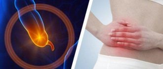
How to check the stomach without swallowing a tube?
- degree of damage to the gastric walls;
- the size of the inflammatory shaft.
The inflammatory shaft can visually lengthen the true size of the niche, as well as completely close it. The cavity itself sometimes turns out to be completely filled with food debris, mucus, and blood clots. That is why it can remain radiologically invisible.
In case of chronic ulcers prone to recurrence, as well as their callous forms, a modification of the relief of the gastric mucosa can be determined in the form of convergence of folds to a niche (scar changes). The combined detection of an inflammatory shaft and cicatricial changes indicate the presence of gastric ulcer (GUD).
Niches come in different sizes - small, medium, large. The acute period of ulcerative pathology is accompanied by the formation of a small notch. The most common size is 5 x 8 mm. Small niches, not exceeding the diameter of a pea, are localized in the duodenal bulb.
Most often, medium-sized niches are identified: 0.5...0.8 x 1.0...1.2 cm.
Large ulcerative niches, the depth and diameter of which exceed several centimeters, accompany chronic forms of the disease with pronounced symptoms. Formations of this size, in most cases, are penetrating ulcers. The penetrating niche is a niche that extends beyond the stomach into an organ located in close proximity.
It can be three-layered - barium, liquid, air - and two-layered - barium and an air layer. The presence of air bubbles on the x-ray is a clear sign of penetration. A niche of ulcerative origin almost always has smooth walls. If there are defects, we can talk about bleeding or the beginning of the process of degeneration.
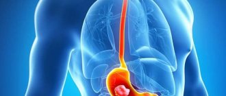
The ulcer can be localized anywhere in the organ
Indirect signs of the presence of ulcers, for which the patient is prescribed an X-ray examination, are recognized as:
- dysphagia;
- stomach discomfort;
- bouts of vomiting;
- sudden weight loss;
- pain syndrome;
- the presence of a palpable compaction in the abdomen;
- determination of occult blood;
- anemic conditions;
- violation of secretory function.
For a more accurate diagnosis of stomach ulcers, the double contrast technique is used - the use of a contrast agent and air. After studying the motor function of the stomach and compensatory capabilities, the doctor prescribes treatment appropriate to the condition.
General condition of a person with peptic ulcer disease
With an ulcer, as with other diseases, a person feels a general physical malaise. It is worth noting the frequency of dizziness - when the disease occurs, blood pressure decreases. Pale skin, black circles under the eyes, poor appetite are the external symptoms of ulcerative lesions. An increase in temperature is observed more often than an increase in heart rate. Poor sleep, feeling unwell during the day, and lack of mood indicate the progression of the disease.
When taste preferences change, a sign cannot be missed. An aversion to sour foods indicates an increase in the acidity of the gastric environment. Aggressive gastric juice eats away the mucous membrane, an ulcer is not far off. Weight loss associated with exacerbation of the disease is observed in frequent cases and becomes an alarming symptom. Anorexia (complete loss of appetite) may occur with severe inflammation.
FGDS
Fibrogastroduodenoscopy (FGDS) is one of the advanced techniques that allows you to recognize peptic ulcers of the stomach and intestines. The procedure is an endoscopic method of examining the stomach and duodenum using a fiberscope.
FGDS helps to obtain a detailed image of the gastric mucosa. The device allows you to assess the condition of the organ: determine the location of ulcers, see the current size of the formation, and obtain tissue for biopsy. The FGDS technique is used to evaluate the ongoing treatment therapy. In approximately 90% of all cases, the study helps confirm the diagnosis and identify concomitant pathologies.
The procedure is generally safe and has virtually no contraindications. But examination of the stomach with a fiberscope will need to be abandoned if the following pathological conditions and diseases are present:
- heart failure in the stage of decompensation;
- acute period of stroke and post-stroke condition;
- myocardial infarction;
- stenosis and burns of the esophagus;
- aortic aneurysm;
- terminal stages of chronic diseases.
General symptoms
In the absence of characteristic and indirect signs, it is almost impossible to identify the pathology at home, since a person’s condition with a peptic ulcer can be relatively satisfactory for a long time. An ulcer often manifests itself with a number of general symptoms that may indicate the development of diseases of a completely different etiology, however, a person should pay attention to them. These symptoms include:
- increased sweating;
- cyanosis and dry lips;
- insomnia;
- various central nervous system disorders;
- bitterness in the mouth;
- prostration;
- constipation
If all or some of the above signs are detected, a person should visit a doctor, and this does not have to be a gastroenterologist. Making a diagnosis may require examination by several specialists at once, since the clinical picture, consisting of general symptoms, does not make it possible to determine a specific disease without carrying out appropriate diagnostic procedures. In this regard, a person should first make an appointment with a therapist, who will issue a referral for tests and tell you which specialists need to be visited.
Biopsy
Biopsy is used as a method of differential diagnosis. Examination of a biopsy allows one to distinguish a classic ulcerative defect from a cancerous lesion. Peptic ulcer of the stomach and duodenum is initially regarded as a potential cancer. But the initial stages of cancer can only be determined by performing a histological examination of living stomach tissue.
The biomaterial is obtained during an endoscopy procedure. Deep ulcers may outwardly resemble cancerous areas, but in reality they are not. To confirm the diagnosis, a multiple biopsy is performed, i.e., the physician takes the marginal and bottom tissues of each ulcerative element. Be sure to take material from scar tissue that forms at the site of the healed injury. In addition, it is desirable to obtain tissue images from potentially hazardous areas.
For reliable diagnosis, it is necessary to study at least 6 samples obtained from different damage zones. This is quite enough to make an accurate diagnosis. If it was not possible to obtain samples of all affected areas of the mucosa, then the patient is prescribed a repeat biopsy.
Attention to your body
To understand whether you have a stomach ulcer, you should monitor your physical condition for some time, listen to the discomfort that arises, determine its location and causes, and clarify the signs of the disease. This will allow you to consult a doctor in a timely manner and avoid long and painful treatment.
Remember when the most severe, aching, pressing, but not cramping pain appears. Pay special attention to pain that occurs half an hour after eating or at night. Often with such persistent pain, relief comes from vomiting, which occurs without a preceding feeling of nausea.
If you have the symptoms described above, then be sure to determine the location of the pain. With a stomach ulcer, it is felt on the right at the level of the lower rib or on the top of the abdomen in the area of the xiphoid process. If the pain lasts for such a long time, you can lie on your stomach, then it will gradually go away. The seasonality of pain, which is characterized by spring and autumn exacerbation, also makes it possible to determine a stomach ulcer.
Pain is the first and main sign that helps to recognize a stomach ulcer - the occurrence of severe pain in or near the epigastric region, sometimes it can be acute. Features of pain:
- pain often occurs on an empty stomach in the morning and at night;
- pain may radiate to the back and lower back;
- often increased pain occurs after eating, especially if the diet contains a large amount of sour, spicy, salty and coarse foods;
- the patient may experience a clear rhythmicity in the occurrence and frequency of pain;
- the intensity of pain may depend on the extent of the lesion, its location and the severity of the complication of this disease;
- Sometimes the pain can be intense, causing a loss of appetite.
Ulcer pain tends to get worse after eating. But still, you shouldn’t make a diagnosis yourself; it’s better to see a doctor.
Don't forget to measure your temperature, which should not be elevated. Normal human body temperature ranges from 36.4 to 36.7 degrees Celsius. Determine your body weight, which is significantly reduced in case of gastric ulcer.
Think about possible stomach problems if you suffer from heartburn or sour belching (but not rotten), as well as frequent stool retention. Most patients with an open ulcerative process have constipation. The causes of constipation in acute ulcers are the following factors:
- Large intestine spasm.
- The gentle nature of the diet, excluding fiber.
- Decreased patient physical activity.
- Taking antacid medications.
These symptoms are often accompanied by an exorbitant feeling of hunger and increased appetite, at the same time with which an intolerance to fatty or spicy foods occurs. There is no disgust or complete refusal of food even after vomiting. If sudden vomiting with pain suddenly occurs, this is another sign that the patient has a stomach ulcer. In this condition, the stomach is freed from acidic contents and the pain becomes much less. Sometimes patients induce vomiting on their own to make the pain much less. At the same time, with ulcerative lesions, damage to the structure of blood vessels is observed. For this reason, the vomit may contain blood clots.
Darkening of stool . Blood clots from stomach ulcers can enter the intestinal cavity. As a result, the color of the stool changes; it becomes darker. Moreover, if intense bleeding occurs, then this symptom will indicate the presence of a complicated gastric ulcer.
Reducing body weight . A decrease in body weight with peptic ulcer may occur due to the fact that the patient stops eating food, which causes severe pain. As a result, weight begins to decrease. But in cases where the patient excluded from his menu spicy, highly salty, sour foods and foods that provoke inflammatory processes in the mucous layer, and replaced them with more gentle foods, weight loss is not observed. It is worth noting that sometimes the opposite situation may occur - the patient has an increased feeling of hunger, but due to intolerance to too fatty, spicy, sour foods, he begins to eat other foods in larger quantities.
The appearance of excessive salivation . In almost a quarter of cases, gastric ulcers do not cause pain. Typically, in these situations, the presence of an ulcer is determined by the occurrence of increased salivation. If they are secreted constantly and in large quantities, then it is necessary to conduct an endoscopic analysis, which will help examine the condition of the stomach cavity.
Indirect signs can also help identify a stomach ulcer: fatigue, increased sweating, dizziness and severe weakness, an unpleasant feeling of bitterness in the mouth in the morning, psychological discomfort (nervous excitability or severe depression, depression).
If you have the above symptoms, to confirm the diagnosis, consult your doctor for a referral for fibrogastroscopy and analysis of gastric juice. An endoscopic examination will not only confirm the presence of an ulcerative defect in the gastric mucosa, but also determine its location.
Video capsule endoscopy
In this way, the presence of an ulcer is checked for people who have contraindications to endoscopy or FGSD or for patients who are afraid of such techniques.
To detect the wound surface, the patient needs to swallow a disposable endoscopic capsule, which is small in size. It is made of biological material, completely safe for the human body, is small in size and weighs 4 grams. The capsule is equipped with several light sources, a radio transmitter and a small camera.
Video capsule endoscopy is performed in the morning on an empty stomach. The person swallows the capsule and pictures are taken as it passes through the stomach. During this time, the patient can be in the hospital or go about his business. The only condition is that any physical activity should be completely excluded.
8 hours after swallowing the capsule, the doctor views all the data that the mini-camera transmits to the computer. Further, the miniature camera is removed from the body naturally, without causing negative effects to the patient.
Despite all its effectiveness and safety, this method has a number of contraindications, these include:
- if a person has a pacemaker installed,
- with intestinal obstruction,
- during the period of bearing a child,
- if the patient is under 12 years old.
It is easiest to identify an ulcer using this technique. It is informative, completely safe and convenient. However, the cost of this type of examination is quite high.
Other signs
In addition to the above signs, there are other specific symptoms that indicate the presence of this defect. These include the following:
- increased feeling of weakness,
- excessive sweating,
- fast fatiguability,
- the appearance of a feeling of lethargy,
- in rare cases, severe excitability is possible,
- insomnia.
Important: If you suspect an ulcer, you must seek qualified help as soon as possible. The doctor will carry out all the necessary therapeutic measures to make an accurate diagnosis.
In addition, the patient is often bothered by a feeling of bitterness, especially in the morning. This sign indicates that a certain gastric disorder is progressing.
In what cases is diagnostics necessary?
Modern methods for diagnosing gastrointestinal ulcers allow gastroenterologists to identify the disease in the early stages and begin therapy. The patient can avoid surgery. Experts name the following risk factors:
- heredity;
- stress;
- improper diet;
- other pathologies of the digestive system;
- alcohol abuse;
- reduced immunity.
All people with such factors should undergo regular examinations. A diagnosed disease must be treated. In the early stages, antibiotic therapy is effective. These drugs destroy Helicobacter colonies in the stomach. Special gastroprotectors will help restore the mucous membranes. To prevent the development of peptic ulcers, you need to adhere to certain nutritional rules and strengthen your immune system.
Weight loss
Another sign that allows you to recognize gastrointestinal pathology is weight loss. With such an illness, the patient reduces the number of meals due to severe pain, which leads to weight loss.
Important: In some cases, a completely different picture is observed. A patient suffering from the disease in question experiences a constant feeling of hunger. And since spicy, sour and fatty foods become intolerable, a person begins to eat other foods in excessive quantities.
Moreover, if the patient eliminates the consumption of fatty, salty and spicy foods, which cause inflammation of the mucous membrane, and switches to a more gentle menu, there will be no weight loss.
X-ray examination
If it is impossible to perform FGDS, an X-ray examination is performed. This method allows you to diagnose gastrointestinal tract defects. The accuracy of this method is 80%. Radiologists describe the radiographic signs of an ulcer as a “niche”, opposite which there are spasmodic muscles (“pointing finger”). X-rays are most often indicated for patients who have the following symptoms:
- dysphagia;
- stomach discomfort;
- vomit;
- sudden weight loss;
- pain;
- lumps in the abdomen;
- detection of occult blood;
- anemia;
- violation of secretory function.
The main methods of X-ray examination are distinguished: traditional, emergency, two-phase, verification and double contrast. Gastric ulcers are more often detected on x-rays using the double contrast method (using a contrast agent and air). Doctors study the motor function of the gastrointestinal tract and the compensatory capacity of the organ in order to provide adequate therapy in the future.
Ultrasound
Depending on the correct preparation for the examination, it depends whether an ultrasound will show a stomach ulcer or not. It is preferable to perform ultrasound using a device that operates in real time, as it produces a high-quality image of the organ. The examination is carried out on an empty stomach in the morning. If there is a stomach ulcer, ultrasound helps to identify places of greatest pain (using palpation under the control of the device), determine the tone and peristalsis, and the evacuation ability of the stomach. During the examination, special attention is paid to the condition of the stomach walls. In some cases, ultrasound shows a stomach ulcer because the walls of the entire organ and its contours are assessed.
Carrying out radiography
An ulcer can be determined using radiography. This method of identifying pathology is carried out using several techniques. And manipulation is indicated for the following conditions:
- anemia,
- the occurrence of severe pain,
- if there is discomfort in the gastric cavity,
- if the patient is experiencing vomiting,
- the occurrence of dysphagia,
- if a lump is detected in the abdominal area,
- weight loss.
The accuracy of radiography data ranges at 80%. As mentioned above, this manipulation is carried out using several techniques. Contrast can be double, two-phase, calibration and emergency. In this case, the most informative is double.
Tests for the presence of Helicobacter pylori
There are invasive and non-invasive tests that help identify the bacterium that causes ulcers - Helicobacter pylori. Invasive tests include tests that examine pieces of tissue that are taken during an FGDS or a blood test. Examination of saliva, urine, feces, exhaled air - non-invasive tests. Using these tests, peptic ulcers are diagnosed and their healing is monitored. There are several non-invasive tests.
Reduced breathing
The principle of the test is based on the fact that the Helicobacter bacterium is capable of decomposing urea, resulting in ammonia and CO2. On an empty stomach, the patient takes 2 samples of exhaled air, then is given a light breakfast and a special substrate that contains a solution of urea with labeled carbon. After taking the test substrate, exhaled air samples are taken every 15 minutes for an hour and the amount of labeled carbon is determined using special equipment. Using this test, the presence of bacteria is reliably determined in 98% of cases. It should be noted that the method is absolutely safe. The test is not performed if the stomach ulcer is bleeding.
Immunological tests
Used when it is not possible to conduct a breath test. The study is based on the fact that the Helicobacter bacterium causes immunological reactions by producing specific antibodies. They are found in saliva, urine, feces, and blood.
Quick stripped down
To conduct the study, a piece of tissue (biopsy) is required, which is tested for the presence of bacteria using a special indicator containing urea and a reagent. The intensity of the reaction will depend on the number of bacteria in the biopsy specimen.
Bacteriological test
This study is the most accurate and gives 100% results. The test allows you to isolate a pure culture and study its properties and sensitivity to drugs. To carry it out, special equipment and expensive reagents are required. The biopsy examination itself takes up to 7 days. For primary diagnosis, shortened respiratory or immunological tests are used. If the picture is clear, special express tests with immunological techniques are used to clarify. For serious studies, shortened respiratory, shortened rapid and bacteriological tests are used.