Lack of awareness
Not every actively practicing physician will immediately agree that in 70% of gastroenterological patients with symptoms of “gastric dyspepsia” or “functional dyspepsia,” no significant pathological signs can be found during endoscopic examination of the upper gastrointestinal tract. Nevertheless, this is an established fact, albeit from Western sources. Hand on heart, we could give us a similar figure, but the logic is brought down by the fact that almost all such patients have dyspepsia, but in the absence of significant visual changes, we prescribe antiulcer treatment, and even eradication therapy (if there is a hint for H. pylori
) .
However, somehow I can’t bear to accept the tactics of blanket treatment for ulcers, when there is not even gastritis...
What is the problem and ways to solve the problem at least in some cases?
Firstly, it is necessary to remember that there is a whole list of diseases that fall under the category of “functional disorders”. With them, no matter how hard we try, we cannot identify any morphological signs that could provide grounds for a specific diagnosis such as “peptic ulcer” or something like that.
However, there is a “second thing”. I dare to assure my colleagues that not all, but many patients who complain of very painful “gastric” symptoms, have the latter caused by gastroptosis. Not recognizing the first or second means dooming a person to a waste of time, money, health, visiting doctors and research. Quite often we see patients who, indeed, losing a lot of strength and resources, walk in circles without any prospect of improvement, because therapeutic measures can have serious success only when the true cause of suffering is accurately established.
Apparently, in the West they also don’t think too much about gastroptosis, and all patients of this kind end up in one cohort, which is called “functional dyspepsia.”
Prevention of displacement of internal organs
The pathology in question can be corrected if all recommendations are followed, bed rest is avoided, and an active lifestyle is not associated with excessive stress.
The main preventive measures are:
- moderate sports training already in childhood - morning exercises, swimming, outdoor games;
- expectant mothers should consider including abdominal exercises in their daily schedule, and during pregnancy do not refuse to wear a bandage;
- Hiking is good in the fresh air for any age;
- adherence to diet; it is necessary to exclude overeating - eat 5-6 times a day, but little by little;
- visiting a massage therapist;
- try to lead a measured lifestyle: eat on time, go to bed on time, do not overwork.
Remember the dangers of increased loads - consider your capabilities!
Concept
According to what is written in the Encyclopedic Dictionary of Medical Terms, “Gastroptosis (gastro- + Greek ptosis, fall, prolapse) is a pathological downward displacement of the stomach; develops, for example, as a result of a sharp weakening of the muscles of the abdominal wall, with significant weight loss, after extraction of ascitic fluid.”
Splanchnoptosis is often associated with gastroptosis (see below), which, in general, is quite understandable, because, in particular, the large intestine is fixed to the stomach by the gastrocolic ligament (lig. gastrocolicum), its fibers connect the greater curvature with the transverse colon gut. The stomach descends, and the transverse colon also descends.
Diagnosis of the disease
If you suspect a pathology, you should contact a gastroenterologist.
- when interviewing and examining the patient, the doctor will reveal weight loss, flabbiness of the abdomen, and sagging; displacement of the stomach can be determined even by palpation;
- EGD will help to see a change in the shape of the stomach, its lengthening, decreased motility, and establish increased acidity of the environment;
- X-rays with a contrast solution will clarify the diagnosis: the contrast agent accumulates at the bottom of the stomach and lingers there for a long time, without moving towards the intestines;
- Ultrasound is prescribed if an abnormal condition is suspected in other abdominal organs at the same time.
Epidemiology, prevalence
With all this, it is pointless to judge the frequency and prevalence of gastroptosis based on literary data, since, firstly, gastroptosis as a cause of human suffering is rarely considered at all: few colleagues come up with the idea of identifying gastric prolapse. If not identified, it means there is no problem. In short, ignorance is the main obstacle.
On the other hand, not all gastroptoses cause symptoms. Naturally, no one will set themselves the task of identifying a disease that, in fact, does not exist. Yes, and there is no particular need to identify asymptomatic gastroptosis.
The third aspect is quite common: there are many patients with pronounced prolapse of the stomach, who have symptoms and happily don’t care about the discomfort - maybe it will go away on its own.
Relevance of the problem
The relevance of the problem can only be judged on the basis of personal experience. And so it turns out: if you show one of your colleagues a patient with pronounced gastroptosis, you see two types of reactions: either true interest and surprise that this is possible; or skepticism and denial of significance. Let’s discard the last option as, to put it mildly, shallow and counterproductive.
Counterproductive, also because the doctor loses the chance to see the bright emotions of the patient, who, looking at her own x-rays, expresses genuine amazement. The effect is enhanced if you first show her what the normal topography of the organ looks like using a picture in a book or the Internet.
Emotions are emotions, but the main thing, of course, is the medical aspects - the ability to identify the anatomical and physiological features to explain the symptoms of the patient. Sometimes, after looking at the picture, a person himself comes to the solution to a clinical puzzle, for example, one patient exclaimed: “So that’s why after eating my lower abdomen becomes enlarged, as if from a baby!” Indeed, in her case, the contrasted stomach – without the slightest exaggeration – “lay on the uterus” with its lower pole.
In order to facilitate the demonstration of visual material, you will have to slightly change the usual order of presentation of the clinical topic, starting from the middle.
Establishment of pathology
Gastroptosis is diagnosed by x-ray, although fluoroscopy is also suitable for its determination. The images obtained as a result of one of the appropriate procedures will allow us to determine the placement of the stomach. If the position of the organ is significantly shifted towards the conventional line (or beyond it), an appropriate conclusion is drawn up.
However, before the x-ray, the doctor will be able to preliminarily determine the presence of pathology at the first, superficial examination. This can be done through normal palpation.
The disease can also be caused by the presence of other pathologies. Therefore, sometimes, if there are additional symptoms, the doctor prescribes other procedures aimed at examining the patient.
X-ray diagnostics
To identify gastroptosis, it is enough to conduct a relatively simple study - an x-ray of the abdomen with contrasting the stomach with barium sulfate. If the clinical diagnosis - gastroptosis - is confirmed by an x-ray picture, it is advisable to repeat the image after 2 and 6 hours (barium passage through the intestines) in order to roughly determine the topography of the small and large intestines.
To begin with, the layout of the stomach is normal. This is what the topography of the stomach looks like in relation to the spine and lower ribs (the picture is taken from the Internet, slightly modified):
Rice. 1. Normally, the lower border of the stomach is located at the level of the body of the 2nd or 3rd lumbar vertebra. The transition of the stomach to the duodenum is slightly higher.
In this aspect, it is not so important from what level the organ begins. The main thing is where its lower pole is.
With the development of gastroptosis, the stomach lengthens and goes down, but the duodenum in any case remains in place. Due to this, the anatomical relationship of organs changes - not far from the development of functional disorders. In Figure 2 – gastroptosis of the II-III degree (closer to the III degree).
Rice. 2. The lower pole of the stomach is clearly not at the level of the upper lumbar vertebrae. Here it is below the level of the iliac crests and even below the L5-S1 articulation (gradation according to the degree of severity of gastroptosis occurs along the line connecting the tops of the iliac crests: below the line is the third degree, on the line is the second, above is the first).
Instrumental diagnosis of gastroptosis is the most impressive section in this topic. Indeed, neither the clinic, the clinic for gastritis, peptic ulcer disease or functional dyspepsia, is anything too striking; Neither drug treatment - pills cannot compensate for anatomical disorders. All that remains is to enjoy the process of making a diagnosis, especially since the visuals are impressive.
So, gastroptosis degree III:
Rice. 3. Gastroptosis stage III. The shadow of the stomach is much lower than the ridges, descends “below the coccyx” (in order not to be misled by the topography, one should take into account the peculiarity of the X-ray rays: leaving the tube, they diverge like a fan, so a certain optical illusion arises, as if the stomach “goes” into the pelvis , but in reality this does not happen). Nevertheless, no one here will argue that the lower border of the stomach is located extremely low.
Another example of advanced gastroptosis. It is difficult to hope that such prolapse and relaxation of the stomach will remain asymptomatic:
Rice. 4. Gastroptosis stage III. (the same case “... as if from a child!”).
Accordingly, the entire small intestine is located very low - the picture was taken shortly after filling the stomach with contrast:
Rice. 5. Gastroptosis stage III. The contrast began to move into the small intestine (this happens almost immediately after taking a dose of barium sulfate, if the contrast is not given on an empty stomach, but while digestion is ongoing). It can be seen that the small intestine is greatly prolapsed.
This means that the mesentery of the small intestine is stretched to the maximum, and stretched, but does not compress the initial sections, and there is no hint of intestinal obstruction, although it could be expected (as happens when the small intestine is full after a long fast).
It is also important that the transverse colon is connected to the stomach by the gastrocolic ligament, therefore, as the stomach descends, the large intestine also goes down. This can be clearly seen in the following picture:
Rice. 6. Gastroptosis stage III. Photo taken 4-5 hours after taking barium sulfate. The shadow of the stomach is much lower than the ridges - its contours can only be guessed. It is important that both the small intestine (a pile of contrast in the middle) and the entire large intestine (with its haustra) are spread out along the edge of the entrance to the small pelvis.
Additional illustration of low position of the small intestine in a patient with gastroptosis:
Rice. 7. Gastroptosis stage III. Pictures taken immediately and about an hour after taking barium sulfate. The shadow of the stomach is below the iliac crests. It can be seen that the partially contrasted small intestine, like the stomach, is located very low.
It must be emphasized that the place of transition of the stomach into the duodenum (more precisely, the beginning of the duodenum) is located mesoperitoneally, has limited mobility and moves only in lateral directions, but not up and down (!).
Rice. 8. Gastroptosis stage III. The transition of the stomach contents into the duodenum is clearly visible. The place where the stomach enters the intestine is fixed. The stomach has to lift each portion of the contents and pass it as if “over the side”.
Another (more visual) demonstration of the degree of prolapse of the transverse colon, with the latter clearly elongated:
Rice. 9. Gastroptosis stage III. Photo taken a few hours after taking barium sulfate. There is a residual amount of contrast in the stomach. Among other things, severe scoliosis of the lumbar spine cannot but attract attention.
Treatment of gastroptosis
Stages 1, 2 and 3 gastric prolapse cannot be corrected surgically. It is impossible to surgically influence the cause of the disease - weakening of the muscle corset. The effectiveness of therapeutic procedures depends on the behavior and lifestyle of the person himself.
Nutrition
- When diagnosing this pathology, it is necessary to often eat in small portions so as not to further stretch the stomach;
- A large amount of fiber is introduced into the diet: whole grain products, vegetable dishes;
- After eating, rest is recommended: lie down for one and a half to two hours;
- Food should be satisfying and easily digestible; A nutritionist will tell you how to restore normal body weight and restore the balance of vitamins and microelements in the body.
Physical training
- In order to train the diaphragm and adjacent muscles, breathing exercises are shown: before going to bed, while lying in bed, breathe deeply - inhale as deeply as possible and exhale the same.
- Therapeutic gymnastics is first performed under the guidance of a specialist, then you can move on to independent training. To help your stomach move into the correct position, do the exercises while lying on your back with your legs elevated. Gymnastics aims to strengthen the abdominal and core muscles. They keep the internal organs at the required level, so physical therapy should be done daily in sufficient quantities to eliminate the possibility of the disease returning.
- Please note: heavy physical activity and carrying bags are strictly contraindicated!
Medicines
To restore the normal functioning of the gastrointestinal tract, drugs that increase appetite, herbal laxatives, gastric juice substitutes, and anabolic hormones are prescribed.
Physiotherapy
- Professional massage plays a significant role in recovery: light circular massage movements lasting a few minutes, which a specialist performs after physical exercise. In the future, you can switch to self-massage.
- Treatment of third-degree gastroptosis is also facilitated by wearing a special bandage, which is worn while lying on your back.
- An excellent way is sanatorium-resort treatment: therapeutic showers, various water procedures, mineral baths.
Clinical picture
No matter how hard you try, it will not be possible to create an unambiguous list of symptoms of gastroptosis, since the clinical picture is variable. The main manifestations, as a rule, resemble gastritis.
Usually, complaints of heaviness in the abdomen immediately after eating predominate, and the patient tends to point his finger not at the epigastric region, but at the lower abdomen. The latter circumstance immediately confuses the cards, since the doctor has a desire to suspect changes at the level of the lower intestines and examine the large intestine. There is nothing wrong with this, especially since the organs of the gastrointestinal tract are never upset individually, but time drags on, and some patients simply disappear, being frightened by the proposed colonoscopy.
“Ulcerative gastric” symptoms include: loss of appetite, nausea, epigastric pain. Belching, regurgitation and heartburn should be added to gastric troubles. Some people complain of “palpable swelling”, “pancreatitis”, severe asthenia and bowel disorders. When all of the above flows into one direction, the life of the patient (and more often the patient) turns out to be unbearable, and he becomes a regular at the clinic.
Abdominal pain is one of the symptoms of gastroptosis. Its appearance is explained by the tension of the ligamentous apparatus of the stomach. If the pain is monotonous, of a pulling nature, it seems that the tension and sprain of the ligaments really affects it. As the stomach empties, this pain, synchronous with the heaviness in the abdomen, should subside. However, patients, as a rule, describe two, or even three, types of pain at the same time. In addition to what is mentioned, wavy or aching spastic pains are often noted. It is possible that such pain is caused by excessively intense work of the stomach muscles. In altered spatial conditions (see below), the stomach is unnaturally active, since the contents of the organ do not descend down like a slide (which happens normally) - the stomach has to strain additionally in order to repeatedly lift the mass up and portion it into the duodenum . Due to the powerful muscular activity of the organ, this picture can be completed by belching, acid reflux, vomiting, and again a feeling of heaviness in the abdomen.
A separate very rare variant is gastroptosis with the simultaneous development of gastroparesis. In the literature there are isolated descriptions of such a combination (for example, here). Here's my observation:
Rice. 10. Gastroptosis stage III. The stomach is dilated due to a large amount of contents that are mixed with contrast. The walls of the stomach are smooth without a hint of peristaltic activity. Gas with haustration also produces a severely prolapsed colon. It is impossible not to notice serious scoliosis of the lumbar spine (see also below).
Spasmodic pain is unlikely to be expected with a prolapsed atonic stomach. Here a new problem appears - possible stagnation of contents in the stomach with the troubles caused by this; It’s time to talk about gastric paresis with its symptoms in the form of epigastric pain, nausea, vomiting, and belching with an unpleasant odor. This patient would require close attention, since the functional disorder would probably progress, but, unfortunately, she fell out of sight.
Objective examination
You can suspect gastroptosis based on the patient’s complaints (indeed, almost all patients with gastroptosis are women): a typical combination of abdominal pain of one kind or another with a feeling of heaviness after eating and tension in the lower abdomen. Suspicion increases even more if the patient is thin and tall.
During examination, you should pay attention to signs of dysmorphism (see below) and a sharp expansion of the presumptive boundaries of the stomach, which are determined by auscultation with percussion: listening is carried out with a phonendoscope (membrane attached to the epigastrium) with light percussion with one finger along the anterior abdominal wall moving from the epigastric areas to the periphery and down (instead of light percussion, you can use a finger to strike along the anterior abdominal wall). A loud sound perceived through a phonendoscope makes it possible to identify the approximate boundaries of the stomach (this effect is partly provided by the gas bubble of the stomach). Outside the organ, the sound becomes less sonorous.
There was one more sign I didn’t have to read about. When percussing in the upper abdomen, tympanitis is usually heard due to gases in the transverse colon and a duller sound in the lower part, especially to the left. If the patient has severe gastroptosis, the ratio changes to the opposite: a dull sound at the top and tympanitis in the hypogastrium (we’ll modestly call this “Demichev’s symptom”). If you set a goal, you can go even further: explain why a person complains of bloating, and the presence of “gas in the abdomen” is very modest - only in the hypogastrium and slightly in the right iliac region. For example, in the case of patients whose photographs are presented in the figures, everything is obvious: there is not a single gas clearing in the upper abdomen, except for very modest gas bubbles in the stomachs.
Since gastroptosis is invariably associated with a change in the configuration of the colon (splanchnoptosis), patients often complain of stool disorders (usually constipation), the previously mentioned bloating and increased discharge of gases.
Psychasthenic symptoms are described in the literature, but attention should not be focused on them. Psychasthenia is not very common and is unlikely to differ in any way in its representation in other populations.
Symptoms
The initial stage of gastroptosis may not be manifested by a deterioration in well-being - the disease occurs in a latent form. The following symptoms should alert you:
- frequent heartburn;
- problems with bowel movements;
- loss of appetite and sudden change in taste;
- constant flatulence - rumbling in the stomach and bloating;
- frequent nausea and vomiting.
In some patients, food preferences and eating habits also change. Suddenly a person feels a craving for spicy and salty foods. In this case, they visit a doctor for diagnostics - the condition cannot be ignored.
With the development of the disease and the transition of gastroptosis to stage 3, the following occur:
- symptoms of vitamin deficiency: pale and dry skin, cracks in the corners of the mouth, frequent colds, constant fatigue and apathy, bleeding from the gums while brushing teeth, exacerbation of chronic ailments (fungal infections, psoriasis, herpes);
- belching with a rotten smell;
- painful sensations in the abdominal area and intensification of the symptom during sudden movements;
- increased diarrhea and constipation, which replace each other.
It is noteworthy that even at the last stage of the disease, the pain goes away as soon as the patient assumes a lying position. As a result, he loses the ability to carry out normal work activities, and sometimes just move around.
This condition affects the mental health of the patient, so signs of neurosis appear:
- dizziness;
- irritability;
- increased heart rate;
- attacks of fear or apathy.
During the development of gastroptosis, signs characteristic of one sex or another appear. The stomach puts pressure on neighboring organs: in women - on the uterus, and in men - on the bladder and prostate gland (on the anus). As a result, the following manifestations arise:
- increased frequency of urination in men;
- increased pain during menstruation in women.
Etiology and pathogenesis
When reading the literature, one gets the impression that the issue of the etiology and pathogenesis of gastro- and splanchnoptosis is discussed little. More precisely, they talk about the reason, but somehow in passing and in the same way, so let’s take part in the discussion.
Previously, a definition was given from the Encyclopedic Dictionary of Medical Terms, which indicates the origin of gastroptosis. However, the definition is clearly limited: “... develops, for example, as a result of a sharp weakening of the muscles of the abdominal wall, with significant weight loss, after extraction of ascitic fluid.”
A large number of patients with proven gastroptosis and the clinical picture associated with it passed through without a hint of ascites; more precisely, from this cohort there was not a single patient with ascites. Many of the examined patients not only are not distinguished by “sharp weakening of the abdominal wall muscles”, on the contrary, they are quite physically developed and are involved in fitness. Although it must be admitted that almost all patients with gastroptosis were thin. Yes, lack of fat in the abdominal cavity, mainly in the mesentery, is one of the factors associated with gastric prolapse. Nevertheless, in first place I would put not the weakening of the anterior abdominal wall and thinness, but primary mesenchymal insufficiency, i.e. genetic defects and connective tissue deficiency.
Mesenchymal failure is a separate issue. When applied to gastroptosis, it is important that this condition is associated with weakness of connective tissue structures. Patients have hypermobility of the joints, some elongation of the limbs (in particular the fingers), flat feet, and certain manifestations of dysmorphism (high palate, altered bite, scoliosis, etc.), although there is no talk of obvious marfanism.
It is reasonable to put mesenchymal failure in first place. Therefore, in the diagnosis of gastroptosis, a very important point is the first glance at the patient. A typical patient is tall, thin, with fingers longer than those of the doctor (if the height is comparable, you can put “palm to palm”). Further examination usually reveals other manifestations: the patient easily brings his thumb to his forearm, clasps his hands behind his back between the shoulder blades, reaches the floor with his entire palm when bending over, etc.
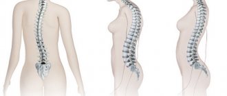
Noteworthy are longitudinal and transverse flat feet, scoliosis, and sometimes funnel chest deformity. By the way, in most of the photographs presented, scoliosis of the spine is clearly visible. This is far from an accident: scoliosis is one of the typical manifestations of congenital mesenchymal deficiency.
I consider mesenchymal deficiency (congenital deficiency of connective tissue) to be the main cause of gastro- and splanchnoptosis.
In this regard, one cannot help but express great doubt that gastroptosis can occur after pumping out ascites or as a result of any other “weakening of the anterior abdominal wall.” I would be grateful if someone could explain why a person should develop gastroptosis due to ascites if there was no gastroptosis before the accumulation of transudate in the abdominal cavity began. In my opinion, formed and pumped out ascites cannot provoke gastroptosis, i.e. Lengthening the ligaments of the stomach: it is not so easy to make the ligaments several times longer. Moreover, in ascitic fluid the organs are weightless, and the gas-containing colon apparently also acquires additional buoyancy. At the moment of fluid removal, even having lost support from the flabby anterior abdominal wall, the gastric ligaments will not suddenly stretch.
The idea creeps in that the position that “flabby anterior abdominal wall” is associated with gastroptosis or is its main cause is simply thoughtlessly copied from one work to another and has even been introduced into the definition printed in the “Encyclopedic Dictionary of Medical Terms.”
However, the truly real cause of gastroptosis should be recognized as long-term gastroparesis or mechanical obstruction at the level of the outlet of the stomach or duodenum, but again, most likely, only in the presence of congenital mesenchymal failure. In this case, prolapse of the stomach is possible as a result of constant overfilling of the organ, as is observed in the patient whose photograph is presented in Fig. 10. Her connective tissue is also clearly defective - this is indicated by scoliosis. The stomach is hypotonic, without peristalsis, distended with a huge amount of contents (the contrast study was done without any preparation).
Causes of prolapsed stomach
Congenital disease:
- congenital weakness of the muscles that support organs;
- asthenic physique;
- high growth;
- lengthening of the mesentery of the large intestine, which leads to displacement of first the intestines, and then the stomach.
Acquired pathology:
- rapid loss of body weight;
- surgical removal of tumors or ascites (pumping out fluid from the abdominal cavity);
- abnormal position of the diaphragm due to the development of a lung tumor or pleurisy;
- postpartum period in women;
- lifting weights;
- physical activity: jumping, running, weightlifting;
- serious illnesses that lead to depletion of the body.