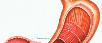
After we have safely swallowed a portion of food treated with lysozyme, having previously taken a small sip of water, and our proteins have been denatured by hydrochloric acid, the pyloric sphincter passes the portion of food further and the digestion process continues in the duodenum, which is located immediately behind the stomach.
It is called so because the length is 12 fingers folded across. In fact, this department is the main compartment of the human digestive system.
Two ducts open into the duodenum: the pancreas and the gallbladder.
Location and structure
This organ, located directly in the abdominal cavity, often envelops the pancreas along its length, namely its head. The duodenum may not be constant in its location and this depends on gender, age, constitution, fatness, position of the body in space, etc.
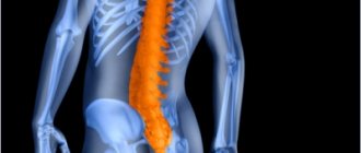
Skeletotopically, taking into account the four sections of the intestine, its upper part starts from the 12th thoracic vertebra, makes the first (upper) bend at the level of the 1st lumbar, then goes down and reaches the 3rd vertebra of the lumbar spine, makes the lower (second) curve, follows from right to left in a horizontal position and finally reaches the 2nd lumbar vertebra.
Sections of the duodenum
This organ lies retroperitoneally and does not have a mesentery. The organ is conventionally divided into four main sections:
- Upper horizontal section. The upper horizontal section may border on the liver, namely its right lobe, and is located in the area of the first lumbar vertebra.
- Descending part (department). The descending section borders the right kidney, bends and can reach the second third lumbar vertebra.
- Lower horizontal section. The lower horizontal section carries out the second bend and begins with it; it is located near the abdominal aorta and the inferior vena cava, which are located posterior to the duodenum.
- Rising department. The ascending section ends with a second bend, rises upward and smoothly passes into the jejunum.
We recommend reading:
Preparation for gastroscopy and procedure
The organ is supplied with blood by the celiac trunk and the superior mesenteric artery, which, in addition to the intestine, also supplies the base of the head of the pancreas.
The structure of the wall of the duodenum
The wall is represented by the following layers:
- serous is the serous membrane covering the outside of the intestine;
- muscular – represented by muscle fibers (located circularly and along the organ), as well as nerve ganglia;
- submucosal – represented by lymphatic and blood vessels, as well as the submucosal membrane, which has a folded shape with crescents;
- mucous - represented by villi (they are wider and shorter than in other parts of the intestine).
Inside the intestine there are the major and minor nipples. The major nipple (Vaterov) is located approximately 7-7.5 cm directly from the pylorus of the stomach. The main pancreatic duct and common bile duct (or common bile duct) exit into it. The minor papilla emerges approximately 8-45 mm from the papilla of Vater, into which the accessory duct of the pancreas emerges.
The most common diseases of the duodenum.
- Duodenitis is the most common ailment of the duodenum of acute or chronic type, manifested in the form of inflammation of the mucous membrane.
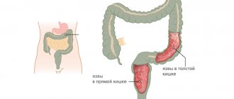
- Ulcer – develops as a result of chronic duodenitis. Chronic damage to the duodenum, in which ulcers form in the mucous layer.
- A cancerous tumor is a malignant neoplasm localized in different layers of the duodenal wall.
Duodenitis
More than 90% of patients develop chronic duodenitis. It can develop due to many factors, including:
- consumption of low-quality products;
- alcohol abuse;
- smoking;
- ingress of foreign bodies and toxic substances;
- other chronic intestinal diseases.
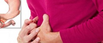
This disease manifests itself in the form of pain in the epigastrium of moderate intensity, weakness, belching, heartburn, nausea, turning into vomiting. Symptoms are often accompanied by fever.
A variation of this inflammatory phenomenon is bulbitis, in which the pathological process occurs only in the duodenal bulb. This form of duodenitis does not occur just like that - it is a consequence of other pathologies of the intestines or stomach. The cause of bulbitis can be:
- gastritis;
- stomach or duodenal ulcer.
If the disease is at an acute stage, the person feels pain and nausea and suffers from repeated vomiting. Acute bulbitis develops against the background of long-term use of a large group of drugs, or poisoning. In the chronic form, there is also an aching pain syndrome, sometimes it can be accompanied by nausea.
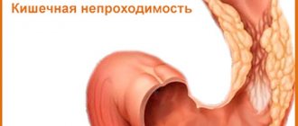
Patients also experience chronic duodenal obstruction, which occurs against the background of tumor processes, developmental anomalies and other disorders in the duodenum. It is expressed in a violation of motor and evacuation functions in this part of the intestine and is characterized by the following symptoms:
- heartburn;
- decreased appetite;
- feeling of heaviness and discomfort in the epigastric region;
- constipation;
- gurgling and bubbling.
The manifestation of this disease is influenced by the causes that caused duodenal obstruction, the stage of progression and how long ago the disease arose.
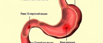
Peptic ulcer
The main cause of this dangerous disease is the reflux of acid from the gastric contents and its destructive effect on the mucous membrane of this part of the intestine. But this pathological process develops only when the surface layers of the intestine fail to cope with their protective functions. The ulcer is localized in the initial part of the duodenum and in the bulb, that is, in that zone of the intestine that is located at a minimum distance from the stomach.
Functions
- Motor-evacuation. It is the process of pushing food through the alimentary canal. The organ also serves as a reservoir; it releases bile acids and various pancreatic enzymes.
- Digestive. The initial stage of digestion occurs in the intestine, due to the action of bile acids and pancreatic enzymes.
- Regulatory. Caused by the regulation of bile acids and pancreatic enzymes.
- Acid-base. In the duodenum, the pH of the bolus of food is brought to optimal levels for its further transformation in other parts of the digestive tract.
Duodenal bulb, its role and causes of inflammation
The first section of the duodenum, just behind the stomach, is called the duodenal bulb, named for its shape.
The process of passing a portion of food through the area where the bulb is located and neutralizing it usually takes 2-3 minutes.
The main function of the duodenal bulb is to convert the acidic reaction of food coming from the stomach to neutral, that is, to remove acidity.
And then, by the end of the DPC, give the entire food mass an alkaline reaction, since all the remaining elements of the digestive system work in an alkaline environment.
If a too acidic environment comes from the stomach, then due to the insufficiency of the main function of the bulb - neutralizing, there is a high predisposition to the development of inflammation in its cavity, leading over time to the formation of ulcers.
Inflammation of the duodenal bulb is called duodenitis, a disease in which the mucous membrane is not only changed, but also subject to structural changes.
Often this is a consequence of diseases of other organs - the liver and biliary tract, gastric ulcer, pancreatitis.
Unfavorable factors for its occurrence are an increase in pepsin and hydrochloric acid in gastric juice, a deterioration in the process of neutralization of hydrochloric acid, and a delay in the evacuation of duodenal contents.
Symptoms of acute inflammation of the duodenum are:
- feeling of fullness in the epigastric region
- pain
- nausea
- salivation
- vomit
- loss of appetite
- increase in body temperature
Diagnostic methods for examining the duodenum
- Gastroscopy (EGDS) is an endoscopic examination that allows you to assess the condition of the duodenum from the inside and is 100% accurate for making a diagnosis. The condition of the organ is displayed on the monitor, the doctor evaluates the patient’s disease: the presence of ulceration, swelling of the mucous membrane, inflammation, the presence of polyps, scar changes, etc.
- Biopsy - during endoscopy, if there are polyps or suspicious neoplasms, the doctor can excise a small area of the intestinal mucosa for histological examination, as well as remove polyps.
- X-ray (including contrast) is performed to make a diagnosis, often with the use of a contrast agent. This allows you to see whether the intestine is passable, what its lumen is, shape, boundaries, etc.
- Ultrasound is not the main diagnostic method, which allows you to determine the topography (location) of the abdominal organs, including the duodenum.
Why do we need the sphincter of Oddi?
Next, the duodenal bulb smoothly passes into the second zone of the duodenum - the large duodenal papilla, or sphincter of Oddi, named after the Hungarian anatomist who first described it.
The sphincter of Oddi is a muscular valve located in the large duodenal nipple of the duodenum, through which the flow of bile and pancreatic juice is controlled through two ducts - the bile and pancreatic, which open in the duodenum.
The sphincter of Oddi works on the principle of preparing the well-known hot dog, that is, by layering products in a strictly defined sequence: the bun with sausage is placed under the tap with ketchup, and then under the tap with mustard.
The same thing happens in the main “kitchen” of the human body - the duodenum: the pancreatic ducts open in turn.
When a portion of food arrives, the pancreatic duct first opens and a portion of pancreatic juice is injected onto the passing food, then the gallbladder duct opens and a portion of bile completes the formation of our “hotdog.”
In the same way, for each piece of food we swallow, a strictly defined portion of pancreatic juice is secreted first and, secondly, bile.
Gastroduodenitis
Inflammation of the lining of the duodenum or duodenitis is the most common pathology of this organ and is quite rarely independent.
Symptoms of this disease include:
- heaviness in the stomach;
- rapid satiety, despite the fact that a small amount of food was eaten;
- decreased or complete lack of appetite;
- attacks of nausea that end in vomiting only during exacerbations of the disease;
- pain in the abdominal area, often patients experience night hunger pains;
- the appearance of blood impurities in feces and vomit;
- severe weakness and dizziness are explained by the presence of anemia.
By the way your stomach hurts, you can recognize the location of the pathological process. For example, when the upper part of this organ is affected, the pain syndrome appears much faster after eating food than when the lower part is involved in inflammation.
This disorder exists in several forms - acute and chronic; depending on the nature of the course, the intensity of the manifestation of symptoms will differ.
The disease consists of the combined occurrence of gastritis and duodenitis. Inflammation begins in the stomach, spreads to its entire surface and spreads to the duodenum.
Similar to duodenitis, it can occur in acute and chronic forms. In an acute course, the main symptom is pain in the right hypochondrium, while in a chronic course, this symptom becomes more intense, atrophy of the membrane occurs, and the motor and secretory function of the affected organs is impaired.
Structure
Where is the duodenum located? Let's consider the location of each of its parts, as well as the structure of the 12 duodenum and its sections. It consists of 4 parts:
- Top part. This is the initial part of the duodenum. You can find it between the last thoracic and 1st lumbar vertebrae; above it you can see part of the liver. The length of this part is about 5-6 cm. First it goes obliquely, from left to right, and then makes an upper bend.
- Descending part. Its length is about 7-12 cm, it can be found to the right of the lumbar region, it gradually forms a lower curve. Reaches the 3rd lumbar vertebra and touches the kidney located on the right.
- Horizontal or bottom part. The length of this part is about 6-8 cm. It is directed from right to left, then passes next to the spine and bends upward. Behind the lower part is the aorta, as well as the inferior vena cava.
- The ascending part. The length of this part is no more than 4 or 5 cm. It can be found on the left side of the lumbar region, where the 2nd lumbar vertebra is located, where it forms a bend.
In one section of the intestine, located near the stomach, there is an expansion called the duodenal bulb or ampulla. It is different from the rest of the intestine. The mucous membrane of the bulb is the same as that of the pylorus of the stomach, with longitudinal folding, whereas in other parts the folding is circular.
IPP
Proton pump inhibitors are one of the most effective groups of drugs for duodenal ulcers. This is explained by the fact that they are highly effective in eliminating the disease compared to other antiulcer substances. Another feature of them is the ability to create favorable conditions for eliminating Helicobacter pylori.
There is a wide range of drugs used in the treatment of not only ulcers, but also inflammation of the duodenum, which are classified as PPIs. The most common of them include:
- Omeprazole;
- Esomeprazole;
- Pantoprazole;
- Lansoprazole;
- Rabeprazole.
In addition to such medications, their equally effective analogues are used.
The mechanism of action of such substances is to block parietal cells, against the background of which the last stage in the production of chloride acid is blocked.
Treatment
Treatment of the duodenum in most cases consists of conservative methods, including:
- taking medications aimed at eliminating symptoms and pathogens;
- maintaining a gentle diet - patients are often prescribed diet tables No. 1 and 5;
- use of traditional medicine.
Surgical intervention is resorted to in cases of ineffectiveness of other treatment methods, as well as in serious condition of the patient.
The treatment regimen is prescribed individually for each patient.
Possible diseases
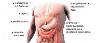
Diseases of the duodenum provoke the development of an inflammatory process in the mucous membrane.
Diseases of the duodenum are inflammatory processes in the mucous membranes of the organ, which affect its functioning and the digestive chain as a whole. Various diseases that affect the performance of the entire body can provoke the development of inflammation. Every year the average age of patients decreases, which is due to the rhythm of life, bad habits, eating on the go and other factors. Atrophy of the mucosa, duodenal hormonal insufficiency, fistulas, bleeding are common complications of inflammatory processes of the duodenum in a neglected state.
Duodenitis of the duodenum
Duodenitis is a disease of the duodenum, which is localized in the transitional section of the intestine. Inflammation can be secondary (accompanying another disease) or primary. This causes spasm of the sphincter of Oddi and thickening of the walls of the organ. Often occurs against the background of secretory insufficiency. An advanced disease can lead to atrophy of the organ mucosa. There are signs of pathology that depend on the neglect of the process and the concomitant disease:
- pain in the epigastrium - just below the stomach of a dull or acute nature;
- nausea;
- gagging;
- spasms;
- burning in the esophagus;
- prostration;
- swelling of the organ mucosa;
- feeling of fullness in the stomach after eating.
Peptic ulcer disease
Duodenal ulcer is an inflammation that is accompanied by the appearance of ulcers on the mucous membranes of the organ. The pathology is chronic and often recurs. The endoscopic picture shows thickening of the intestinal wall. The disease can spread to other parts of the gastrointestinal tract. If the disease is not treated, fistulas, mucosal atrophy and severe bleeding may appear, which is life-threatening for the patient. In the absence of adequate medical care, complications can lead to death.
The most common cause of ulcers is Helicobacter. This type of pathological microorganisms affects the mucous membranes of the digestive organs with toxins, the release of which occurs during their vital activity. They enhance the secretion of enzymes in the organ. Peptic ulcer disease is often secondary, and occurs as a consequence of gastritis and duodenitis. Other reasons:
- genetic predisposition;
- stress and psycho-emotional problems;
- drinking alcohol and smoking;
- poor nutrition.
Symptoms:
- sharp pain in the epigastric region that shoots into the back and ribs;
- nausea and vomiting due to stagnation of food;
- pain on the right under the ribs due to stagnation of bile;
- blood impurities in vomit and feces (sometimes).
Erosion of the duodenum
Erosion is an inflammatory process on the surface of the mucous membrane of an organ, which does not penetrate the muscle layer, and is accompanied by the appearance of eroded areas. Ultrasound shows thickening of the organ wall. The disease can be provoked by:
- stress and psycho-emotional stress;
- smoking;
- Helicobacter;
- poor nutrition;
- medicines.
Erosion of the duodenum is accompanied by a number of symptoms.
Signs of the pathological process:
- belching;
- burning in the esophagus;
- problems with stool;
- pain syndrome.
Duodenostasis
Duodenostasis is also called dyskinesia - a disease that affects the motor function of the duodenum, which is why food gruel (chyme) cannot be evacuated from the small intestine, which causes long-term stagnation of food. Dysfunction is accompanied by the following symptoms:
- loss of appetite;
- pain in the stomach area and on the right under the ribs;
- be sick;
- I'm worried about constipation.
Causes of the disease:
- endocrine disorders;
- problems with the autonomic and central nervous system;
- parasites;
- surgical intervention;
- diseases of the duodenum or adjacent organs.
Parasitic infection
Parasites enter the body through food, if basic hygiene rules are not followed. They can affect any organ and not make themselves felt for a long time. One of the parasites that is often found in the duodenum is nematodes (crooked head or necator). The larvae can be transmitted by the fecal-oral route or through skin pores. They can affect not only the duodenum, but also enter other organs through the blood. Over time, mucosal atrophy occurs. Signs:
- rash;
- itching of the skin;
- diarrhea;
- pain (pain in the peritoneum);
- heartburn.
Neoplasms
Malignant tumors of an internal organ do not occur often.
Duodenal cancer is diagnosed extremely rarely, usually in older people. Its development is preceded by dysplasia. There are 3 degrees of pathology. With stage 3 dysplasia, the development of cancer can rarely be avoided. With dysplasia, the histological structure of the epithelial tissue of the organ is disrupted.
Symptoms are similar to other diseases of the organ:
- painful sensations that are enhanced by palpation;
- lack of appetite up to aversion to food;
- prostration;
- sudden weight loss;
- obstructive jaundice due to impaired excretion of bile.
Lymphofollicular hyperplasia is a submucosal lesion of the duodenum, which can spread to all digestive organs and peritoneal lymph nodes. It is also considered a precancerous condition. If lymphofollicular hyperplasia extensively affects intestinal tissue, external signs appear. But if it is limited to a small area of the duodenum, there may be no symptoms at all. With any tumor, uniform thickening of the intestinal walls is visible.
Obstruction
Chronic organ obstruction develops for many reasons. Among them:
- malrotation of the intestine;
- inverted and mobile intestine;
- congenital malformations;
- vascular compression.
It is possible for gallstones to enter the stomach through a fistula between the organ and the duodenum or stomach. The stone migrates through the alimentary canal and gets stuck in the small intestine. This type of obstruction is diagnosed extremely rarely. Before the onset of pathology, the patient has been bothered by pain on the right under the ribs for a long time. Gallstone obstruction of the small intestine is usually diagnosed in older females.
Developmental defects
Duodenal diverticula are a congenital disease.
Abnormal development of the organ is rare. One of the pathological conditions is congenital stenosis, which is diagnosed in the first hours of a child’s life (vomiting, regurgitation, lack of stool). Congenital anomalies include diverticulum (protrusion of the wall). Lymphangiectasia belongs to this group of diseases. The cause of development is unilateral lymphedema. Lymphangiectasia can develop due to other malformations of the gastrointestinal tract, for example, against the background of Crohn's disease, ulcerative colitis.
If you want to preserve your gallbladder, eat in the morning!
The sphincter of Oddi opens only to food; it does not open to liquid, with the exception of milk.
Why is this happening?
Milk is the first product that a person tries, and even in infancy, when it is consumed, all digestive systems are activated - lysozyme and gastric juice are released in response to it, the process of protein denaturation begins and portions of pancreatic juice and bile are released.
Therefore, those who have breakfast only with tea or coffee are acting imprudently, since this liquid quickly passes through the walls of the stomach and duodenum, and the main sphincter reflex does not work.
That is, the digestive system is asleep, which in itself does not bring any value to the body and is fraught with stagnation in the pancreas and gall bladder.
With various disturbances in the patency of pancreatic juice and bile ducts, a so-called spasm of the sphincter of Oddi occurs.
It involves attacks of repeated severe or moderate pain for more than 20 minutes, for more than 3 months, neurotic disorders, and dyspepsia. This often happens after cholecystectomy and with disorders of the structure of the biliary tract.
Dietary recommendations for gastric and intestinal ulcers
If you have a stomach or duodenal ulcer, special attention should be paid to a proper diet, because this is an important component of proper treatment.
You can eat foods that are gentle on the mucous membranes of the stomach and duodenum. These dishes include:
- soups, dairy, cereal or vegetable, soups with weak meat and chicken broth are also allowed;
- liquid porridge (with water or milk);
- thoroughly cooked meat;
- boiled or steamed fish.
Steam soufflés made from meat, chicken and fish are also often included in the diet.
Milk and dairy products are allowed, you can eat eggs - they are eaten in the form of a steam omelet, or you can eat them soft-boiled. They eat bread that was baked yesterday. Drinks allowed include weak tea and jelly.
Diagnostics
To establish the correct diagnosis, the patient must undergo a comprehensive diagnosis, consisting of:
- examination by a specialist of medical history and life history;
- performing a thorough physical examination and questioning of the patient;
- laboratory studies of blood, urine and stool tests;
- performing breath tests for the presence of the bacterium Helicobacter pylori;
- FGDS is an endoscopic examination for examining the internal surface of the gastrointestinal tract. During this procedure, a biopsy is indicated, which is necessary if the doctor suspects cancer;
- radiography using a contrast agent.