Anemia is a fairly common diagnosis in the modern world. This disease can be seen in the medical history and people often wonder what hypochromic anemia is? Hypochromic anemia is a complex name for all types of anemia, caused by a decrease in the level of hemoglobin in red blood cells.
This disease is characterized by two development paths: the first is a lack of iron in the blood - it is explained by the nutritional characteristics of the person who sought medical help; the second is a violation of the absorption of iron by the cells of the body.
In order to understand what causes this disease, you need to see a doctor who will prescribe the appropriate diagnosis.
What is hypochromic anemia
Hypochromic anemia is a collective term for several types of anemia. Characterized by a decrease in the level of hemoglobin (iron-containing protein present in blood cells). This leads to oxygen starvation of the organs and tissues of the body. As a result of lack of oxygen, they cease to perform their functions, which is fraught with serious complications.
Anemia is translated from Greek as anemia. It can occur with any disease that is in any way associated with blood damage.
Hypochromia is characterized by a decrease in hemoglobin levels and the number of red blood cells (erythrocytes)
Important! People with low hemoglobin levels suffer from intestinal infections and acute respiratory infections 2 times more often.
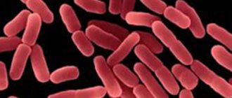
With anemia, not only the level of hemoglobin and red blood cells decreases, but also the color indicator changes. Blood cells also change size and shape; they take on the appearance of a ring with a clear area in the middle and a dark rim around the edges.
With hypochromic anemia, the color index of red blood cells changes. They become discolored and have a dark edging around the edges.
The problem of anemia is often encountered in pediatric practice. At birth, the baby produces a certain amount of iron. Subsequently it must be replenished. If this does not happen, the likelihood of developing the disease increases.
How to increase a child’s hemoglobin at home:
Causes of the disease
In order to make a diagnosis, specialists examine red blood cells, hemoglobin and color indicators, and with this type of disease, not only the hemoglobin level decreases, but also the color indicator.
Causes of hypochromic anemia include:
- Various types of bleeding (they can occur due to injuries, after childbirth, or as a consequence of abortion or any surgical intervention).
- Internal hemorrhages – this pathology is most often chronic, but can also be caused by various diseases. Anemia may occur with large blood loss.
- Poor nutrition is also one of the causes of anemia. In order to prevent this disease, it is necessary to eat foods rich in iron - liver, dried apricots, apples, prunes, and drink pomegranate juice.
- Often pregnant women are diagnosed with this because they need to consume much more iron due to the development of the fetus, which is fed from the mother's body.
- Diseases of the gastrointestinal tract provoke anemia, because as a result the stomach loses its ability to absorb many useful substances, including iron. Hypochromic anemia can also be caused by worms.
- This disease can also appear in people who frequently donate blood (donors).
Causes
The causes depend on the type of anemia. However, the most common causes of the disease are:
- heavy bleeding (menstruation, post-operative or severe injuries);
- unbalanced diet with insufficient amounts of vitamins and proteins. Typical for people who adhere to a strict diet, as well as for vegetarians;
- internal hemorrhages. Blood loss may be minor, but constant (frequent). These include bleeding gums, hemorrhoids, and gastrointestinal diseases. With fibroids (benign tumors) of the uterus and ovarian cysts, their cavities are filled with blood, hemoglobin is converted into other compounds and gradually dissolves. In medicine, this phenomenon is called “pseudo-blood loss”;
- chronic infectious diseases - with tuberculosis, enterocolitis, hepatitis, redistribution of iron occurs or its poor absorption. In older people, anemia is often caused by liver and kidney diseases;
- intoxication, poisoning with chemical elements - occurs in iron-saturated anemia;
- pregnancy - during this period the body requires an increased amount of iron;
- worms;
- blood diseases;
- autoimmune diseases provoke the death of red blood cells, which leads to a decrease in hemoglobin.
Anemia in newborns and premature babies appears when:
- Rhesus conflict;
- infection of the fetus during gestation with herpes and rubella viruses;
- improper nutrition of the expectant mother;
- birth trauma.
In infants, the disease can develop due to poor nutrition. This mainly happens with artificial feeding.
Hypochromic anemia often occurs in adolescence, when hormonal changes occur.
Why does it occur
Hypochromic anemia is caused by:
- Chronic bleeding (gastrointestinal, uterine, pulmonary, menstrual). The causes may be peptic ulcer, gynecological diseases (endometritis, endometriosis, tumors, erosion), anal fissures and hemorrhoids.
- Helminthiases.
- Exudative diathesis. This is a common cause of anemia in children.
- Diseases of the blood system (blood incoagulability, hemorrhagic diathesis).
- Frequent blood donation.
- Carrying out hemodialysis.
- Chronic renal failure.
- Impaired absorption of iron. This element is part of hemoglobin. The causes of insufficient absorption of iron are parasitic infections, hypoacid gastritis, malabsorption syndrome, chronic inflammatory bowel diseases and removal of part of the stomach.
- Insufficient intake of iron from food. It is observed when there is a deficiency in the diet of meat and meat products. Vegetarians are at risk.
- Anorexia (exhaustion).
- Artificial feeding of a child.
- Liver dysfunction.
- Increased need for iron. It is observed during puberty, during infectious pathology and during pregnancy.
- Genetic mutations.
- DIC syndrome.
- Hypovitaminosis of ascorbic acid.
- Sepsis.
- Radiation sickness.
- Mechanical damage to blood vessels.
- Extensive myocardial infarction.
- Rupture of the fallopian tubes.
- Aneurysm rupture.
With hypochromic anemia, the cause may lie in systemic diseases (lupus erythematosus), vasculitis, prolonged alcohol consumption and irrational use of medications (NSAIDs, antacids, iron-binding drugs).
Important information: How to treat hyperchromic anemia and its symptoms
Classification
Doctors distinguish several types of hypochromic anemia:
- iron deficiency (microcytic) – occurs most often. The cause may be frequent bleeding, lack of iron and its poor absorption, physiological processes (lactation, pregnancy). Seen more in children and young women;
- iron-saturated (sideroachrestic) - characterized by a normal level of iron in the blood, but this element is not absorbed, as a result of which hemoglobin is not produced. More common in older people. Pathology is observed with alcohol intoxication, poisoning with poisons or chemicals, long-term use of medications;
- iron redistribution - occurs when there is a high concentration of iron in the blood after the breakdown of erythrocytes (red blood cells). The disease often occurs in tuberculosis and purulent infectious processes;
- mixed - develops due to deficiency of vitamin B12 and iron. The main symptoms include fatigue, decreased immunity, and swelling of the upper extremities.
The disease can be hereditary or acquired.
- The acquired form occurs after operations, infectious diseases, and poisoning.
- Congenital anemia occurs with blood diseases.
According to WHO, every third woman and every sixth man suffers from a chronic form of the disease. The fact is that chronic diseases, unbalanced nutrition, and diets lead to a lack of iron in the body and a decrease in hemoglobin levels. Patients get used to weakness and poor health, attributing this to overwork and stress.
Anemia in children
Hypochromic anemia in children is observed in the same way as in adults. It is characterized by low hemoglobin in the blood, detected during a general blood test. In children, anemia most often develops as a result of poor nutrition or insufficient iron in the body.
At risk are children born from multiple pregnancies, or premature babies, or suffering from any intestinal diseases. Children under three years of age are also quite vulnerable to this disease, because all their organs and systems are not yet sufficiently developed, and receiving an unbalanced diet disrupts the natural balance of nutrients in the body. Most often, such children have iron deficiency anemia.
To prevent your child from developing hypochromic anemia, you often need to go for walks in the forest or field, lead an active lifestyle and give him food rich in all microelements and vitamins.
Foods High in Iron
Causes of iron deficiency in children
- The baby’s body may lack the minerals that are responsible for sufficient levels of iron in the blood.
- Pathologies that are observed due to diseases of the stomach and intestines, when absorption functions in the stomach or intestines are disrupted.
- Disorders of hematopoietic function in the bone marrow.
- The organs of the hematopoietic system are not yet mature enough to produce the required amount of hemoglobin in the blood.
- Living in areas with an unfavorable environment for humans.
- The presence of helminths in the body.
- Frequent illnesses with various colds.
- Dietary disorders, lack of important vitamins and microelements.
- If the child does not receive vitamins C and B, folic acid, manganese, copper, iron, cobalt, nickel.
- Premature babies often suffer from this disease because they were born prematurely and did not receive the necessary nutrition in the mother's body.
Symptoms in a child
Symptoms of hypochromic anemia depend, first of all, on the severity of the disease and the causes that caused it. Usually they talk about the general symptoms of anemia, which are always observed in the patient, and about special cases that are caused by completely different factors and have slightly different symptoms. You need to know that only a doctor can diagnose pathology and establish its cause, so you should not self-medicate in this case.
The following symptoms are distinguished:
- Dyspnea.
- Pale skin in children.
- Tachycardia.
- The child may complain of dizziness, headache, and tinnitus.
- Weakness, lethargy.
- Your baby gets tired quickly, even from minor physical activity.
- The child has a poor appetite.
- Hair and nails are dry and brittle.
- The child has a taste disorder (may eat chalk).
- The appearance of the skin deteriorates - it may become rough.
- Muscle weakness.
- The skin takes on a yellow tint.
- Nosebleeds are common.
- The child is constantly sick.
- The baby's liver and spleen may be enlarged and thickened.
Characteristic pale skin
Types of anemia in children
Scarce
Most often, deficiency anemia occurs in children under one year of age and is the result of a lack of certain substances in the body that contribute to the normal production of hemoglobin in the blood. Malnutrition in this case is the child’s insufficient intake of iron or protein.
As a rule, such a pathology can occur as a result of prematurity of the child, if the substances necessary for the production of hemoglobin are not absorbed in the intestines or due to an incorrect or unbalanced diet.
Asthenovegetative
Asthenovegetative anemia is a consequence of impaired brain function. It is characterized by the following symptoms: bedwetting, distracted attention of the child, poor memory, muscle pain.
Epithelial
With this type of disease, fragility of hair and nails, degeneration of the mucous membranes and skin are noted. Epithelial anemia may also cause pallor of the skin, the child’s swallowing process is impaired - all these pathologies arise as a result of disruption of the intestines, which do not absorb substances necessary for the child’s body.
Immunodeficiency
In this case, there are constant infectious or colds in children, because the immune system of the child’s body is in a weakened state. Immunodeficiency anemia is diagnosed by a low level of plasma in the blood.
Cardiovascular
This is a more severe type of anemia, which is characterized by increased fatigue, decreased blood pressure and rapid heartbeat in the child.
Anemia B12
B12 anemia occurs when there is not enough of the corresponding vitamin in the child’s body. The pathology is characterized by a decrease in the level of platelets in the blood, the formation of large cells that are far from normal in the bone marrow area, which leads to bleeding and decreased blood clotting. This condition occurs as the inability of the stomach or intestines to absorb the substances necessary to maintain hemoglobin levels and may be accompanied by nervous disorders.
If the child has recently suffered from diseases such as enteritis and hepatitis, then the absorption of B12 in the small intestine may be impaired, which leads to its complete removal from the liver. Moreover, the level of bilirubin in the patient’s blood increases, and the skin becomes yellowish.
Aplastic
Aplastic anemia often requires surgery or blood transfusion. They are characterized by a decrease in the level of red blood cells, platelets and leukocytes in the blood, dysfunction of the bone marrow and hematopoietic system.
Symptoms are pale skin, bruising when touched, and fever. If you observe such signs in a child, you should urgently call an appropriate specialist to your home.
Hemolytic
Hemolytic anemia can be either congenital or acquired. It is characterized by the fact that red blood cells are destroyed, their destruction occurs directly in the spleen. Symptoms of the disease can be observed in the patient both from birth and at an older age.
A sign of this pathology is a yellow color of the skin and pale skin, delayed mental and physical development, and compactions in the area of the liver or spleen are observed. To treat this disease, the spleen must be surgically removed.
Degrees of hemolytic anemia in newborns
Treatment methods for children's bodies
To treat hypochromic anemia, it is necessary to know the reasons that caused it. If this is a lack of iron, then the child is prescribed medications containing it and a certain diet that will contain the necessary minerals and vitamins.
Iron supplements are taken after meals or during meals. It is better to wash them down with citrus juice if there is no allergic reaction to it. Herbal preparations can also be used to treat anemia. It is also possible to receive treatment in a complex that includes vitamins and minerals necessary for the patient’s body.
Symptoms of hypochromia
For a long time, patients do not pay attention to the deterioration of their condition, attributing their poor health to stress and fatigue.
Symptoms depend entirely on the severity of the anemia. First, all patients complain of:
- general malaise;
- rapid fatigue;
- violation of attention;
- decreased physical endurance;
- drowsiness.
Signs of the disease depending on the severity - table
| Degree | Signs |
| I - light | Symptoms are mild. There is weakness and malaise. |
| II - average |
|
| III - heavy |
This degree of disease can lead to death. |
Features of manifestation in children
In children, especially infants, symptoms are mild. The disease can often be diagnosed only after a blood test. Parents need to pay attention to the following signs:
- pale skin;
- poor sleep and appetite;
- lethargy;
- frequent colds;
- cracks in the corners of the mouth;
- retardation in physical and psychomotor development.
Prolonged neglect of the disease can lead to death, so the pathology requires mandatory treatment.
Establishing diagnosis
Hypochromic anemia can be diagnosed using laboratory blood tests. If during the initial analysis the indicators of red blood cells and hemoglobin are reduced, the doctor determines the contributing factors and, if they are absent, a repeat analysis is prescribed. This allows you to track changes in hemoglobin levels and, accordingly, highlight a trend.
The diagnostic process itself is simple, but it should be noted that determining the true causes of the development of hypochromic anemia is not so simple. Only after the diagnosis is made and the causes of its development are identified, effective treatment is prescribed.
Diagnostics
First of all, the doctor conducts an external examination, studies clinical manifestations and hereditary pathologies. The diagnosis is confirmed by a complete blood count, which shows the level of hemoglobin and the number of erythrocytes (red blood cells), which determine the type of anemia and the color index.
Interpretation of the results of a general blood test - table
| Hemoglobin norm, g/l | Pathology, g/l | ||
| Men | 130–160 | < 130 | |
| Women | 120 -150 | < 120 | |
| Pregnant women | 110–150 | < 110 | |
| Children under 5 years old | from birth and during the first day of life | 145 | < 145 |
| 1–14 days | 130 | < 130 | |
| 15–28 days | 120 | < 120 | |
| 1–6 years | 110 | < 110 | |
Normally, the hemoglobin content in red blood cells ranges from 0.85 to 1.15. With hypochromic anemia, the color index becomes below 0.85.
Additionally, differential diagnosis is carried out:
- stool occult blood test;
- gastroscopy - examination of the gastrointestinal tract using a flexible hose that is inserted through the mouth;
- colonoscopy - examination of the large intestine with a special probe;
- Analysis of urine;
- Ultrasound of the kidneys;
- chest x-ray;
- gynecological examination (women);
- study of blood serum for iron content and bone marrow samples.
To confirm anemia, patients undergo a clinical blood test
Content:
- Features of the disease
- Causes
- Types and symptoms
← Hyperchromic anemia: causes, symptoms, treatment methods
Iron deficiency anemia in adults →
A disease such as hypochromic anemia can pose a serious danger to human health if it is not treated in time. In order to understand the features of this problem and determine ways to eliminate it, let us consider in more detail the symptoms, causes and main types of this type of anemia.
Characteristic appearance of red blood cells in hypochromic anemia
Treatment
Therapy depends entirely on the clinical manifestations and severity of hypochromia. First of all, treatment is aimed at eliminating the cause of the disease.
Drug therapy
The main emphasis is on taking medications that compensate for the deficiency of iron and vitamin B12.
It is necessary to take into account that vitamins are quickly excreted in the urine, so it is impossible to achieve quick results only from taking vitamin-containing preparations.
- Iron supplements are taken for 4 to 8 weeks until hemoglobin levels normalize. These can be: Ferrum Lek, Fenyuls, Hemofer. Drugs that need to be administered by drip or injection are recommended to be used only in a hospital setting to exclude allergic manifestations.
- If insufficient intake of vitamin B12 is detected, subcutaneous injections of Cyanocobalamin are prescribed. The course of treatment is 1–2 months until the condition normalizes.
- Vitamin B deficiency is often accompanied by folic acid deficiency. In this case, additional medications are prescribed. The course of treatment is up to 30 days.
It is recommended to take medications in the form of syrup, capsules or tablets. Injections are prescribed if the patient has gastrointestinal diseases or acute blood loss.
It should be borne in mind that therapy for moderate and severe forms of anemia is carried out only in a hospital under the supervision of a doctor.
Increasing hemoglobin with drugs and folk remedies:
Drugs for the treatment of the disease - gallery
Ferrum Lek Fenyuls Hemofer
Folk remedies
Traditional medicine is taken only as maintenance therapy and in combination with medications.
- In the morning, it is recommended to eat 100–150 g of grated carrots with sour cream.
- Eat several pieces of boiled pumpkin throughout the day.
- Nettle improves blood composition, increases the level of hemoglobin and red blood cells. To prepare the infusion, you need to grind 10 g of leaves, steam with a glass of boiling water and leave for 10 minutes. Liquid take 1 tbsp. l. 3 times a day.
- Dried fruit mixture. Grind dried apricots, prunes, raisins, figs, rose hips in equal proportions, add honey. Take 3-4 times a day, 1 tbsp. l.
- Rose hip decoction.
Diet therapy
Diet therapy is recommended for patients in combination with drug treatment.
Basic principles:
- Every day the diet should contain at least 130–150 g of animal proteins (beef, veal).
Proteins contribute to the production of hemoglobin and red blood cells.
- It is recommended to limit fat intake, since anemia causes obesity of the bone marrow and liver, which inhibits hematopoiesis.
- In patients with anemia, there is a decrease in appetite, which is explained by a deterioration in secretory function. To improve your condition, you should include fish, meat, and mushroom decoctions in your diet.
- The menu must include foods rich in vitamin B - eggs, fish, cottage cheese, brewer's yeast. Take 100 g of liver daily or every other day.
Recommended Products:
- fish, meat, liver (low-fat);
- cottage cheese;
- mushrooms;
- eggs;
- black and white bread;
- yeast;
- pomegranate juice;
- legumes (peas, beans, lentils);
- berries and fruits with a high content of vitamin C - rose hips, black currants, pomegranate juice.
It is not recommended to consume pomegranate juice in its pure form; it is better to dilute it with beet juice in a 1:1 ratio.
Featured Products - Gallery
Beef is good for blood formation. High content of vitamins B, C and minerals have a beneficial effect on blood quality. With one apple, about 480 mg of iron enters the body, which helps maintain normal hemoglobin levels. Fish is a source of vitamin B12. Pomegranate juice contains a large amount of ascorbic acid, which promotes the absorption of iron.
Foods that interfere with iron absorption:
- oatmeal, millet;
- tea;
- leafy greens;
- dairy products;
- coffee;
- dishes high in fat.
Foods that should be excluded from the diet - gallery
Alcohol contributes to pathological processes in anemia Dairy products should be consumed in limited quantities, as they delay the absorption of iron Coffee and tea wash out iron, so you should not abuse these drinks
Sample menu - table
| I breakfast | II breakfast | Dinner | Afternoon snack | Dinner |
|
|
|
|
|
What foods increase hemoglobin levels:
Recipes
- Millet porridge. Soak 250g of flakes in 500ml of apple juice overnight. In the morning, boil the mixture in a thick-bottomed saucepan for 10 minutes. Millet contains microelements that are necessary to strengthen the immune system.
- Baked beets. Wash the vegetable, cut the shoots to a height of 1.5 cm from the root crop. Brush generously with olive oil and season with sea salt. Bake in the oven at 175°C for about 45 minutes.
- Drink made from yeast. Grind 10 g of live culture in 100 ml of water, leave for 50 minutes. Drink in small sips.
- Berry salad. To prepare, you will need to take 50 g of raspberries, blackberries, and strawberries. Add 1 banana and apple. Mix the fruits, sprinkle with lemon juice and pour over liquid honey (1 tbsp.).
- Rowan fruit drink. Grind 100 g of dried berries, add 300 g of rowan flowers and 10 g of mint. Pour boiling water in the proportion of 2 tbsp. l. for 200 ml of water. Cool.
Treatment method
To clarify the diagnosis, a general blood test and, if necessary, a number of other studies should be performed. After determining the type of hypochromic anemia, a course of treatment is established. Thus, with iron deficiency, the patient’s condition can be improved with the help of special diets and iron-containing medications, while with sideroachrastic and iron redistribution forms such measures will not give any result.
Treatment of anemia is largely related to the cause of its occurrence.
Important! The main efforts must be directed towards eliminating the root cause. That is, measures may be limited to adjusting the diet or leading to emergency surgery to eliminate internal bleeding.
We also recommend studying this topic:
Symptoms of chronic posthemorrhagic anemia
There are such approaches in the treatment of hypochromic anemia:
- stopping bleeding and inflammatory processes;
- normalization of the condition of the digestive tract;
- elimination of intoxication;
- treatment of the underlying disease;
- introducing foods rich in iron and B vitamins into the patient’s diet;
- taking medications to compensate for iron deficiency.
Iron-containing preparations are used in the form of tablets and capsules, intravenous injections. In severe cases, red blood cell transfusion is required. When iron levels are normal, the emphasis is on vitamin therapy with an emphasis on vitamins B6 and B12.
We recommend studying an article on a similar topic: Treatment of aplastic anemia within the framework of this material.
Medicines used for hypochromic anemia
Advice: for hypochromic anemia, combine diet and vitamin supplements to quickly create an iron depot in the body.
Competent therapy and early initiation of treatment will quickly solve the problem.
← Hyperchromic anemia: causes, symptoms, treatment methods
Iron deficiency anemia in adults →
We recommend studying similar materials:
- 1. Gunther's disease - all about Porphyry's disease
- 2. Autoimmune hemolytic anemia in children: what is missing and how it manifests itself
- 3. Causes and dangers of increased basophil levels in children
- 4. Reasons for an increase or decrease in neutrophils in a blood test in children?
- 5. Functions and possible causes of pathologies of segmented neutrophils
- 6. Low level of total bilirubin in the blood: reasons for the decrease
- 7. Normal bilirubin levels in newborns and children: reasons for the increase
Treatment prognosis and possible complications
If you consult a doctor in a timely manner, the prognosis is favorable in most cases. If left untreated, hypochromic anemia can lead to:
- decreased immunity;
- growth retardation, mental and psychomotor development (in children);
- development of cardiomyopathy - with insufficient oxygen supply, the heart begins to work with redoubled force. This leads to heart failure;
- liver enlargement;
- chronic anemia.
Low hemoglobin levels negatively affect the nervous system.
How to treat anemia in adults
Of course, for the treatment of hypochromic anemia, iron supplements are the best remedy. Here are the main medications that a doctor can prescribe for this pathology:
- Iron gluconate.
- Iron fumarate.
- Ferrous ferrous sulfate.
- Ferrogrademet.
- Tardiferron.
- Ferroplex.
- Ferkoven.
- Artiferrin.
- Ferrum lek.
Almost all of these drugs contain ascorbic acid, which promotes the absorption of iron into the blood.
Patients usually take medications for anemia orally. But it happens that they can cause nausea, vomiting or intestinal colic in the patient (which is observed in half of the patients). Then it is recommended to administer iron intravenously. But at the same time, a number of side effects are also observed, such as pain in muscles and joints, anaphylactic shock and damage to the walls of blood vessels.
When this drug is administered intramuscularly, inflammation may be observed at the injection site and the color of the skin may also change (and this color can last for up to two years).
Thus, it should be understood that you should not take iron supplements without a doctor’s prescription, because they are mainly prescribed for iron deficiency anemia and they can have a number of side effects that can only be avoided by consulting a specialist.
Prevention
The main preventive measure is a proper and balanced diet. The diet should contain foods rich in iron.
- Beef liver, kidneys.
- Quail and chicken eggs.
- Fresh vegetables and fruits - Brussels sprouts, persimmons, pomegranates, beets, dried apricots.
Women should pay special attention to the prevention of anemia, since they lose twice as much blood as men due to menstruation.
How to cure
For hypochromic anemia, treatment is complex. The goals of therapy are:
- elimination of etiological factors (treatment of helminthiases, diseases of the gastrointestinal tract, infectious pathology);
- normalization of color indicator, hemoglobin level, iron and red blood cell count;
- elimination of symptoms;
- prevention of complications.
The main aspects of treatment are:
- Following a strict diet.
- Taking medications.
- Blood transfusions (transfusion of blood components). This is required in case of low blood pressure, decreased hemoglobin and damage to vital organs.
- The use of folk remedies.
- Splenectomy. Indicated for thalassemia and increased spleen function (hypersplenism).
For thalassemia, a bone marrow transplant may be needed.
Drug therapy
When treating hypochromic anemia, the following can be used:
- Antianemic agents (Sorbifer Durules, Ferro-Folgamma, Ferrum Lek). These drugs are indicated for hypochromia in combination with iron deficiency.
- Glucocorticoids. Can be used for thalassemia.
- Anti-inflammatory drugs. Indicated for enteritis and enterocolitis.
- Antiparasitic agents (Biltricid, Vermox, Nemozol). Used for hypochromia caused by helminthiasis.
- Hemostatics (Vikasol). Indicated for hemorrhagic syndrome.
- Complexing compounds (Exjade, Desferal). Prescribed for thalassemia.
Folk remedies
Hypochromic anemia in adults and children can be treated using folk remedies. For low iron levels, beets, fermented yeast and vegetable juices are effective. For thalassemia, herbs rich in zinc and folic acid (oats, red clover, nettle, burdock root, dandelion and parsley) are used. You can prepare decoctions and infusions from them and take them orally.
Important information: Can hypochromic hemolytic autoimmune anemia and its symptoms be cured?
Diet therapy
Patients need to enrich their diet with meat, offal (liver), eggs, cottage cheese, prunes, seaweed, baked beets (helps increase hemoglobin), cereals (buckwheat), apples and peaches. Vitamin C, succinic and citric acids improve the absorption of iron, so it is recommended to eat currants, cabbage, rose hips and citrus fruits.
For hypochromic anemia with iron deficiency, you need to reduce the consumption of strong tea, coffee, rice, soy, fish and seafood, milk and chocolate. Animal products are the most beneficial. This diet must be followed for at least a month until the color index normalizes.
Prevention
Methods for preventing hypochromic anemia are:
- good nutrition (including meat in the diet, enriching the menu with fruits and berries);
- refusal of vegetarianism;
- treatment of existing intestinal diseases;
- thorough hand washing before eating and heat treatment of water, meat and fish (reduces the risk of helminth infection);
- prevention of intoxication;
- treatment of vascular diseases;
- maintaining hemoglobin and red blood cell levels at the proper level;
- treatment of blood diseases;
- taking vitamins (ascorbic acid).
There is no specific prevention.
Characteristics of red blood cells
In shape, red blood cells resemble biconcave discs with a diameter of 7-8 microns, their volume averages 80-100 femtoliters, and their color is normochromic. In the case of pathological changes in red blood cells with microcytosis, macrocytosis, normocytosis, hypochromia and hyperchromia, anemic conditions are most often found. The concept of “microcytosis” is characterized by the presence in erythrocytes of a large number of small midget cells. This indicates the development of microcytic anemia.
What symptoms should you pay attention to?
Any form of the disease, including microcytic hypochromic anemia, is accompanied by very characteristic symptoms. Of course, the clinical picture largely depends on the stage of development of the disease - in the initial stages, the disease can occur without any symptoms at all:
- Patients, as a rule, suffer from constant weakness, frequent dizziness, and the appearance of “floaters” before the eyes. Such symptoms can appear both at rest and after a sudden change in body position.
- The patient's skin often takes on a pale tint. The outer skin becomes too dry and flaky. Painful cracks appear between the fingers, on the feet, and in the corners of the lips, which slowly heal.
- Damage and inflammation of the mucous membranes of the oral cavity are observed.
- Patients' teeth are much more likely to undergo carious processes.
- Patients often complain of changes in the perception of tastes and smells.
- Anemia is often associated with a lack of appetite and, accordingly, weight loss.
- Digestive disorders are also possible, in particular nausea, diarrhea or, conversely, constipation.
- The condition of nails and hair also worsens - they become brittle.
- Patients are lethargic, suffer from constant fatigue and drowsiness, and their performance is significantly reduced.
- Even minimal physical activity leads to severe shortness of breath, which, again, is associated with oxygen starvation of the tissues.
- If we are talking about anemia in children, then children with a similar diagnosis are usually weak, often cry, are capricious, and sleep poorly.
- Severe stages of anemia in children (especially if the disease is congenital) in the absence of therapy can lead to delayed physical and psycho-emotional development.
Difference between macrocytic and microcytic anemia
Macrocytes and microcytes in a blood smear
The main difference between micro- and macrocytic anemia is the size of the red blood cells. If with microcytosis the size of red blood cells is less than 8 microns, then with macrocytic anemia it will be 8-10 or more.
Also, a distinctive feature of some forms of macrocytic anemia will be the detection of megalocytes - red blood cells with a size of more than 12 microns.
The conditions differ from each other in their causes, course, and clinical manifestations. Macrocytosis is most often caused by a lack of vitamins B12 or B9, and microcytosis is caused by a lack of iron.
In general, each of these conditions requires constant monitoring, assessment of the treatment, the dynamics of the underlying disease and the implementation of all medical recommendations.
Algorithms for diagnosis and treatment of iron deficiency anemia
AND
iron deficiency anemia (IDA) is a clinical and hematological syndrome characterized by impaired hemoglobin synthesis as a result of iron deficiency, developing against the background of various pathological (physiological) processes, and manifested by signs of anemia and sideropenia.
The importance of rational and effective therapy for IDA is due to the medical and social significance and high prevalence of this condition among the population.
, especially women of childbearing age, because:
- women of childbearing age constitute the main risk group for developing IDA
- Iron reserves in the body of women are 3 times less than in men
- iron intake in girls and fertile women in developed countries (USA) is 55–60% of the required level
- IDA accounts for 75–95% of all anemias in pregnant women
- in Russia, about 12% of women of childbearing age suffer from IDA
- Latent iron deficiency in some regions of Russia reaches 50%.
Diagnosis of iron deficiency anemia
Stages of diagnostic search:
1 – diagnosis of hypochromic anemia
1 – diagnosis of hypochromic anemia
2 – diagnosis of iron deficiency anemia
3 – diagnosis of the cause of IDA.
1. Diagnosis of hypochromic anemia
. All IDA are hypochromic. Therefore, the hypochromic nature of anemia is a key sign that allows one to suspect, first of all, IDA and determine the further direction of the diagnostic search.
When interpreting the results of a blood test, a clinician must pay attention not only to the color indicator (it may be calculated incorrectly if the laboratory assistant makes an error in counting the number of red blood cells), but also to the morphological picture of red blood cells, which is described by the laboratory assistant when viewing the smear (for example, hypochromia , microcytosis, etc.).
2. Diagnosis of iron deficiency anemia (differential diagnosis of hypochromic anemia)
.
Not all hypochromic anemias are iron deficiency. Taking this into account, the presence of hypochromic anemia does not exclude hypochromic anemia of another origin. In this regard, at this stage of the diagnostic search, it is necessary to carry out a differential diagnosis between IDA and the so-called sideroachrestic (achresia - non-use) anemia. In sideroahrestic anemia
(a group concept), also referred to as iron-saturated anemia, the iron content in the body is within normal limits or even there is an excess of it, however, for various reasons, iron is not used to build heme in the hemoglobin molecule, which ultimately leads to the formation hypochromic red blood cells with low hemoglobin content. Unused iron enters reserves and is deposited in organs and tissues (liver, pancreas, skin, macrophage system, etc.), leading to the development of hemosiderosis.
Correctly recognizing IDA and distinguishing it from sideroachrestic anemia is extremely important, since an erroneous diagnosis of IDA in patients with iron-saturated anemia can lead to unjustified prescription of iron supplements to such patients, which in this situation will lead to an even greater “overload” of organs and tissues with iron. In this case, the therapeutic effect of iron supplements will be absent.
The main hypochromic anemias with which the differential diagnosis of IDA should be made
, are the following:
- anemia associated with impaired heme synthesis
, resulting from inhibition of the activity of certain enzymes (heme synthetase), which ensure the inclusion of iron in the heme molecule. This enzyme defect may be of a hereditary nature (hereditary sideroachrestic anemia) or occur as a result of exposure to certain medications (isoniazid, PAS, etc.), chronic alcohol intoxication, contact with lead, etc.; - thalassemia
, belongs to the group of hereditary hemolytic anemias associated with impaired synthesis of globin - the protein part of hemoglobin. The disease has several variants and is characterized by signs of hemolysis (reticulocytosis, increased levels of indirect bilirubin, enlarged spleen), high iron content in the serum and in the depot, and hypochromic anemia. In fact, with thalassemia we are also talking about sideroachresia, i.e. about the non-use of iron, but not as a result of defects in the enzymes involved in heme synthesis, but due to a disruption in the process of constructing the hemoglobin molecule as a whole due to the pathology of its globin part; - anemia associated with chronic diseases
. This term is usually used to designate a group of anemias that occur in patients against the background of various diseases, most often of an inflammatory nature (infectious and non-infectious). An example is anemia in suppurative diseases of various locations (lungs, abdominal cavity, osteomyelitis), sepsis, tuberculosis, infective endocarditis, rheumatoid arthritis, malignant tumors in the absence of chronic blood loss. With all the diversity of pathogenetic mechanisms of anemia in these situations, one of the main ones is considered to be the redistribution of iron into the cells of the macrophage system, which is activated during inflammatory and tumor processes. Since true iron deficiency is not observed in these anemias, it is more justified to talk not about IDA, but about iron redistribution anemia. The latter are, as a rule, moderately hypochromic in nature, the iron content in the serum may be slightly reduced, the life-sustaining blood pressure is usually within the normal range or moderately reduced, which distinguishes this variant of anemia from iron deficiency anemia. An increase in ferritin levels in the blood is typical. Understanding and correct interpretation of the pathogenetic mechanisms of anemia development in the above diseases allows the doctor to refrain from prescribing iron supplements to these patients, which are usually ineffective.
Thus, we can talk about the presence of IDA in cases of hypochromic anemia, accompanied by a decrease in serum iron content, an increase in PVSS, and a decrease in ferritin concentration. To avoid errors when interpreting the results of determining the iron content in serum, the following rules and recommendations must be taken into account:
- the study should be carried out before starting treatment with iron preparations. Otherwise, even when taking the drugs for a short period of time, the resulting values do not reflect the true iron content in the serum. If iron supplements were prescribed, the study can be carried out no earlier than 7 days after their discontinuation;
- red blood cell transfusions, often carried out before the nature of anemia is clarified (marked decrease in hemoglobin levels, signs of heart failure, etc.), also distort the assessment of the true iron content in the serum;
- To test serum for iron content, special test tubes should be used, washed twice with distilled water, since the use of tap water containing small amounts of iron for washing affects the results of the study. Drying cabinets should not be used to dry test tubes, since a small amount of iron gets into the dishes from their walls when heated;
- Currently, for the study of iron, it is customary to use bathophenanthraline as a reagent, which forms a colored complex with iron ions with a stable color and a high molar extinction coefficient; the accuracy of the method is quite high;
- blood for analysis should be taken in the morning, since there are daily fluctuations in the concentration of iron in the serum (iron levels are higher in the morning);
- serum iron levels are influenced by the phase of the menstrual cycle (immediately before and during menstruation, the level of serum iron is higher), pregnancy (increased iron levels in the first weeks of pregnancy), taking oral contraceptives (increased), acute hepatitis and cirrhosis of the liver (increased ). Random variations in the studied parameters may be observed.
3. Identifying the cause of IDA
. After confirming the iron deficiency nature of anemia, i.e., verification of IDA syndrome, an equally important task is to establish the cause of this anemic syndrome. Recognizing the cause of the development of IDA in each specific case is the final stage of the diagnostic search. Focus on nosological diagnosis is very important, since in most cases, when treating anemia, it is possible to influence the underlying pathological process.
Algorithm for diagnosing iron deficiency anemia
The development of iron deficiency anemia is based
on various reasons, among which the following are of primary importance:
– chronic blood loss
various locations (gastrointestinal, uterine, nasal, renal) due to various diseases;
– impaired absorption of dietary iron
in the intestines (enteritis, resection of the small intestine, malabsorption syndrome, “blind loop” syndrome);
– increased need for iron
(pregnancy, lactation, intensive growth, etc.);
– nutritional iron deficiency
(malnutrition, anorexia of various origins, vegetarianism, etc.).
Causes of iron deficiency anemia
Treatment of IDA
When identifying the cause of the development of IDA, the main treatment should be aimed at eliminating it (surgical treatment of stomach and intestinal tumors, treatment of enteritis, correction of nutritional deficiency, etc.). However, in a number of cases, radical elimination of the cause of IDA is not possible (for example, with ongoing menorrhagia, hereditary hemorrhagic diathesis, manifested by nosebleeds, in pregnant women, as well as in some other situations). In such cases, pathogenetic therapy with iron-containing drugs becomes of primary importance.
When identifying the cause of the development of IDA, the main treatment should be aimed at eliminating it (surgical treatment of stomach and intestinal tumors, treatment of enteritis, correction of nutritional deficiency, etc.). However, in a number of cases, radical elimination of the cause of IDA is not possible (for example, with ongoing menorrhagia, hereditary hemorrhagic diathesis, manifested by nosebleeds, in pregnant women, as well as in some other situations). In such cases, pathogenetic therapy with iron-containing drugs becomes of primary importance.
Iron medications (IF) are the treatment of choice for correcting iron deficiency and hemoglobin levels in patients with IDA. Pancreas should be preferred to foods containing iron.
Iron medications for the treatment of iron deficiency anemia
Currently, the doctor has at his disposal a large arsenal of medicinal prostates, characterized by different composition and properties, the amount of iron they contain, the presence of additional components that affect the pharmacokinetics of the drug, and dosage form.
In clinical practice, medicinal pancreatic drugs are used orally or parenterally. The route of administration of the drug in patients with IDA is determined by the specific clinical situation. In addition, all iron-containing preparations can be divided into two groups - preparations of iron salts and preparations in the form of iron-containing complexes, which have some distinctive properties (composition, pharmacokinetics, tolerability, etc.), see table. 1.
Making a decision about prescribing iron supplements
1. Route of administration of iron supplements
1. Route of administration of iron supplements
Clinical situation
In the vast majority of cases, iron supplements should be prescribed orally.
Parenteral iron supplements can be used in the following clinical situations:
– malabsorption due to intestinal pathology (enteritis, malabsorption syndrome, resection of the small intestine, Billroth II gastric resection including the duodenum);
– exacerbation of gastric or duodenal ulcer;
– intolerance of the pancreas for oral administration, which does not allow continued treatment;
– the need to more quickly saturate the body with iron, for example, in patients with IDA who are undergoing surgical interventions (uterine fibroids, hemorrhoids, etc.).
2. Choosing an iron-containing drug for oral administration
- Amount of ferrous iron
- The presence of substances in the preparation that improve iron absorption
- Tolerability of the drug
When choosing a specific drug and optimal dosage regimen
It must be borne in mind that an adequate increase in hemoglobin levels in the presence of IDA can be ensured by the intake of 30 to 100 mg of divalent iron into the body. Considering that with the development of IDA, iron absorption increases compared to the norm and amounts to 25–30% (with normal iron reserves - only 3–7%), it is necessary to prescribe from 100 to 300 mg of ferrous iron per day. The use of higher doses does not make sense, since iron absorption does not increase. Thus, the minimum effective dose is 100 mg, the maximum is 300 mg of ferrous iron per day. Individual fluctuations in the amount of required iron are determined by the degree of iron deficiency in the body, depletion of reserves, the rate of erythropoiesis, absorption, tolerance and some other factors. Taking this into account, when choosing a medicinal pancreas, you should focus not only on the total amount contained in it, but mainly on the amount of ferrous iron, which is absorbed only in the intestine.
PZh should be taken with meals. At the same time, iron absorption is better when taking medications before meals.
Preferably preparations containing ascorbic acid (Sorbifer Durules)
. It is not recommended to drink iron supplements with tea, since tannin forms poorly soluble complexes with iron. You should not take calcium supplements, tetracycline and fluoroquinolone antibiotics at the same time as iron supplements.
3. Assessing the effectiveness of the prescribed drug
- Reticulocyte count 7–10 days after drug administration
- The amount and rate of increase in hemoglobin every week
Substances affecting the absorption of iron supplements
When prescribing PZH in a sufficient dose, an increase in the number of reticulocytes is observed on the 7-10th day from the start of treatment. Normalization of hemoglobin levels is observed in most cases after 3–4 weeks of treatment, but sometimes the period of normalization of hemoglobin levels is delayed up to 6–8 weeks and a sharp abrupt increase in hemoglobin may be observed. These individual manifestations and characteristics may be due to the severity of IDA, the degree of depletion of iron stores, as well as an incompletely eliminated cause (chronic blood loss, etc.).
4. Assessment of drug tolerability when administered orally
- Monitoring patients, monitoring treatment
Among the side effects
When using pancreatic acid orally, nausea, anorexia, a metallic taste in the mouth, constipation, and, less commonly, diarrhea most often occur. The development of constipation is most likely due to the binding of hydrogen sulfide in the intestine, which is one of the stimuli of intestinal peristalsis. In most cases, modern pancreatic products cause minor side effects that require their cancellation and switching to the parenteral route of administration.
Assessment of tolerability of oral iron supplementation
Dyspeptic disorders may improve when taking medications after meals or reducing the dose.
Using modern technologies, pancreases with a slow release of iron from them (Sorbifer Durules) are currently produced due to the presence of inert substances, from which iron is gradually released through small pores. This ensures a prolonged absorption effect and reduces the incidence of gastrointestinal disorders.
5. Duration of saturation therapy
- Rate of increase in hemoglobin level
- Time frame for normalization of hemoglobin levels
- Clinical situation
The duration of so-called saturating therapy with iron supplements is determined by the rate of increase in hemoglobin, and, consequently, by the timing of normalization of hemoglobin levels. This, in turn, may depend on the activity of the iron preparation and on the degree of depletion of iron reserves in the body. According to our data, a high rate of increase in hemoglobin is observed during treatment with the drug sorbifer-durules. The average increase in hemoglobin per day is about 2 g/l, which makes it possible to complete the course of saturating therapy in 2–3 weeks.
6. The need for maintenance therapy
- Clinical situation (pregnancy, unrecoverable menorrhagia and nosebleeds, etc.)
Treatment with oral iron supplements
In most cases, to correct iron deficiency in the absence of special indications, pancreas should be administered orally.
Currently, the Russian pharmaceutical market has a large number of iron preparations for oral administration - in the form of various iron salts or in the form of iron-containing complexes. Preparations differ in the amount of iron salts they contain, including ferrous iron, the presence of additional components (ascorbic and succinic acids, vitamins, fructose, etc.), dosage forms (tablets, dragees, syrups, solutions) and cost.
The main iron-containing preparations are presented in the form of ferrous sulfate, gluconate, chloride, ferrous fumarate, glycine sulfate, and ferrous sulfate preparations have the highest degree of absorption, and glycine sulfate has the lowest.
Table 2 presents the main medicinal pancreatic products registered in Russia in the form of salts and iron-containing complexes for oral administration. The reasons for the ineffectiveness of therapy with oral iron preparations and methods of correction are given in Table 3.
Treatment with iron preparations for parenteral administration
Unlike pancreas for oral administration, in injection preparations iron is always in the trivalent form.
Unlike pancreas for oral administration, in injection preparations iron is always in trivalent form.
During parenteral treatment of the pancreas, especially with intravenous use, allergic reactions often occur in the form of urticaria, fever, and anaphylactic shock. In addition, with intramuscular injection of the pancreas, darkening of the skin at the injection sites, infiltrates, and abscesses may occur. With intravenous administration, the development of phlebitis is possible. If pancreatic acid for parenteral administration is prescribed to patients with hypochromic anemia not associated with iron deficiency, there is an increased risk of severe disorders due to “overload” of iron in various organs and tissues (liver, pancreas, etc.) with the development of hemosiderosis. At the same time, with the erroneous administration of pancreatic acid orally, hemosiderosis is never observed.
Table 4 shows pancreas used for parenteral administration.
Tactics of treatment of IDA in various clinical situations
Treatment of patients with IDA has its own characteristics depending on the specific clinical situation, taking into account many factors, including the nature of the underlying disease and concomitant pathology, the age of the patients (children, the elderly), the severity of anemic syndrome, iron deficiency, tolerance Pancreas, etc. Below are the most common situations in clinical practice and some features of the treatment of patients with IDA.
Treatment of patients with IDA has its own characteristics depending on the specific clinical situation, taking into account many factors, including the nature of the underlying disease and concomitant pathology, the age of the patients (children, old people), the severity of anemic syndrome, iron deficiency, pancreas tolerance, etc. The following are the most situations often encountered in clinical practice and some features of the treatment of patients with IDA.
IDA in newborns and children
. The main cause of IDA in newborns is considered to be the presence of IDA or hidden iron deficiency in the mother during pregnancy. In young children, the most common cause of IDA is a nutritional factor, in particular, feeding exclusively with milk, since the iron contained in human milk is absorbed in small quantities. Among the dietary supplements that are indicated for newborns and children, along with appropriate nutritional correction (vitamins, mineral salts, animal protein), oral medications containing small and medium doses of ferrous iron (10–45 mg) should be prescribed. It is preferable to administer PZ in drops or in the form of syrup. In young children, it is convenient to use a polymaltose iron complex in the form of chewable tablets (maltoferfol).
IDA in teenage girls
is most often a consequence of insufficient iron stores as a result of maternal iron deficiency during pregnancy. At the same time, their relative iron deficiency during the period of intensive growth and with the appearance of menstrual blood loss can lead to the development of clinical and hematological signs of IDA. For such patients, oral pancreatic therapy is indicated. It is advisable to use ferrous sulfate preparations containing various vitamins, since during the period of intensive growth the need for vitamins A, B, C increases. After hemoglobin levels are restored to normal values, repeated courses of treatment should be recommended, especially if heavy periods occur or there are other minor blood losses (nasal, gingival).
IDA in pregnant women
is the most common pathogenetic variant of anemia that occurs during pregnancy. Most often, IDA is diagnosed in the 2nd–3rd trimester and requires correction with pancreatic medications. It is advisable to prescribe ferrous sulfate preparations containing ascorbic acid. The content of ascorbic acid should be 2–5 times higher than the amount of iron in the preparation. Daily doses of ferrous iron in pregnant women with mild forms of IDA may not exceed 100 mg, since higher doses are likely to cause various dyspeptic disorders, to which pregnant women are already prone. Combinations of pancreas with vitamin B12 and folic acid, as well as pancreas containing folic acid, are not justified, since folate deficiency anemia in pregnant women occurs rarely and has specific clinical and laboratory signs.
The parenteral route of administration of pancreas in most pregnant women without special indications should be considered inappropriate. Treatment of the pancreas when verifying IDA in pregnant women should be carried out until the end of pregnancy. This is of fundamental importance not only for the correction of anemia in a pregnant woman, but mainly for the prevention of iron deficiency in the fetus.
According to WHO recommendations, all pregnant women during the II–III trimesters of pregnancy and in the first 6 months of lactation should receive breastfeeding.
IDA in women suffering from menorrhagia
. Regardless of the cause of menorrhagia (fibroids, endometriosis, ovarian dysfunction, thrombocytopathy, etc.) and the need to influence the corresponding factor, long-term therapy of the pancreas for oral administration is necessary. The dose, dosing regimen and specific pancreas are selected individually, taking into account the iron content in the drug, its tolerability, etc. In case of severe anemia with clinical signs of hyposiderosis, it is advisable to prescribe drugs with a high content of ferrous iron (100 mg), which allows, on the one hand, adequate compensation for iron deficiency, and on the other hand, makes it easier and more convenient to take iron-containing drugs (1-2 times per day). After normalization of hemoglobin levels, it is necessary to carry out maintenance therapy for the pancreas for 5–7 days after the end of menstruation. With satisfactory condition and stable hemoglobin levels, breaks in treatment are possible, which, however, should not be long, since ongoing menorrhagia in women quickly depletes iron reserves with the risk of relapse of IDA.
IDA in patients with malabsorption
(enteritis, resection of the small intestine, “blind loop” syndrome) requires the appointment of pancreas for parenteral administration along with treatment of the underlying disease. The pancreas is prescribed in the form of an iron-polymaltose complex for intramuscular or intravenous administration. You should not use more than 100 mg of iron per day (the content of 1 ampoule of the drug). You should remember about the possibility of developing side effects with parenteral administration of pancreas (phlebitis, infiltrates, darkening of the skin at injection sites, allergic reactions).
IDA in elderly and senile people
may have a polyetiological nature. For example, the reasons for the development of IDA in this age group may be chronic blood loss due to a tumor process in the stomach, large intestine (the localization of the tumor is difficult to detect in old people), malabsorption, and nutritional deficiency of iron and protein. There may be cases of a combination of IDA and B12 deficiency anemia. In addition, signs of IDA may appear in patients with B12-deficiency anemia (the most common anemic syndrome in late age) during treatment with vitamin B12. The resulting activation of normoblastic hematopoiesis requires increased consumption of iron, the reserves of which in old people may be limited for various reasons.
If, for objective reasons, it is not possible to verify IDA in the elderly (severity of the condition, decompensation of concomitant pathology, refusal of examination, etc.), then it is legal to prescribe a trial treatment of the pancreas in the form of iron salts (ferrous sulfate, ferrous fumarate) or in the form of iron – polymaltose complex orally (in the absence of signs of malabsorption) with a high iron content. A guideline for the correctness of the chosen tactics and further continuation of treatment of the pancreas can be an increase in the number of reticulocytes compared to the initial one 7–10 days after the start of treatment. Treatment tactics for IDA in various groups of patients are presented in Table 5.
Folk remedies and prevention
Fruit and vegetable juice. Mix the same amount (100 ml) of lemon, apple and carrot juices with a glass of pomegranate juice and add 75-80 ml. honey Shake everything well and put it in the refrigerator. Use 1-2 tbsp. l. 3-4 times a day. Take the juice only warm.
https://www.youtube.com/watch?v=oXZjXP07gQg
The best prevention of anemia is a proper diet rich in iron. Iron is found in the following foods: meat, eggs, fruits, tomatoes, nuts, seeds, cereals. Mostly a lot of iron is found in dark meats, as well as in the liver.
Causes of hypochromia
Hypochromic anemia can be triggered by a variety of phenomena:
- Impaired absorption of iron during the recovery period after surgery or during enteritis;
- Bleeding is chronic or does not stop for a long time;
- Conditions that are accompanied by an increase in the body's need for iron (pregnancy and breastfeeding);
- Intoxication with industrial chemicals;
- Poor nutrition with insufficient meat content in the diet;
- Taking certain medications.
Causes of hypochromia
Possible complications of anemia
Low iron levels lead to weakened immune defenses
Lack of timely diagnosis leads to late initiation of treatment. In addition, non-compliance with doctor’s prescriptions, self-correction of treatment and chaotic use of iron-containing drugs “do” their job.
As a result, patients develop complications that are often incompatible with life.
Most often, the result of microcytic anemia is:
- depletion of immune system resources, which is reflected in frequent, severe infectious diseases,
- skin lesions in the form of eczema, which is dangerous due to the addition of a secondary infection,
- deterioration in the absorption of all vitamins, microelements and other nutrients in the mucous membranes of the stomach and intestines,
- cardiomyopathy,
- congestive heart failure,
- cirrhosis, liver cancer, liver failure,
- neurological pathology,
- massive bleeding, most of which cause death in patients of any age.
Special diet for patients
During therapy, it is extremely important to adhere to the correct diet - this is the key to a quick recovery:
- It is necessary to provide the body with sufficient iron. This substance is found in large quantities in meat. It is recommended to include turkey, rabbit, chicken, and beef in your diet. In addition, eggs, buckwheat and oatmeal, porcini mushrooms, legumes, and cocoa will be useful.
- The daily menu must include fresh juices and fruit drinks, fruits (apples are considered especially healthy). The fact is that these products are sources of organic acids, in particular succinic, ascorbic and malic. In turn, acids improve the absorption of iron by the intestinal walls.
- You should also include fish, cottage cheese, liver and other foods that contain B vitamins in your diet.
Prevention and prognosis
Correction of diet in the treatment of hypochromia
In most cases, good prevention of hypochromic conditions will be to control the body's condition with the help of a well-balanced diet. It is better to create a balanced diet, taking into account all the characteristics of the body, under the supervision of a nutritionist.
But even by making independent amendments to your daily diet, you can be sure of the absence of the most common type of anemia (iron deficiency). You need to regularly eat iron-containing foods, as well as fortified foods that improve iron absorption: fruits (especially apples and pomegranates), whole grain bread, meat, freshly squeezed red juices, beets, etc.
A good addition to the diet would be to take multivitamin preparations with a balanced content of all microelements. As a rule, multivitamins do not require a medical prescription, but it is better to check with your doctor about the benefits of a certain composition of vitamins for your body.
Hypochromia does not pose a particular danger to the body if you apply preventive measures yourself, monitor your well-being for symptoms, and also check your condition with a doctor at least six months regularly.
Causes
Diet without iron-containing foods – risk of developing anemia
Hypochromia does not occur as an independent disease. It always develops as a result of certain deviations in the functioning of the body:
- the most common cause of iron deficiency is an unbalanced diet with low nutritional value, neglect of healthy foods rich in iron, lack of intake of vitamins B6 and B12;
- physical excessive blood loss (due to trauma with external bleeding, menstruation, internal bleeding);
- inflammatory processes that change the properties of tissues and complicate the absorption of iron;
- malignant or benign formations and other oncological diseases that change the properties of cells inside tumors;
- intestinal diseases that impede the absorption of microelements;
- taking medications of chemical origin that cause disruption of the bone marrow;
- viral and infectious diseases.
It will not be possible to independently determine the root cause of the development of a hypochromic state, so a complete diagnosis of the body should be carried out under medical supervision.