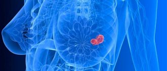
Oleogranuloma or granuloma of the adipose tissue of the mammary gland is a benign nodular formation that occurs in the subcutaneous fatty tissue of the breast, under the influence of various traumatic factors or the penetration of foreign objects or substances under the skin.
Signs of the appearance of a pathological neoplasm do not appear immediately, but 2-3 months after the introduction of irritating agents under the skin of the breast. By its nature and cellular composition, oleogranuloma is not a benign or malignant tumor growth.
How does it arise
There can be two reasons for the occurrence of granuloma, the first is the presence of any infectious disease in the body, the second is the non-infectious nature of the appearance, which includes various infiltration penetrations of exogenous foreign bodies into the tissue, as well as the presence of chronic and specific diseases. The resulting inflammatory reaction to such damage (alterations) and penetrating agents is a protective-adaptive reaction of the body, which is aimed at destroying the penetrating agent that caused micro or macro trauma, and further restoration of damaged tissues.
The process of the appearance of oleogranuloma begins at the moment when human blood cells and tissues begin to capture and digest infiltrated hostile microorganisms and solid particles (the phenomenon of phagocytosis). At the same time, they actively divide and grow, transforming into some kind of neoplasm and forming a future focus of inflammation. The spread phagocytic cells are transformed into epithelial cells, forming an infiltrate consisting of a shaft of leukocytes and lymphocytes. Then, around the accumulation of these cells, a dense epithelioid cell granuloma (capsule) is formed, which is a limited focus of productive inflammation.
Causes of breast granuloma formation
The reasons for the development of formation in the mammary gland can be divided into two types: infectious and non-infectious. The first type includes inflammatory diseases caused by pathogenic microorganisms, the second includes mechanical damage, foreign bodies or substances found in the tissues of the mammary gland.
Often, a lump forms after an injury or bruise to the chest. As a result of ruptures of small blood vessels, blood circulation in the damaged area is disrupted. Necrosis begins in this area of tissue, and inflammation grows around it. Often it goes away on its own. Sometimes a capsule of fluid forms in the affected tissue. If the contents of this capsule become infected, suppuration may occur and a compaction may form as a result. A photo of a diseased mammary gland shows reddish or bluish areas of skin at the site of formation of the node.
The development of the disease is promoted by:
- Long-term use of silicone implants. Studies have shown that after 8-10 years of use, silicone is capable of releasing substances that are toxic to tissues, provoking inflammation and triggering the formation of granuloma nodes.
- Surgical interventions. In this case, the neoplasm is localized in the area of the postoperative suture. The node can be either on the surface or inside the scar. The formation does not cause pain, fever or other alarming symptoms.
- Actinomycosis is a fungal infection of tissues. As the disease progresses, purulent granuloma nodules may form.
- Sarcoidosis of the breast. The disease affects the connective tissue of the mammary gland and in rare cases can provoke the appearance and growth of a neoplasm. Sarcoidosis often does not have severe symptoms and goes away without treatment within 2-3 years, without causing discomfort to the patient.
- Rapid decrease in body weight.
- Hormonal imbalances.
- Use of radiation therapy methods.
Women with large breasts and the elderly are at risk. After a bruise, granuloma develops rapidly. In other cases, several months may pass from the beginning of the growth of the node to the pronounced manifestation of symptoms.
Classification
Mammologists classify lipogranuloma according to the reasons that led to its appearance:
- artificially created or injected - arise at the site of plastic surgery on the mammary gland, performed using materials unsuitable for implantation: paraffin, petroleum jelly, various vegetable oils. Spreading between the fatty layers and lobules of the glandular body of the breast, foreign substances lead to the formation of multiple foci of inflammation and granulation, which subsequently cause the appearance of serious purulent processes;
- after injuries to the mammary gland (post-traumatic) - appear as a result of falls, blows, compression in everyday life or transport, or unprofessionally performed massage;
- previous surgical interventions on this breast (near inflammatory) - a granuloma is formed near the place where there is an inflammatory focus and, as it were, surrounds it. The causes of inflammatory processes can be: lactation mastitis, mastectomy, non-healing postoperative sutures, remnants of unremoved ligature stitching threads or their untimely removal;
- spontaneous (idiopathic), which appeared without obvious reasons, with little noticed, small, local skin infections or minor injuries to the mammary gland;
- as complications after radiation therapy, hormonal imbalance, rapid loss of normal weight;
- with long-term therapy with anticoagulants;
- with Weber's panniculitis.
Varieties
In modern medical science, depending on the cause and speed of the inflammatory process, the following types of disease are distinguished:
- injection (artificial) oleogranuloma (injection of oils, fats, threads under the skin);
- post-traumatic (traumatic chest injuries, consequences of an unsuccessful massage);
- periinflammatory (formation near the site of inflammation);
- spontaneous (reasons unknown).
On this topic
- Breast
Do lymph nodes enlarge with mastopathy?
- Natalya Gennadievna Butsyk
- November 29, 2020
This classification is generally accepted. Regardless of what caused the disease, it is important to pay attention to the symptoms in time and seek medical help.
Injection, post-traumatic, peri-inflammatory forms of the disease are formed in the place that was injured or injections were made. Spontaneous tumor affects any part of the body, sometimes symmetrically on the body, limiting the vital activity of the organ located in the immediate vicinity.
Stages of the disease, characteristic symptoms
- The initial stage is characterized by the following symptom complex - a lesion or lesions form in the tissues of the gland in the form of nodular tubercles of oval or round shape (accumulation of monocyte cells), they are colorless, do not hurt or bother, do not rise above the surface of the skin, but are easily visualized and palpated.
- As the lipogranuloma grows, the following symptoms appear - lymphatic edema, the gland becomes reddish or cyanotic in color, may lose its sensitivity, tuberous formations (consisting of fused macrophage cells) on it noticeably grow and increase in size. The inflammatory process involves the tissues of the mammary gland adjacent to the focal formation, the skin becomes compacted, rough, wrinkled, foci of decomposition of glandular and adipose tissue appear, a capsule-shaped infiltrate appears, which over time becomes overgrown with granulation tissue.
- The last (advanced) stage of lipogranuloma is when, due to poor circulation in the breast tissue, necrotization (death) occurs in the thickness of its tissue. The body temperature rises, discomfort occurs, the pain syndrome gradually intensifies, and soreness of the adjacent lymph nodes appears. The areas of skin above the lump are retracted, the shape of the breast and nipple changes, areas with wet cracks, trophic disturbances with the formation of ulcers appear, and fistula openings with purulent contents appear.
Symptoms of oleogranulomas
The development of the presented pathological condition is characterized by certain stages, depending on which the symptoms vary.
In the first stage, nodules or minor formations disappear after a certain period of time.
They leave behind only a small tissue compaction. Next, the oleogranuloma begins to transform into a solid formation. At this stage of development of the presented phenomenon, women most often identify it, because it is obvious.
At the last stage, a progressive increase in oleogranuloma is identified, which resembles a malignant neoplasm, however, in fact, it is not one and will never degenerate into an oncologically dependent tumor.
The symptoms of the condition are directly related to numerous nodules and plaques, which are identified by their round or oval shape. They also have different sizes, smooth or bumpy surface. The consistency that is characteristic of the lesions is dense or cartilaginous. In the initial stages, the neoplasm is painless and colorless, but after a certain number of months it suddenly begins to turn a bright copper shade. At the same time, the oleogranuloma begins to seriously hurt, almost without interruption.
It should be noted that the histological type analysis demonstrates a granuloma with a significant proportion of fat cells. They appear as numerous cavities. A possible, although rare, but nevertheless very real transformation into sarcoma is possible. Mammologists note that injection oleogranulomas should be considered the most common type.
Activation of the inflammatory process is characterized by general symptoms:
- an increase in body temperature;
- significant weakness;
- constant malaise.
All this indicates to the woman that the disease is entering an advanced stage and it is necessary to attend to diagnosis.
Diagnosis of oleogranuloma
- Self-examination by feeling and palpating the breast by the woman herself. If any dense neoplasm is detected, you need to contact a doctor who will prescribe clarifying examinations.
- X-ray examination (mammography) - it will clearly show the location, area of the lesion and its size. Evidence of the presence of oleogranuloma will be thin, dense capsule-shaped lesions visible in the photo with clearly defined boundaries. Another indicator visible in the image indicates the presence of a granuloma - a site of accumulation of calcite (calcium salts).
- Ultrasound (layer-by-layer) scanning or ultrasound allows you to determine neoplasms that were not visible on the mammogram, as well as the nature of soft tissue lesions, their consistency, and fluid filling. In the scan results, lipogranuloma will be defined as a lesion with high density (increased echogenicity). Simultaneously with the research process, material can be taken for histology and biopsy.
- Aspiration biopsy or cytological examination of the composition of cellular structures affected by fatty necrosis of breast tissue. The study will allow us to accurately determine whether the neoplasm is benign or malignant.
- Histological examination of a biopsy of glandular tissue (trepanobiopsy or fine-needle puncture). It is carried out under X-ray or ultrasound control.
Diagnostic measures
The diagnosis of oleogranuloma can be made based on visual examination and palpation, ultrasound and mammography, as well as cytological and histological examinations.
When performing any type of mammography, a round formation of low density is identified; most often it is characterized by smooth and clearly defined contours.
The structure is heterogeneous, in addition, the capsule may be partially inhibited by calcium particles.
Treatment of the mammary gland for nodular mastopathy
In some situations, the boundaries of the formation are unclear and the contour is uneven. All this can create certain difficulties in terms of differential diagnosis with oncologically dependent tumors or initial processes of the mammary gland. Therefore, an additional examination is recommended, within the framework of which the oleogranuloma and its nature will be 100% clear.
An important element of diagnosis should be considered examination after the rehabilitation course. In some cases, the presented pathological condition may have recurrent forms. In order to avoid this or facilitate another stage of treatment, it is recommended to perform an ultrasound scan every six months, as well as undergo tests.
Removal of oleogranuloma
Oleogranuloma of adipose tissue is not amenable to conservative treatment; it cannot be cured by various physiotherapy procedures or medications that are not able to penetrate the inflamed infiltrate of lymphogranuloma and act there. Lipogranuloma can only be removed surgically.
Surgical removal of granulosa formation is performed using the method of low-traumatic, sectoral organ-preserving resection (excision) of the tumor, together with adjacent areas of adipose and glandular tissue. Excision is carried out to the extent of healthy tissue and without removing lymph nodes. If fluid is detected in the necrotic area, then aspiration (pumping out) of the liquid contents is carried out. The volume and nature of surgical intervention depend on the location of the tumor-like nodule and its size.
Analyzes during preparation
In preparation for surgery, the following tests are prescribed:
- blood tests: biochemical, general, blood clotting time;
- blood test for tumor markers;
- determination of blood group and Rh factor;
- blood test for syphilis, HIV, hepatitis B and C.
The intervention is carried out under general (if the formation is located deep in the tissues) or local anesthesia. The breast surgeon first (marks) marks the incision lines on the skin, then dissects the breast tissue, under hand control, with two arcuate incisions radially towards the nipple. In small areas of pathological formations, all affected (necrotic) fragments are removed and the wound cavity is cleaned.
After stopping the bleeding (hemostasis), the edges of the wound are sutured while simultaneously grasping its bottom, so sutures are first placed on the subcutaneous area. And cosmetic or interrupted sutures are applied directly to the skin.
If the oleogranuloma is in an advanced state and has a large area of coverage, up to 1/4 of the breast lobe can be removed (quadrantectomy). In this case, the surgeon will perform plastic surgery simultaneously with the excision operation. It will consist of covering the open area with a skin flap and performing a complex of reconstructive mammoplasty procedures. The skin flap is transplanted from the back or forearm.
During the operation, a histological rapid analysis of the biopsy material taken from the infiltrate is necessarily performed. This will make it possible to verify the good quality of the contents of the granuloma capsule and exclude suddenly starting low-quality processes. Postoperative sutures are usually removed after a week or on the 10th day.
Possible complications
After surgery for sectoral removal of oleogranuloma, it is extremely rare, but the following complications may appear:
- suddenly appeared purulent discharge from the wound - occurs due to infections;
- bloody subcutaneous hematomas appear - occur when bleeding is poorly stopped during surgery or due to existing blood clotting disorders;
- high fever and pain usually disappear completely 2-3 days after surgery.
Modern methods and methods of treatment
Granulomatous breast lesions in women begin to be treated only after excluding the oncological nature of the pathology. The choice of treatment method depends on the cause of granuloma formation.
So, if the disease is caused by long-term wearing of a silicone implant, then the doctor, first of all, recommends removing or replacing it.
In medical practice, treatment of granulomatosis is carried out using the following methods:
Conservative treatment
Women are prescribed a course of antibiotics, corticosteroids, immunomodulators and vitamins. The goal of such treatment is to eliminate individual signs of inflammation and activate the body's resistance.
Surgery
Radical intervention is carried out under local anesthesia or general anesthesia. Before surgery, doctors must determine whether the patient has an allergic reaction to painkillers. During the operation, the surgeon excises the granulomatous tissue and sutures the postoperative wound.
Innovative treatment technologies include:
- Cryotherapy, in which breast granuloma is removed by deep freezing with liquid nitrogen.
- Laser therapy - in this case, treatment is based on the destructive effects of a laser beam.
- Diathermocoagulation - doctors recommend that patients with small single granulomas undergo a procedure for excision of the tumor using an electric current.
Most leading specialists use a wait-and-see approach to therapy, which consists of sequential conservative, minimally invasive and surgical treatment technologies.
Recovery period
The complex of postoperative therapeutic measures includes a course of drug therapy, consisting of: immunomodulatory, anti-inflammatory, antibacterial medications and vitamin preparations, as well as a rehabilitation course that accelerates rehabilitation, and physical procedures.
You can prevent the occurrence of unpleasant complications in the postoperative period if you follow these rules:
- timely removal and treatment of sutures until they are completely healed;
- wear post-operative compression garments (support top) for as long as the doctor requires;
- you need to inform your doctor about any allergies you have to antiseptic drugs and synthetic materials;
- timely treatment of infectious chronic foci with a full course of antibiotics;
- For at least 30 days it is prohibited: visiting saunas and baths, lifting and carrying heavy objects, not playing sports, not actively sunbathing, not visiting a solarium, not taking baths with hot water.
Treatment of complications
Treatment of various complications leading to deformation of the postoperative scar is carried out with the help of surgery, but sometimes conservative treatment methods can also help. So, hematomas in most cases go away on their own, leaving no traces. But in some cases, their removal can be carried out using punctures (a needle is inserted into the postoperative scar and excess fluid is removed from it through the needle) or through surgery (if the hematoma grows, but during a repeat operation the source of bleeding is identified and stopped). To speed up the resorption of the hematoma, you can use Arnica cream after obtaining permission from your doctor.
Physiotherapy methods are used to treat infiltration, as well as antibacterial therapy and bilateral novocaine blockade according to Vishnevsky. Complete resorption of the infiltrate with adequate treatment should occur in 10-12 days. If this does not happen, the abscess is opened, and pus is removed from it using a double-lumen tube or cotton swab.
In order to cure suppuration of a postoperative scar, you need to remove the sutures from it and thoroughly clean the wound of pus and dead tissue, rinse and drain it. If the suppuration has spread greatly, all dead tissue must be excised. After such a procedure, the wound requires especially careful care.
In case of formation of a granuloma of a postoperative scar, the scar tissue is excised, all granulomas and non-absorbed suture material are removed. In the first three months after removal of the granuloma, it is necessary to ensure that the wound is clean and dry. Subsequently, after consultation with a doctor, you can use Contractubex or Mederma cream, which will speed up the resorption of the scar.
Postoperative scars with seroma can be treated with puncture, when excess serous fluid is sucked out through an inserted needle. After this, a bandage is applied to the scar, and after 3-5 weeks, repeated punctures may be required.
Treatment of endometriosis in post-surgical scars can be done with hormones, surgery, or a combination. Synthetic progestins are used for hormonal treatment. Surgical treatment is often combined with preoperative hormonal therapy.
Prevention
As preventive measures to prevent the appearance of such granuloma formations of the breast, breast surgeons recommend:
- timely treatment of any form of mastopathy, periodic monitoring of your hormonal levels (status) and adjusting it if necessary;
- if lactostasis or mastitis is detected, they must be treated promptly and fully with antimicrobial drugs;
- do not perform plastic surgery, especially in places where fat or oil emulsions will have to be injected into the glandular tissue of the breast;
- after surgery, you should strictly adhere to the doctor’s recommendations for the entire rehabilitation period;
- avoid bruises, blows to the chest, try not to injure it during sports;
- regular breast self-examination;
- annual preventive ultrasound or mammological examination of the breast.
Similar articles
No similar articles
Causes
- A small lump may appear due to recent surgery. The neoplasm is localized both outside and inside the scar. The size of such a compaction rarely reaches large sizes; often the granuloma on the scar feels like a pea or a bump.
- The reason for the appearance of granulomatous compaction lies in the use of non-absorbable materials when suturing the wound. There are no other symptoms, such as pain and fever. In this case, there is no reason to worry. The lump is benign and will disappear on its own within a short period of time.
- Another cause of granuloma can be the entry of foreign particles into the wound, such as talc and starch, which are used for rubber gloves. If sanitary rules are not followed during surgery, microbes can enter the body and can trigger the onset of an inflammatory process.
- Infection of tissues by microorganisms will certainly have its consequences. There is an increase in body temperature, general malaise, and pain in the operated area. In this case, an urgent consultation with a doctor will be required to select the necessary treatment. You should not resort to home treatment using traditional methods. There is a risk of developing serious complications.
Redness of the skin, high temperature, acute pain - all these signs may indicate the appearance of pus. It is recommended to undergo an examination and additional examinations to differentiate a granuloma from a seroma or fistula.
Lipogranuloma of the mammary gland, the so-called steatogranuloma, can develop as a consequence of surgical intervention, be it breast surgery or sectoral resection. It is fat necrosis of lipocytes. Lipogranuloma of the mammary gland can also appear due to injury. Injury leads to impaired blood circulation inside the gland, which causes the development of the inflammatory process.
Granulomatous formations may appear asymptomatically. For this reason, it is necessary to regularly palpate the suture area to be able to diagnose the seal in the early stages.


