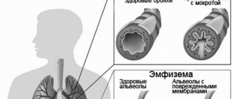
What is pulmonary fibrosis? Fibrosis is scarring; Pulmonary fibrosis is the irreversible formation and replacement of the lung parenchyma with fibrous tissue. Lung fibrosis always leads to impaired respiratory function, because the elasticity of the lung tissue noticeably decreases.
As a result, this makes it difficult to exchange oxygen in the alveoli. Namely, it is in the alveoli that gas exchange occurs: upon contact with blood, oxygen from the air passes into it, and carbon dioxide leaves. Recovery from pulmonary fibrosis does not occur because regeneration of fibrosis into the lung tissue does not occur.
What is pulmonary fibrosis?
Pulmonary fibrosis is a chronic lung disease characterized by progressive scarring of the tissue that covers the organ: the lung parenchyma.
As the wall of the pulmonary alveoli (the sac-like structures in which the smaller bronchial branches terminate) thickens and is reinforced by this progressive scarring, their ability to exchange gases decreases (that is, the ability to carry inhaled oxygen into the bloodstream and exhale much of the carbon dioxide into the air, which secreted by blood). As a result, the body no longer receives sufficiently oxygenated blood (i.e., free of carbon dioxide and saturated with oxygen).
Over time, scar tissue closes the capillaries of the pulmonary alveoli, further impairing gas exchange and causing circulatory and cardiac consequences (causing a disease known as chronic cor pulmonale).
Description
Pulmonary fibrosis is a process characterized by the replacement of lung tissue with fibrous (scar) tissue, which is accompanied by impaired respiratory function.
Thanks to the work of our lungs, the blood is saturated with oxygen necessary for energy consumption, as well as the release of carbon dioxide, which is formed as a by-product during the life of cells. The development of pulmonary fibrosis leads to a decrease in the volume of normally functioning lung tissue, and as a result, breathing efficiency decreases.
Replacement of pulmonary connective tissue can occur in one lung or in both at the same time. Depending on this, unilateral and bilateral fibrosis are distinguished. In addition, pulmonary fibrosis is divided into focal, in which a small area of lung tissue is affected, and total, in which the pathological process spreads to almost the entire lung.
The reasons for the development of pulmonary fibrosis are varied:
- diseases of the respiratory system (chronic bronchitis with broncho-obstructive syndrome, pneumonia, tuberculosis, chronic obstructive pulmonary disease);
- connective tissue diseases (rheumatoid arthritis, systemic lupus erythematosus, systemic scleroderma);
- exposure to production factors that negatively affect the respiratory system. For example, with prolonged inhalation of silicate dust in production, silicosis occurs. Occupational diseases also include asbestosis, which occurs due to inhalation of asbestos dust;
- long-term use of certain medications (antiarrhythmic drugs, drugs used to treat malignant tumors);
- the presence of vasculitis (a disease accompanied by inflammation of the walls of blood vessels);
- idiopathic or primary pulmonary fibrosis, the exact cause of which has not been established.
Relevance of the problem:
This disease is not so common in the modern world, but it still occurs. So, if treatment and diagnosis are not timely, the problem can become serious.
At present, scientists have not yet fully elucidated
the exact reasons for the development of such a disease. There are many predisposing factors to the development of fibrosis, the most important of which are constant contact with environmental dust. A consequence of silicosis or sarcoidosis changes in the lungs. This is usually due to harmful factors in silicon production.
In addition, a predisposing factor is chronic low-grade pneumonia, which exists for a long time.
The combination of the following factors influences the development of the disease:
- Exposure to various chemicals that are in the air. These may include various pesticides and toxic fumes.
- Regular and prolonged intake of foreign particles such as dust, pollen and flour into the upper and, accordingly, lower respiratory tract.
- Long-term exposure to an infectious factor on the body, with the development of an inflammatory process.
- An allergic reaction of the body to a chronic or long-term exposure agent. As a result, the lungs are exposed to aseptic inflammation for a long time.
- Exposure to radiation energy. This may be due to the treatment of cancer or radiation exposure, for example among liquidators of the Chernobyl disaster.
In addition, the following conditions contribute to the development of the disease:
- Smoking, for a long time and in large quantities.
- Age threshold: People aged about 50 years are most often affected by the disease.
- Environmental conditions. The disease often develops in people living in industrial cities or densely populated cities.
- People with a family history of similar diseases are more likely to develop pulmonary fibrosis.
- Constant contact of an irritant from the outside.
Diagnosis of fibrosis
If pathological symptoms appear, the patient goes to the clinic. Treatment and diagnosis of this disease is carried out by a gastroenterologist or general practitioner.
In order to make a diagnosis, the doctor conducts an examination. At the initial stages it will not be informative. But if the patient has fibrosis of the 3rd or 4th degree, then upon palpation the liver will noticeably increase in size, as well as its denser structure. The patient may complain of pain when pressing.
Additionally for diagnosis use:
- general blood test - decreased hemoglobin, red blood cells, increased ESR;
- general urine analysis - the presence of protein, casts, bilirubin in it;
- biochemical blood test - increased activity of all liver parameters (ALT, AST, bilirubin, alkaline phosphatase, etc.);
- Ultrasound of the liver - during the examination, it is possible to detect an increase in the size of the organ and a change in its structure: strands of connective tissue, foci of fibrosis, parasitic cysts, dilation of the bile ducts and liver vessels;
- indirect elastometry - performed using a fibroscan, allows you to evaluate the structure of the liver without compromising the integrity of the skin. The device evaluates tissue elasticity: fibrous tissue is denser than normal liver parenchyma;
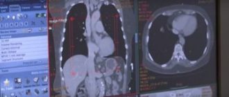
- MRI, CT – the quantity and quality of fibrous lesions are determined.
But to make a diagnosis of liver fibrosis, a biopsy is necessary. During the examination, a piece of affected liver tissue is taken for analysis using a thick trepanation needle (under ultrasound guidance).
To assess the stages of fibrosis, the following scale is used:
Formation of fibrous tissue
- 0 degree – no fibrosis;
- fibrosis of the 1st degree – liver functionality is impaired. The portal tracts are stellate in shape. If the disease is detected on time and treatment is started, the prognosis is favorable;
- fibrosis of the 2nd degree – the number of fibrotic foci increases. Single septa appear in the hepatic lobes. With the help of medications, normal liver functioning is possible;
- fibrosis of the 3rd degree - the liver is penetrated by strands of connective tissue, its size is increased, the bile ducts are dilated. The prognosis is unfavorable. Drug therapy provides little relief.
- Stage 4 – the disease progresses to cirrhosis, which cannot be treated. The only way to survive with such a diagnosis is liver transplantation.
If the patient has contraindications to a biopsy (reduced blood clotting, parasitic cysts, serious condition of the patient), the diagnosis of liver fibrosis can be made on the basis of elastometry.
Symptoms of pulmonary fibrosis
Pulmonary fibrosis tends to present with the following symptoms:
- shortness of breath, wheezing (shortness of breath occurs at the beginning due to physical exertion, then it begins to appear more and more when a person gets dressed, walks around the house or talks);
- chronic “dry” cough that does not produce sputum;
- weakness, asthenia;
- chest discomfort;
- loss of appetite and weight.
Less commonly, the following symptoms may also be present:
- fever;
- pain in muscles and joints;
- hemoptysis (the release of blood from the respiratory tract, usually through coughing).
Pulmonary fibrosis - symptoms
In the early stages of the pathology, signs of pulmonary fibrosis are completely absent. However, at this time the disease progresses rapidly. One of the possible signs of the disease at this stage may be shortness of breath. At first, it appears only after physical activity (climbing stairs, long walking), but after a short time it begins to accompany the patient constantly.
Many people learn about pulmonary fibrosis, what kind of disorder it is, only at the stage of dry cough. The substance periodically coughed up from the lungs contains mucus.
If pulmonary fibrosis is suspected, symptoms may include the following:
- constant pain in the chest area;
- audible wheezing when breathing;
- cyanosis of the lips (blue discoloration);
- weight loss;
- increase in the size of the phalanges of the fingers;
- decreased performance;
- increased fatigue.
Types of disease
The classification of fibrosis varies among specific organs. In the liver, the type of disease depends on the location of the scars in its lobules:
- focal;
- perihepatocellular;
- zonal;
- multibular;
- bridge-like;
- periductular;
- perivenular.
Pulmonary fibrosis can be local and diffuse. Fibrosis of the prostate gland can be focal and with nodose hyperplasia, with cyst transformation and parenchymal atrophy. Sometimes a congenital form occurs.
Local and focal fibrosis is the initial stage of the disease, when isolated areas of tissue are damaged. With a diffuse disease, the damage covers most of the organ. Cystic fibrosis is characterized by damage to the exocrine gland, the ducts become blocked and cysts form. This leads to the development of disorders in the respiratory organs and gastrointestinal tract.
Among the sensory organs, epiretinal fibrosis of the eye occurs, when changes of varying degrees occur in the structures of the vitreous body and retina. Men may develop cavernous fibrosis of the penis. Women in some clinical situations may develop linear breast fibrosis.
Characteristic symptoms and types of disease
In the primary stages, the disease is extremely difficult to diagnose. Possible progressive development of fibrosis without any manifestations for a long time. Depending on the degree of spread of the disease, one can distinguish:
Local or focal fibrosis results in an increase in connective tissue, usually as a result of inflammation. In this case, older people are at increased risk.
As a result of the inflammatory process, a small area of the lung is affected. There is a compaction of lung tissue and a decrease in lung volume due to an increase in connective tissue. These minor changes do not inhibit the general functions of gas exchange of the lung, so the course of the disease can occur unnoticed by the patient.
- Diffuse fibrosis is a large-scale lesion of large areas of the lung. Destructive processes cover most of the lung, which leads to a significant decrease in its volume. The mechanical parameters of the lung deteriorate. In the area of the lung susceptible to fibrosis, significant areas of collagen fibers are observed.
In some cases, this process can also affect the blood vessels of the lungs. Linear fibrosis can also be unilateral or bilateral.
One of the main symptoms of the disease is shortness of breath. Initially, its manifestation is noticeable only during significant physical exertion, and only over time does it become a permanent ailment. Subsequently, another symptom appears - a dry cough, often with mucous discharge.
Such an unpleasant phenomenon appears as cyanosis of the skin, which primarily affects the phalanges of the fingers and the oral mucosa. People with fibrosis often begin to suffer from bronchitis and pneumonia. When coughing, purulent discharge is also observed.
Doctors distinguish two stages of the disease.
- At an early stage, sharp pain in the chest, wheezing (especially in a horizontal position and during sleep), frequent and profuse sweating appears. Since the process of formation of connective tissue has begun, the lungs feel a lack of oxygen, usually during physical exertion.
In the secondary stage, there is a significant shortness of breath. The mucous membrane and skin are characterized by an unpleasant bluish tint. The nails become deformed and changes in the outer phalanges of the fingers can be noticed.
Linear fibrosis leads to thickening of the roots of the lungs as the amount of connective tissue increases. As pathological changes increase, the patient experiences elevated temperature and abnormal breathing rhythm. Breathing becomes shallow and rapid.
Symptoms of heart failure may occur. In the further development of the disease, the clinical picture may extend beyond the respiratory system. You may notice swollen veins in the neck and swelling of the lower extremities. Significant reduction in body weight is possible. Working capacity decreases, lethargy and apathy begin to appear. At this stage, qualified medical assistance from a specialist is extremely necessary.
Complications
In the long term, a number of complications may arise that will negatively affect the patient’s clinical picture and quality of life. The most important are:
- hypoxemia and hypercapnia (decreased O2 and increased CO2 in the blood), which can be assessed using blood gas analysis;
- pneumothorax (abnormal presence of air in the pleural cavity, preventing normal expansion of the lungs);
- cyanosis (bluish discoloration of the skin and mucous membranes);
- hypertrophy of the accessory muscles of respiration;
- pulmonary hypertension;
- Hippocratic fingers (flask-shaped thickening of the terminal phalanges of the fingers and toes).
These complications, along with chronic respiratory failure, can then lead to the patient's death after several years of illness.
Medicines
The main drugs used in the treatment of pulmonary fibrosis are:
- glucocorticoids;
- cytostatics;
- antifibrotic agents.
The administration of systemic glucocorticoids (prednisolone, dexamethasone) alleviates the general condition of the body. It is also believed that drugs in this group can influence the replacement of normal lung tissue with connective tissue, inhibiting this process. However, there is also a negative side to taking glucocorticoids. In the treatment of fibrosis, glucocorticoids are prescribed for a long period of time; in some cases, the course of treatment can reach up to 3 months. Such prolonged use is dangerous for the development of side effects. These include:
- increased blood pressure, which is especially dangerous for people suffering from arterial hypertension. While taking glucocorticoids, resistance to early antihypertensive therapy may occur;
- exacerbation of peptic ulcer of the stomach or duodenum;
- osteoporosis, which leads to increased bone fragility;
- weight gain;
- hyperglycemia. Therefore, when taking glucocorticoids, it is important to monitor glycemic levels. Particular attention should be paid to people with diabetes.
The appearance of these symptoms is the reason for contacting your doctor, who, in turn, either adjusts the dosage of the drug or discontinues it.
The use of cytostatics (azathioprine, cyclophosphamide) is also accompanied by some side effects: the function of the gonads is disrupted, hematopoiesis is inhibited, undesirable effects from the gastrointestinal tract, nephro- and hepatotoxicity are observed. The most gentle drug in this group in this regard is azathioprine. This drug is able to block cell division and degeneration of tissue into fibrous tissue, which has a significant role in the treatment of pulmonary fibrosis. Taking the drug is strictly prohibited during pregnancy and lactation, and is also undesirable if you have existing renal or liver failure.
Colchicine also belongs to antifibrotic drugs, which can inhibit the production of fibronectin. With long-term use, a picture of myelosuppression can be detected, that is, a decrease in leukocytes in the blood, and thrombocytopenia (decrease in platelets), temporary alopecia, myopathy, peripheral neuritis, etc. is not a rare case. The drug is contraindicated in cases of severe impairment of liver and kidney function, cardiovascular pathology, purulent infections, pregnancy and lactation. In all other cases, the use of the drug is justified.
How to treat
Diagnostic tests are carried out by a pulmonologist. He appoints:
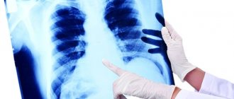
- X-rays of light. The main symptom of the disease is a pronounced pattern and change in the lung. In the picture you can see the vessels and the shadows extending from them. At an advanced stage of the disease, shadows in the form of honeycombs and tissue scarring will be visible.
- Study of the functioning of external respiration. Lung capacity is checked. Its low rate indicates a disorder in the organ and the development of pulmonary diseases.
- Laboratory Used to exclude other diseases, such as tuberculosis.
- Study of the bronchi. A part of the lungs is taken and examined using a camera. This test will show what stage the disease is at and how far it has spread.
Recent and chronic lung diseases of the patient must be ascertained. The doctor makes an oral interview with the patient and studies his medical record.
The disease can be confused with cancer
Therefore, it is very important to conduct a comprehensive examination of the body.
Since the process of proliferation of connective tissue is irreversible, treatment is considered ineffective. It is aimed, first of all, at eliminating the concomitant disease. That is why it is important to study the exact causes of the pathology. Incorrect treatment is a consequence of an incorrect diagnosis.
If the main factor is pneumonia, then a course of treatment with antibiotics is prescribed in combination with other medications and physical therapy. Inflammation must be treated until it disappears completely. After therapy, the patient is observed by a doctor for another year.
In the absence of pulmonary insufficiency, treatment with ions and ultrasound can be performed. If the organ does not have enough air, oxygen therapy is prescribed. Cardiac medications are required to prevent heart attacks.
At a severe stage, pleural drainage may be prescribed. If fibrosis progresses rapidly, surgery will likely be required. The patient is prescribed a special diet, limited physical activity and complete rest.
Symptoms of the disease
Fibrosis develops slowly and at first the patient does not have any complaints. In rare cases, people experience health problems and consult a doctor. There may be regular fatigue. Then disturbances in the functioning of organs appear, in some cases blood flow worsens.
With liver fibrosis, general malaise is initially observed. After a slight blow, bruises appear on the skin. Liver destruction lasts six to eight years, after which critical symptoms occur. Liver function is significantly impaired as scar tissue cells grow and close together. Further, the spleen increases in size. Other complications include varicose veins of the esophagus and bleeding from them. Then either anemia, thrombocytopenia or leukopenia develops.
At the first stage of development, clinical tests show that fibrotic changes in the liver are insignificant. The disease can be determined by the fact that splenic and portal pressure has increased. Ascites may sometimes appear and disappear. There is also a feeling of heaviness in the right hypochondrium and problems with digestion. Sometimes itching and rashes occur on the skin.
Pulmonary fibrosis can be signaled by shortness of breath, which worsens over time and is accompanied by a dry cough. Then chest pain and rapid shallow breathing occur. Cyanosis is noted on the skin. Frequent bronchitis and heart failure may indicate the progressive development of the disease.
Women may develop focal fibrosis of the mammary gland during hormonal changes. It can be felt by palpation only when the compaction reaches a size of 2–3 millimeters or more. The skin over the affected area will change color. Over time, discomfort in the chest occurs, and then the pain increases. As the disease progresses, there may be a clear or pale discharge from the nipple. There is a feeling of fullness in the chest and heaviness in it. Then the pain intensifies, becomes aching and constant, radiating to the armpit and shoulder.
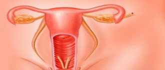
The danger of uterine fibrosis is that fibroids can be a complication. Pain in the lower abdomen and prolonged menstruation, as well as discomfort during sexual intercourse, may signal the development of the disease.
Symptoms of pancreatic fibrosis are decreased appetite and decreased body weight, diarrhea and vomiting, pain in the hypochondrium on the left side and flatulence.
Cardiac fibrosis is characterized by changes in blood pressure and shortness of breath, as well as disturbances in the rhythm of the heart. Initially, aortic valve fibrosis does not show any symptoms. Over time, pain in the heart and dizziness occur, and then the heartbeat quickens, shortness of breath occurs and the patient may lose consciousness.
In men, pain in the perineum and lower abdomen, discomfort during intimacy and urination may indicate prostate fibrosis. Then erection problems arise and libido decreases. Complications may include pyelonephritis, renal failure and hydronephrosis.
Diagnostics
When diagnosing pulmonary fibrosis, the patient's medical history is very important, understood as family history (other cases of pulmonary fibrosis in close relatives) and occupational exposures or taking medications and therapies.
The doctor, based on an assessment of the patient’s characteristics, may also prescribe:
- chest x-ray , high-resolution computed tomography of the chest , biopsy to visualize and evaluate lung characteristics;
- Blood gas test : This is a test that is done on a sample of arterial blood taken from an artery in the wrist. This test helps measure the amounts of oxygen and carbon dioxide in arterial blood;
- oximetry : performed with a small pincer instrument that is attached to a finger or earlobe. Indicates the amount of oxygen present in arterial blood (it is normal for the reading to be around 95-100%);
- pulmonary function test (spirometry and studies of lung diffusion capacity): the study provides information on the exchange capacity of lung gases, oxygen and carbon dioxide;
- 6-minute walk test (6 MW). The patient is asked to walk for 6 minutes while monitoring his oxygen saturation and measuring the meters walked. Evaluates the degree of functional limitation that defines the disease.
Pulmonary fibrosis – what is it?
The most common questions from patients who first learned about pulmonary fibrosis: what is it, is it dangerous or not, how does it manifest? Fibrosis refers to the process of replacing healthy tissue of internal organs with fibrous or scar tissue, as it is also called. By its nature, it is connective tissue that is capable of rapid growth. As a result, after a short time it can absorb the entire organ, disrupting its normal functioning.
With pulmonary fibrosis, there is a marked decrease in the elasticity and extensibility of the lung tissue. As a result, the passage of oxygen and carbon dioxide through the alveolar walls becomes difficult. Gas exchange is disrupted: the blood is poorly saturated with oxygen, the concentration of carbon dioxide in the bloodstream increases, which leads to chronic hypoxia.
Types of pulmonary fibrosis
Having understood the term pulmonary fibrosis and what kind of pathology it is, it is necessary to note the possible types of the disease. When pathology occurs, doctors always carry out comprehensive diagnostic measures that are aimed at establishing the cause of the disease. Depending on this, the following types of disease are distinguished:
- Idiopathic pulmonary fibrosis
is diagnosed when a specific cause cannot be identified. - Interstitial pulmonary fibrosis
- diagnosed when a factor that provoked the pathology is detected.
By volume of lung tissue damage:
- linear pulmonary fibrosis - small areas of tissue are affected;
- focal – a small area, of which there may be several, is involved in the pathological process;
- diffuse pulmonary fibrosis – a large volume of the organ is damaged.
Pulmonary fibrosis - causes
The causes of pulmonary fibrosis are directly related to the inflammatory process in the alveoli and interstitial tissues. In most cases, they are a trigger for the development of pulmonary fibrosis.
Among the possible causes of pathology, experts identify:
- heredity (the presence of lung pathology in relatives increases the risk of developing fibrosis);
- complications of pulmonary diseases (asbestosis, alveolitis, sarcoidosis);
- prolonged inhalation of particles of organic substances and minerals (work in chemical production);
- radioactive irradiation of the chest;
- complicated course of diabetes mellitus, rheumatoid arthritis, systemic lupus erythematosus;
- smoking.
Doctors at risk for developing pulmonary fibrosis include:
- persons over 50 years of age;
- people living near industrial enterprises;
- smokers;
- workers of hazardous production workshops.
Why is pulmonary fibrosis dangerous?
Prolonged absence of therapy leads to rapid progression of the disease. Over time, the risk of developing life-threatening complications only increases.
According to experts, pulmonary fibrosis can provoke the following pathologies:
- emphysema;
- pulmonary circulation disorder;
- heart failure;
- pulmonary hypertension.
How to diagnose pulmonary fibrosis?
Diagnosis of this disease is often delayed and treatment begins late, when irreversible processes have begun in the lung tissues and in the body as a whole.
First of all, the patient usually comes to see a therapist.
- The role of the initial conversation, clarification of all complaints, and the sequence of their occurrence is very important. It is necessary to clearly identify the time of onset and the degree of progression of the disease.
- In addition, you need to collect a detailed medical history. The presence of negative environmental factors, bad habits, household history. Professional history also deserves special attention. Place of work, presence or absence of hazards, length of work experience.
- It is also important to find out what diseases your closest relatives suffered from. Especially those that concerned the respiratory system.
- The main symptoms can be identified by visual examination. Changes in the skin are visible, their color is not healthy. The chest also takes on a different appearance; it becomes barrel-shaped and expanded.
- Lung auscultation and percussion are widely used.
In addition, laboratory and instrumental examination methods are widely used.
First of all, these are laboratory indicators.
- Such as a general blood test. There are no specific changes in it, but you can see the appearance of leukocytosis, due to the addition of infection, as well as the level of hemoglobin and red blood cells.
- A method called pulse oximetry is used. Which allows you to assess the degree of oxygen saturation in the blood.
- To confirm the diagnosis, chest radiography is used, but in most cases it is not sufficiently informative; computed tomography is performed for this purpose. It will show structural changes in the lungs more clearly and understandably. The only disadvantage of this method is the price.
- In addition, lung function and its possible tidal volumes are assessed. It is carried out using spirography. This is a portable device that is used for non-invasive assessment of lung function.
- To assess the degree of conductivity of the respiratory tract, the bronchoscopy method is used, but it allows one to assess the condition of the respiratory system, the severity of inflammation of the bronchial tree, as well as the nature of sputum deposition.
Additional Information . To make a clearer diagnosis and identify the main component in the development of the process, an invasive method is used, this is a biopsy of lung tissue. To do this, take a small piece of tissue, possibly from several places. Especially those that aroused suspicion after radiation research methods.
Diagnostic methods
First, auscultation and percussion of the lungs;
- X-ray - fibrous areas appear darkened on the fluorogram.
- With fibrosis of the root part, the root is stringy due to the enlargement of local lymph nodes, so symptoms are taken into account here.
- After radiography, MRI and CT are prescribed;
- Carrying out bronchoscopy - inserting a probe into the bronchi and examining them.
Spirography – reveals different aspects of respiratory function and determines vital capacity (VC):
- the respiratory rate (RR) is determined - the number of respiratory cycles per minute;
- DO – tidal volume – the amount of air inhaled at one time;
- MOD – the same volume, but per minute;
- Vital capacity – after a maximum quiet inhalation, the volume of exhaled air;
- forced vital capacity (FVC) – the same thing, but after a full exhalation;
- maximum ventilation (MVV) - RR is multiplied by the amplitude of respiratory movements.
A biopsy is also prescribed - a biopsy sample is taken from the area of fibrosis on an x-ray. Cystic fibrosis requires analysis of sweat, stool for chymotrypsin and fatty acids, and PCR diagnostics.
Treatment of pulmonary fibrosis
There are no specific treatments for this disease, which usually has a progressive course. Several experimental treatments for pulmonary fibrosis are currently being studied in some experimental clinical trials.
In younger patients, the most effective therapeutic option is lung transplantation.
However, there are a number of treatment methods aimed at slowing the progression of the disease and improving the patient’s quality of life (oxygen therapy, cortisone and immunosuppressants, pulmonary rehabilitation programs):
- Cortisone : an anti-inflammatory drug that suppresses the immune system's response. With long-term use, cortisone can also cause severe side effects (this drug exposes the patient to the risk of infections, can cause metasteroid diabetes, weight gain, osteoporosis, peptic ulcers, mental changes);
- Cyclophosphamide , Azathioprine : These drugs suppress the immune system's response;
- N-acetylcysteine : This is a mucolytic, expectorant, and antioxidant that also helps sick patients with pulmonary fibrosis;
- Oxygen therapy : serves to replenish the lack of oxygen caused by decreased lung function. The requirement is established based on blood gas analysis and oximetry. It can be administered only at night, during physical activity, or throughout a 24-hour period;
- Respiratory rehabilitation programs : its task is to teach the patient to breathe better and tolerate stress;
- Lung transplantation : This is the definitive therapy for pulmonary fibrosis. It is intended in selected cases, for patients without other concomitant diseases. Lung transplantation is a very complex procedure and is performed only in highly specialized centers.
All patients with pulmonary fibrosis should combine therapy with a healthy lifestyle, which includes:
Treatment of linear fibrosis
The above material provides a complete description of the diagnosis of linear pulmonary fibrosis, what does this give the patient? The answer is the correct choice of treatment method.
It is no longer possible to get rid of the connective tissue that has formed as a result of the disease. Therefore, the treatment of this disease includes measures that stop the process of creating new connective tissues.
First of all, it is necessary to exclude the possibility of inhaling harmful substances that stimulate the development of the disease. If the patient smokes, then getting rid of this addiction is simply necessary. It is much more difficult to protect yourself from infectious diseases, but it is still necessary to take all preventive measures.
Conservative therapy is effective only in the early stages of the disease. Drug treatment can eliminate the symptoms of the disease and improve the patient’s quality of life. To achieve the greatest effectiveness in the early stages, it is advisable to use therapeutic breathing exercises, oxygen therapy and a moderate diet in combination with medications.
Causes and mechanism of development of pneumofibrosis
The causes of the disease are different:
- infectious and inflammatory diseases - mycoses, tuberculosis, syphilis;
- prolonged exposure to allergens;
- mechanical injuries;
- high doses of radiation;
- exposure to chemical compounds;
- hereditary disorders in which fibrillar protein is intensively produced - collagen, fibronectin;
- severe liver damage due to drug addiction;
- long-term use of drugs that have a toxic effect on the lung parenchyma.
The risk group includes people with a history of COPD, bronchial asthma, chronic bronchitis, and pneumonia. More often, men whose professional activities are associated with the risk of negative effects of production processes on the respiratory system - miners, miners, metallurgists - get sick.
With pulmonary fibrosis, chronic inflammation is accompanied by a violation of the outflow of lymph from the affected organ. It accumulates in the interalveolar septa and lung vessels. The movement of blood along the capillary bed is disrupted, the vessels become sclerotic and hypoxia develops. This process activates the production of fibroblasts and collagen, which further aggravates the process of blood circulation in the pulmonary circle.
Against the background of increased pulmonary pressure, dystrophic changes occur in the right half of the heart, it increases in size. The patient develops cor pulmonale syndrome. This is an extremely serious condition that leads to disability and ultimately death.
With the rapid progression of pneumosclerosis (pneumofibrosis, pulmonary fibrosis), healthy parenchyma is quickly rebuilt. The structure of the acini itself, the structural unit of the lung, changes. Glandular pseudostructures form, capillaries weaken, decrease in volume, bronchioles become sclerotic. The growth of connective tissue in the lungs deforms the alveoli, they are destroyed, and fibrous and cystic neoplasms appear in their place.
Etiology of the phenomenon
The growth of connective tissue in the lungs can be caused by various factors:
- contact with organic and inorganic particles of fine structure (anthracite dust, mineral wool, mold, flour, asbestos, silicon);
- chronic pulmonary inflammation (sarcoidosis, TB, pneumonia);
- vasculitis;
- cirrhosis, allergies;
- radiation therapy (ionizing gases), upon completion of which post-radiation pulmonary fibrosis is diagnosed;
- immunodeficiency states and autoimmune processes (lupus, rheumatism);
- cystic fibrosis is a hereditary pathology in which cystic fibrosis develops;
- smoking;
- long-term use of certain drugs.
All of the above applies to cases of interstitial fibrosis. But in more than half of the cases of fibrosis, its etiology is unknown, so some generally consider fibrosis an idiopathic disease.
Idiopathic cases cannot be treated at all. They occur more in men aged 50-60 years. Although the exact causes are unknown, the role of heredity and poor etiology has been proven.
Different types of pulmonary fibrosis in the medical literature may be called differently: diffuse alveolitis, fibrotic, cryptogenic, fibrosing idiopathic, chronic pneumonia, etc.
The most common fibrosis associated with silicosis is inhalation of quartz dust. It contains salicylic acid, which has the ability to cause the growth of connective tissue in the lung tissue.
This occupational pathology is found among workers at metallurgical plants. Construction professions involve inhalation of aluminum dust, talc, welding gases, and cement.
Recent studies show that the appearance of fibrosis is facilitated by: lack of sleep, which causes fatigue to constantly accumulate and a constant lack of oxygen. Patients with pulmonary fibrosis almost always have cardiovascular pathologies.
Prevention and recommendations
The causes of pulmonary fibrosis are unknown. However, there are risk factors that contribute to its occurrence. Therefore, early intervention regarding these risk factors reduces the likelihood of developing the disease.
To prevent pulmonary fibrosis you must:
- refrain from smoking (including passive smoking);
- take protective measures to avoid inhalation of dust in the workplace (asbestos, silica, heavy metals, sawdust);
- take protective measures if you work in poultry or agriculture;
- In case of gastroesophageal reflux disease, consult your doctor to determine appropriate therapy.
Symptoms
Scars on the lung tissue make it thick and tough. As lung tissue scars, it becomes increasingly difficult to carry oxygen from the lungs into the bloodstream. As a result, the brain and other organs do not receive enough oxygen.
Symptoms of pulmonary fibrosis depend on the intensity of scarring and the extent of damage to the lung.
Symptoms of pulmonary fibrosis:
- shortness of breath, especially during or after physical activity;
- persistent dry cough;
- fatigue;
- weight loss and loss of appetite;
- rounded and swollen fingertips;
- fever;
- chills;
- increased night sweats.
In cases of idiopathic fibrosis, scarring usually begins at the edge of the lungs and gradually develops towards the center.
Symptoms of idiopathic pulmonary fibrosis usually develop gradually and worsen over time. Often people don't notice any signs until the disease is diagnosed.
Because idiopathic pulmonary fibrosis is more common in older people, it may be more difficult to determine what is causing the symptoms. However, if light activity causes a person to have difficulty breathing, they should see a doctor as soon as possible.
Life expectancy with pulmonary fibrosis
Life expectancy varies greatly depending on many factors, such as physiological characteristics, health status, stage of the disease, cause of the disease, etc.
There are many variables with interstitial fibrosis and it is not practical to talk about a specific life expectancy.
With idiopathic pulmonary fibrosis, the approximate lifespan is 2 to 4 years. However, with the right treatment and following the doctor’s recommendations, this period can be extended.
Is pulmonary fibrosis cancer?
Lung scarring is not cancer. Although scar tissue grows uncontrollably, it is not cancerous. However, the consequences and threat to life of this disease are comparable to cancer.
other methods
Healing aloe
Traditional healers and their healing methods are a great assistant to therapists, but only as an addition to the main treatment.
The specialist himself will prescribe, if the diagnosis reveals pulmonary fibrosis, herbal treatment, and they will help improve breathing, ease shortness of breath, and strengthen the immune system.
Among the medicinal indoor plants is the popular aloe vera. Its leaves contain a lot of vitamins and microelements. Tinctures, mixtures and rubbings are made on its basis.
In combination with the use of medications, having received medical approval, and only scars after tuberculoma remain on the lungs, the condition of pulmonary fibrosis will improve if you include in the nutritious diet (3 times a day, 25 grams) a mixture consisting of the following ingredients:
- domestic lamb or any animal fat – 100 gr.
- walnut – 100 gr.
- honey – 100 gr.
- aloe leaves – 100 gr.
It is very easy to make a homogeneous mass; all solid elements are twisted in a meat grinder. Melt the internal fat and honey over low heat, do not bring to a boil and mix thoroughly.
The prepared tincture cleanses the respiratory tract well:
- you will need red wine -1 glass
- honey – 2 tbsp. l.
- aloe leaves – 6 large leaves
The leaves are chopped, added to wine, mixed with honey. All received products are stored in a cold place, used before eating, but no more than three times a day and no more than 1 tablespoon.
Eucalyptus is used as an antiseptic. It has many therapeutic effects for various pulmonary diseases, heart ailments, and nervous breakdowns.
It is very easy to make a medicinal potion with its leaves. You need to take boiling water and put the crushed plant there; you can use it immediately after 20 minutes. To enhance the effect, add honey to the solution.
Application does not require time restrictions, but to avoid accumulative formations, it is better to change the herbs after a month of use.
The use of common pikulnik and creeping thyme also has a beneficial effect on the body and respiratory tract. The manufacturing method is identical to eucalyptus; the herbs are simply brewed like tea.
A decoction of oat grains is considered a therapeutic method for pulmonary pathology. It is also consumed before meals. To do this, pour the cereal (1 glass) with water (1 liter) at night, and before breakfast the solution is heated to a boil and continued to be heated until the water is reduced by half. You can drink the strained broth warm.
There are many tips to alleviate the plight of a sick person, but the surest one is turning to a professional.
About idiopathic pulmonary fibrosis - on video:
Prevention of liver fibrosis
- If a patient has chronic liver inflammation, he should reduce psycho-emotional and physical stress.
- It is necessary to carry out prevention and timely therapy for hepatitis C and other hepatitis.
- An unbalanced nutritional diet has a harmful effect on the body. A healthy lifestyle and moderate physical activity will reduce the likelihood of developing liver fibrosis and promote good health. Drugs, smoking and alcohol not only negatively affect the liver, but also lead to a reduction in human life expectancy in general.
- Environmental factors also influence the condition of the organ. Air containing excess amounts of toxins negatively affects human health. You need to spend more time in nature - in the forest, mountainous areas.
- You should undergo regular medical examinations.
Deviations in IVS fibrosis
Hemodynamic intracardiac disorders begin to manifest themselves 3-5 days after the baby is born.
Diagnosis at such an early age is very difficult. There is no extraneous noise in the heart, since blood pressure is proportional in both ventricular sections.
This effect has a name – neonatal pulmonary hypertension. Over time, the pressure begins to decrease. Then the gradient necessary for diagnostics is created. And the blood begins to circulate from the area of high pressure to the area of low pressure (from left to right).
Due to its pumping through the pathological channel, which occurs under pressure, hypertrophy of the walls of the ventricular cavity occurs. Then the main artery, which connects the heart to the lungs, expands.
The pressure rises rapidly, and a reflex spasm of the lung tissue occurs. Due to the blood discharge of oxygen-depleted fluid, hypoxia occurs in all muscle tissues and organs. The result is oxygen starvation.
How does the disease manifest itself?
There are no specific symptoms for liver fibrosis. Most often, the disease is diagnosed accidentally, during an examination of neighboring organs.
The following manifestations are possible with the disease:
- heaviness and aching pain in the right hypochondrium;
- decreased appetite;
- digestive disorders (nausea, vomiting);
- stool disorders;
- weakness and drowsiness;
- irritability;
- headache.
All these symptoms appear after stage 2-3 of fibrosis. They may be manifestations of another disease, which is why diagnosing liver fibrosis is significantly difficult.
In advanced cases (stage 3-4), jaundice, ascites (accumulation of fluid in the abdominal cavity), itching of the skin, discoloration of urine and feces, and disturbances of consciousness (hepatic encephalopathy) occur.
Types of fibrosis: focal and total
The disease affects the organ partially or completely. Depending on the location, there are two types of fibrosis: focal and total. Both of them are found in the lungs, liver, prostate gland, pancreas, heart, female and male genital organs. Focal fibrosis may not manifest itself for a long time. For 6-8 years, patients often do not develop any symptoms. They can lead a normal lifestyle, even play sports.
The total form usually appears at the last stage of development of the disease. With it, many dense knots cover the tissue. Usually such a formation is benign, although hospital patients are often prescribed a biopsy.
- cardiac;
- congenital;
- idiopathic.
In most cases, radiation exposure leads to the appearance of focal fibrosis. It is most often typical for women being treated for malignant tumors in the mammary glands. They are usually diagnosed with pulmonary fibrosis. As a result, two ailments appear. You can reduce the risk of radiation exposure by taking multivitamin complexes and leading a healthy lifestyle.
Causes of fibrosis
Interstitial lung disease is characterized by inflammation of the air sacs or tissue surrounding the air sacs (interstitium) in the lungs. Inflammation can sometimes lead to the accumulation of scar tissue in the lungs, which further leads to fibrosis.
Pulmonary fibrosis due to tuberculosis
Interstitial lung diseases are most often idiopathic, meaning that their exact cause is unknown. Idiopathic pulmonary fibrosis is the most common form. The disease usually affects people aged 70-75 years and is very rare in people under 50 years of age.
One in 20 people with idiopathic pulmonary fibrosis have a close relative who also suffered from the disease.
Risk factors for idiopathic pulmonary fibrosis include:
- viral infection;
- acid reflux from the stomach into the lungs;
- smoking;
- gender, since the diagnosis is more common in men.
The causes of interstitial pulmonary fibrosis can be different. Previous infections can cause scarring in the lungs. These infections include:
- pneumonia (bacterial, fungal or viral);
- tuberculosis.
Other conditions that can trigger fibrosis:
- sarcoidosis;
- cystic fibrosis;
- asbestosis;
- lung injuries;
- allergies;
- mycoses of the lungs;
- alveolitis;
- diabetes.
Pulmonary fibrosis can also be a side effect of some cancer treatments. Chemotherapy drugs can damage the lungs. Irradiation can provoke pathology if an organ in the chest cavity is irradiated.
Characteristics of the disease and sources of its occurrence
As the disease progresses, scarring of the lung air sacs is observed, making normal gas exchange difficult. The alveoli (air sacs) gradually lose their elasticity, lose the ability to fully contract and extract oxygen out. Breathing becomes constricted, partial oxygen starvation is observed.
Connective tissue gradually replaces lung tissue. But connective tissue is denser in structure and does not allow air to pass through. As a result, there is an increase in lung volume due to connective tissue.
In the early stages of the disease, it is still possible to overcome its manifestations by relying on therapeutic treatment and eliminating the underlying causes of the disease itself. This disease is considered progressive, and in its final stages it is characterized as fatal.
The root cause of this disease does not depend on age. This diagnosis is made to both children and adults.
Changes in the lungs that are fibrotic in nature most often occur against the background of:
- past infectious disease;
- proneness to allergies;
- the use of radiotherapy;
- prolonged contact with dust.
Factors that cause the development of linear fibrosis can be triggered by external pathogens. The degree of the disease directly depends on the state of the environment. The worse the environment, the greater the likelihood of linear pulmonary fibrosis.
Smoking, of course, is one of the factors that inevitably provokes the destruction of lung tissue and the destructive functioning of the alveoli. The phenomenon of pulmonary fibrosis is 80% more common in regular smokers.
Medicine has not fully identified all the reasons for the occurrence of this disease, but in addition to the above factors, we can add the following:
- interaction with toxic secretions;
- hereditary predisposition;
- low ecological level of the environment;
- age barrier (after 45 years).
Among the assumptions of doctors is the hypothesis that lack of sleep may be a factor provoking the disease. An insufficiently restored body constantly feels a lack of oxygen. The active functioning of the lungs is constantly deteriorating, and as a result, the onset of the disease.
Possible complications and prevention
The result is:
- HLN;
- increased pressure in the pulmonary circulation;
- “pulmonary heart”;
- pneumonia as a result of secondary infection;
- malignant degeneration (fibrosis increases the risk of lung cancer 12 times).
For prevention it is necessary:
- use protective respirator masks in hazardous industries; strict adherence to TB;
- complete treatment of inflammatory pulmonary pathologies;
- avoiding smoking;
- control of the lungs during treatment and long-term use of drugs;
- daily half-hour walks;
- moderate activity and exercise therapy.
Pulmonary fibrosis: how long do they live and life expectancy? The prognosis directly depends on the duration of fibrosis, age, degree of damage, and immune status. In general, patient survival does not exceed 3-5 years.
Patient Reminder
Every person faced with fibrosis of any type, form and stage must understand: in order to maintain quality of life, as well as to avoid deterioration of the condition, he will have to adjust his lifestyle and habits.
You will need to give up alcohol and nicotine, completely eliminate coffee, strong tea, cocoa and energy drinks from your diet. They need to be replaced with decoctions of medicinal herbs that have a positive effect on the immune system.
You will also have to take greater care of your health. Infections must not be allowed to enter the body! They negatively affect the body's condition and also weaken the immune defense.
For the same reason, in spring and winter you will need to take additional mineral and vitamin complexes. This is necessary in order to increase the body's resistance to harmful bacteria and viruses. The doctor will tell the patient which complexes will need to be taken after the examination.
And finally, a person needs to avoid overexertion, stress and physical overload. And if he gets sick with an infectious or viral disease, he will need to urgently consult a therapist. It is important to clarify the presence of fibrosis - the specialist will definitely take this fact into account when prescribing treatment.
Diagnosis and treatment
The early stage of damage to any organ occurs without obvious signs and complaints about health. First of all, blood and urine tests are taken for diagnosis, and an ultrasound examination should also be performed. Specialists also perform a biopsy - they take tissue from a specific organ for analysis with a special needle and examine it under a microscope. All other diagnostic techniques depend on the specific organ in which fibrosis is suspected.
If there are complaints about liver function, the patient should be examined by a gastroenterologist. He is obliged to prescribe an ultrasound and fibrotest, fibromax, fibroelastography. To detect pulmonary fibrosis, a chest x-ray should be performed. Magnetic resonance or computed tomography and spirography are also performed. If you have pain in the mammary gland, you need to do a mammogram, ultrasound, cytological and histological examination.
The Metavir scale is often used for diagnostic purposes. It helps determine not only the degree of development of the disease, but also clinical indicators. The scale determines the degrees: F0, F1, F2, F3, F4.
Treatment of fibrosis is prescribed by a specialist who has studied the patient’s medical history and reviewed the results of his examination. The doctor may prescribe one or more types of treatment:
- exclusion of influences. It is necessary to give up bad habits and normalize hormonal levels;
- treatment is conservative. In this case, techniques are used to slow down the development of pathology. Oxygen therapy may be one of these;
- treatment with medications. To treat the disease effectively, the doctor prescribes medications that the patient must take according to the regimen. Over time, the pain decreases and the symptoms of the disease disappear;
- surgical intervention. Surgery is necessary if the situation is critical and excision of the affected tissue is required.
Treatment for fibrosis depends on the organ affected and the type of disease. Inpatient treatment is often required. You need a healthy diet and an optimal amount of physical activity, avoid stress and perform breathing exercises. In addition, you need to take anti-inflammatory and antibacterial drugs. Vitamin therapy and physiotherapeutic procedures are recommended.
In general, the treatment plan looks like this:
- treatment of the underlying disease;
- slowing down the production of scar tissue cells - inhibiting the development of the disease;
- reduction of inflammation;
- destruction of seals and scar tissue;
- prevention.
As soon as characteristic symptoms appear, you need to go to a medical facility for diagnosis and examination of the body’s condition. Qualified specialists will conduct numerous studies, make an accurate diagnosis, determine the causes of the disease and prescribe comprehensive treatment. Fibrosis is a disease that should not be treated with traditional medicine. It is better to trust professionals - people with education and experience. You should absolutely follow all the doctors’ instructions and set yourself up for a successful early cure, and then carry out fibrosis prevention.
Significant thickening of connective tissue is called fibrosis. With this disease, cicatricial changes begin in organs, which leads to disruptions in their functioning. For example, pulmonary insufficiency develops in the respiratory system, which prevents a person from leading a normal lifestyle. Often this condition is provoked by infections or allergens from the external environment. Overgrowth of connective tissue in the lungs and liver is often observed in smokers and those who frequently drink alcoholic beverages. Frequent stress, which weakens the body's defense mechanisms, is also considered a risk factor.
Causes
There are a number of factors that influence the occurrence and development of such a disease as liver fibrosis. In this section of the article we will talk about them.
- Inflammatory processes in the liver tissues, which are caused by viral forms of hepatitis B, C and D.
- Alcohol. In itself, drinking alcohol in moderation is not dangerous, but if you do not give the body a break and do not allow it to remove toxins from the body, then liver cells will begin to die under the influence of ethyl alcohol. An example of such a disease is alcoholic liver disease (ALD).
- Infectious diseases. For example, mononucleosis is an infection caused by a type of herpes virus that severely affects the liver.
- Heredity.
- Blockage of the bile ducts.
- Toxic hepatitis is when a chemical enters a person's body and begins to destroy liver cells.
- With autoimmune hepatitis, a malfunction occurs in the body, and the immune system begins to attack liver cells.
- Medicines that have a strong effect on the liver, an excess of vitamin A.
- Venous congestion in the liver.
- Accumulation of copper in the liver due to metabolic failure.
Reasons for appearance
The main causes of fibrotic changes are inflammatory processes and chronic diseases. The disease also occurs after injury, radiation exposure and allergic reactions, infections and due to weakened immunity.
Different organs may have specific causes for the development of the disease. For example, in the liver this disease develops as a result of:
- hereditary diseases;
- immune system disorders;
- inflammation of the biliary tract;
- viral and toxic hepatitis;
- portal hypertension.
Pulmonary fibrosis develops as a result of the following factors:
- pneumonia;
- inhalation of dust microparticles for a long time;
- chemotherapy procedures;
- irradiation of the chest area;
- granulomatous diseases;
- tuberculosis;
- smoking;
- long-term use of antibiotics;
- living in an environmentally polluted area.
Fibrosis in the prostate gland develops due to:
- hormonal imbalances;
- irregular sex life or lack thereof;
- chronic prostatitis;
- vascular atherosclerosis affecting potency.
Fibrous changes in the mammary gland are caused by fibrocystic mastopathy and hormonal imbalance. Uterine fibrosis develops with chronic endometritis. Age-related changes in the myocardium or infarction can lead to cardiac fibrosis. Connective tissue scarring is a complication of diabetes, rheumatoid arthritis and obesity.
Pathology of the heart valves
Another common type of disease. Fibrosis of the heart valves (more precisely, the mitral valve) is a pathology that occurs as a result of rheumatic processes or infections. How is it characterized? The sealed mitral valve leaflet. As a result of fibrosis, it loses its elasticity. Scars often form on the valves.
With this pathology, the valve loses its ability to open the hole located between the ventricle and the atrium, and then close it.
If this condition is not treated in time, diffuse myocardial fibrosis or insufficiency of coronary blood supply may occur.
The symptoms are the same as in the case of other types of pathologies. What about diagnostics? In this case, a more effective method than ultrasound performed for cardiac fibrosis is two-dimensional echocardiography.
This procedure allows you to study the mitral valve in the projection of transverse and longitudinal sections. A two-dimensional image makes it possible to assess changes in the valve structure, detect regurgitation, and also calculate the pressure in the pulmonary artery.
Also, in case of fibrosis of the heart valves, radiography is required. The resulting image demonstrates congested pulmonary roots and pleural lines along the costal and interlobar pleura.
Treatment
Focal pulmonary fibrosis, which does not manifest itself clinically and does not cause discomfort to the patient, does not require treatment. Often, the focus of fibrosis is detected by chance during a preventive X-ray examination of the lungs. If it is detected, it is recommended to monitor the dynamics of the process and not delay contacting a specialist if symptoms from the respiratory system appear.
Total fibrosis requires the prescription of drugs with antifibrotic activity. The use of these drugs makes it possible to reduce the proliferation of fibrous tissue, which has a beneficial effect on the course of the disease.
In addition, we should not forget about the importance of specially designed physical training, which can improve lung function as much as possible, resulting in increased oxygen saturation in the blood.
Oxygen therapy (oxygen therapy) is used to eliminate oxygen deficiency. Oxygen is indispensable in the process of cellular respiration necessary for human life. With pulmonary fibrosis, blood oxygen saturation is significantly reduced. To compensate for this condition, courses of oxygen therapy are prescribed. However, it is also important to know that with frequent use of pure oxygen or inhalation mixtures with high oxygen concentrations, oxygen toxicity can develop. This condition is manifested by dry mouth, dry cough, chest pain, and in some cases there is a convulsive syndrome, which indicates hypertoxic damage to the brain. To prevent oxygen intoxication, it is important to strictly follow accepted standards and carefully monitor changes in the patient’s condition during an oxygen therapy session.
Physiotherapy
Physiotherapy in this case consists of oxygen therapy and breathing exercises. They improve blood flow, gas exchange, and increase vital capacity (vital capacity of the lungs).
Breathing exercises are an auxiliary type of treatment. In addition to this, cycling, morning jogging, and brisk walking are also recommended. Fibrosis (of the lungs) doubles the load on the intercostal muscles and the remaining parenchyma, which causes rapid fatigue of the patient.
Therapeutic breathing exercises for pulmonary fibrosis are aimed at minimizing tension and fatigue. In addition, exercise helps clear phlegm.
Breathing exercises consist of sequential alternation of abdominal, pulmonary and full breathing:
- While standing, inhale and exhale, but only with the stomach. The chest is held for control by the hand and is motionless. On inhalation - deep retraction of the abdomen.
- Only the chest works, the stomach is motionless. You need to breathe deeply and smoothly.
- Full breath. This is the final exercise. Inhalation begins with protrusion of the abdomen; With maximum protrusion, you need to take a few more breaths until failure. Then a smooth exhalation is made through the chest.
Repeat each exercise 5-6 times daily. Gymnastics prevents congestion in the lungs.
To normalize gas exchange, exhale with resistance. Inhale through the nose, exhale through the mouth into a glass of water through a straw - repeat 10 times a day.
To remove pulmonary mucus, squeezing is done while lying down. While lying down, inhale, and as you exhale, press your knees to your chest and squeeze them. Cough when finishing the exercise.
Pulmonary fibrosis - treatment
Hearing about possible pulmonary fibrosis and what kind of pathology it is, many people panic. However, in order to confirm the assumptions and establish that the detected lung disease is fibrosis, doctors prescribe a comprehensive examination of the patient.
The clinical picture of this disease is similar to a number of other disorders of the respiratory system, therefore, for differential diagnosis the following are prescribed:
- chest x-ray;
- CT scan of the lungs;
- MRI of the respiratory tract;
- lung biopsy;
- respiratory function tests.
Only after making a diagnosis, doctors begin to treat the pathology. It’s worth noting right away that existing fibrotic changes cannot be completely eliminated with medication, so the efforts of doctors are aimed at limiting the lesion and preventing its spread. This is called symptomatic treatment of the disease.
Pulmonary fibrosis - clinical guidelines
Treatment of pulmonary fibrosis requires an individual approach. The doctor prescribing therapy carefully studies the patient’s medical history and identifies chronic inflammatory processes in the body. Only in this case it is possible to achieve positive results and stop the progression of the pathology.
In most cases, doctors, when telling patients about pulmonary fibrosis and what kind of disease it is, adhere to the following clinical recommendations:
- treatment of pathologies that provoke fibrosis;
- suppression of foci of fibrous growths;
- conducting breathing exercises aimed at improving respiratory function.
Pulmonary fibrosis - drugs
Treatment of pulmonary tissue fibrosis involves the use of the following groups of drugs:
- Systemic glucocorticoids
– Prednisolone, Dexamethasone. These drugs help alleviate the patient's general condition. It has been established that drugs of this group are capable of inhibiting the process of replacement of lung connective tissue. - Cytostatics
– Azathioprine, Cyclophosphamide. These drugs actively block the process of cell division and degeneration of tissue into fibrous tissue.
Cytostatics must be used with caution and in strict accordance with doctor's prescriptions. These medications often cause side effects such as:
- dysfunction of the gonads;
- inhibition of hematopoiesis;
- hepatotoxicity.
Pulmonary fibrosis - treatment with folk remedies
Treatment of pulmonary fibrosis with folk remedies is often considered an excellent addition to the main course of therapy. There are many recipes that alleviate the manifestations and symptoms of this disease. In order not to harm your body, you should consult your doctor before using this folk remedy. Among the popular recipes for fibrosis are the following.
Decoction of rosehip and elecampane
Ingredients:
- rose hips – 50 g;
- elecampane – 50 g;
- water – 1 l.
Preparation, use
- The raw materials are mixed and filled with water.
- Place on low heat.
- Bring to a boil and simmer for 15 minutes.
- The broth is poured into a thermos and left for 3 hours.
- Take 100 ml 3 times a day for 1–2 months.
Anise seed decoction
Ingredients:
- anise seed - 1 tbsp. spoon;
- water – 200 ml.
Preparation, use
- The raw materials are poured with cold water and put on fire.
- Bring to a boil and remove from heat.
- Take half a glass once a day.
Breathing exercises for pulmonary fibrosis
Lung disease fibrosis is always accompanied by impaired breathing. This negatively affects the patient’s body and internal organs. To normalize breathing, doctors recommend regularly performing breathing exercises. Exercises are selected individually.
To restore breathing in the acute phase of fibrosis with shortness of breath, doctors recommend the following:
- Inhale as much air as possible and hold your breath for 5 seconds. In this case, it is necessary to clench your teeth tightly, without puffing out your cheeks. The first portion of air is exhaled sharply and forcefully, then the rest of the air is gradually exhaled.
- Practice free breathing while walking and jogging quickly (avoiding fatigue).
To improve overall well-being, doctors recommend practicing the following types of breathing:
- abdominal breathing
- when inhaling, the stomach inflates, when exhaling it deflates; - sternal breathing
- only the chest is involved in the respiratory act, the stomach is motionless; - full breathing
- inhale through the abdominal muscles, exhale through the chest.
Lung dissipation in cystic fibrosis
Fibrosis of the lung tissue spreads rapidly in the absence of supportive therapy. As a result of the replacement of lung tissue with fibrous tissue, severe respiratory impairment is observed. Shortness of breath becomes permanent and is often so severe that the patient is forced to be on artificial ventilation. This is not uncommon when fibrosis develops diffusely, gradually affecting the entire organ.
Nutrition for pulmonary fibrosis
Patients with pulmonary fibrosis need special nutrition. To exclude digestive problems that can aggravate the course of the disease, doctors recommend eliminating conditions that cause a feeling of hunger. However, this does not mean that you can include anything in your diet.
Among the products allowed for fibrosis:
- ground porridge (oatmeal, buckwheat, millet);
- yeast-free bakery products;
- vegetables rich in starch (potatoes, beets, pumpkin);
- cabbage;
- dried fruits;
- lean meat;
- sea fish;
- dairy products (low-fat);
- vegetable oils.
During the treatment of pulmonary fibrosis, it is necessary to avoid the following foods:
- pearl barley porridge;
- sausages and smoked meats;
- canned food;
- semi-finished products;
- baking;
- fatty meat and fish;
- margarine;
- butter.
Pneumofibrosis of the lungs - life prognosis
The development of this lung pathology leads to the replacement of lung tissue with connective tissue. The result is deformation of the bronchi and a decrease in lung volume (shrinkage).
The prognosis and rate of development of the disease largely depends on the severity of the main diagnosis that led to the appearance of the pathology.
For most forms of pulmonary fibrosis, the life prognosis is relatively unfavorable, since its complete cure is impossible.
The addition of a secondary infection and the rapid development of pulmonary failure becomes a common cause of death.
Life expectancy with pulmonary fibrosis
Fibrous changes can occur in 2 forms: acute and chronic. The acute course of the disease is uncommon, progresses rapidly and ends in death within 2 months.
The chronic nature of the disease is divided into types: rapid, persistent, slowly developing:
- Rapid, aggressive development shortens the patient's life to 1 year. Shortness of breath, sudden weight loss, severe respiratory failure progresses;
- The second type of chronic fibrosis is less pronounced. Life expectancy is about 5 years;
- A slowly progressive pathology in which changes and respiratory failure develop over a long period of time. The average life expectancy is about 10 years.
The main complications of the disease include hypoxemic coma, respiratory failure, in severe form, pleurisy, thromboembolism, chronic cor pulmonale.
The causes of death may be: heart failure, respiratory failure, lung cancer, thromboembolism. According to research by scientists, patients with pulmonary fibrosis develop cancer several times more often than healthy people.
Consequences and complications
The progressive course of pulmonary fibrosis has the following complications:
- Respiratory failure.
- Pulmonary hypertension with a gradual increase in pressure in the pulmonary circulation. Secondary pulmonary hypertension always develops with fibrosis and appears in the first years of the disease. In the early stages it is asymptomatic. Then there is an increase in shortness of breath, the severity of which depends on the degree of pressure in the pulmonary artery.
- Emphysema.
- Lung cancer. Inhalation of asbestos fibers induces the development of not only fibrosis, but also pleural and lung cancer. Lung cancer can also develop against the background of idiopathic alveolar fibrosis.
- The formation of fibrous cavities and cysts, which are complicated by pulmonary hemorrhage, pneumothorax (entry of air into the pleural cavity due to damage to the lung) and pneumomediastinum (accumulation of air in the mediastinal tissue due to rupture of the lung, which leads to compression of the heart and large vessels).
Diagnostic methods
This pathology is diagnosed by a pulmonologist. First, he collects the patient’s complaints, after which he carries out certain diagnostic measures to identify the disease itself.
These are:
- Radiography;
- Fluorography of the lungs;
- MRI (magnetic resonance imaging);
- CT (computed tomography);
- Tissue analysis of the affected area (biopsy);
- Bronchoscopy;
- General blood analysis;
- Spirography measures the volume of respiratory function.
X-ray Fluorography MRI
CT Biopsy Bronchoscopy Spirography
Breathing exercises
Carrying out breathing exercises helps relieve tension from the lungs and bronchi, which alleviates the patient’s condition.
- Exhale with resistance. Improves lung mechanics and helps normalize gas exchange. Pour water into a container and prepare a straw for a cocktail. Take a deep breath and exhale through the straw. Repeat several times. Track. To avoid dizziness.
- Breathing is diaphragmatic. On a count of three, exhale deeply, engaging your abdominal muscles. At four, inhale using the diaphragm. The exercise can be done either lying down or in motion.
- Lying on your back, bend your knees sharply, while inhaling through your diaphragm. The final step is to make a forced cough to contract the muscles.
What to pay attention to
It is very difficult to detect the presence of such a disease at an early stage, even if there is a basal type of disease (causing pain when inhaling in later stages). While the disease has just begun to develop, its signs practically do not appear at all. The changes that occur in the respiratory organ can occur for a long time hidden from the person himself. According to statistics, it is possible to identify this pathological process in its initial stage only in 20% of patients.
The first symptoms of the development of this disease are severe shortness of breath, as well as coughing when inhaling. At first it will be dry, but after a certain time it will become wet.
After this, breathing will become increasingly heavy, frequent and not very deep (cystic fibrosis also has similar symptoms, when it is difficult to take a lot of air into the chest). Such manifestations should not be ignored; this can cause serious consequences.
Complications of the disease
Cystic formation of the lungs
It is worth knowing that one of the complications of this disease is the appearance of a lung cyst. Over time, the patient’s body temperature begins to rise, and the breathing rhythm becomes disrupted. When the pathology is in the later stages, the fingers and mucous membrane of the oral cavity will have a bluish tint. Snoring appears and increasingly severe pain occurs when inhaling. Then the patient will only feel worse, his ability to work will decrease, and weakness will quickly appear after light exertion.
If the patient does not receive proper medical care, then with a high probability he may also develop cystic fibrosis, when multiple cysts begin to form inside the lung.
Procedures and operations
During any exercise, the heart and lungs work actively. Special breathing exercises are aimed at increasing the functionality of the lungs, reducing shortness of breath and increasing exercise tolerance. The complex should include exercises for training diaphragmatic breathing and exercises with forced exhalation.
You can take breathing exercises according to the method of P. A. Buteyko as a basis. The duration of breathing exercises is 10 minutes, and you need to perform it 3-5 times during the day. The ideal option is individually selected gymnastics. If the condition of the respiratory system allows, you can engage in race walking.
Among the procedures, oxygen replacement therapy is indicated, which is carried out when the blood oxygen pressure decreases to less than 60 mm Hg. Art. In case of chronic respiratory failure, oxygen therapy is carried out at home for a long time (18 hours a day) in a low-flow (2-5 l/min) oxygen supply mode. In case of severe respiratory failure, helium-oxygen mixtures are used. Oxygen concentrators are used at home.
Hemosorption is used to remove circulating immune complexes from the blood (especially in bronchial asthma).
Fibrosis classification
Taking into account the causes of fibrosis and its localization, several forms of the disease are distinguished in medicine. This can be portal, pericellular, perivenular, periductal, septal fibrosis.
Portal (periportal)
Portal fibrosis accompanies viral, alcoholic and toxic hepatitis. In this form of the disease, fibrous tissues form in the area of the portal tracts, for example, they accumulate in the intracranial ducts, branches of the portal zone, and lymphatic vessels. Portal fibrosis is diagnosed in cases of poisoning with toxic substances or infection with parasites.
Pericellular
Pericellular fibrosis develops around hepatocytes. The pathological condition is associated with chronic hepatitis, long-term alcohol abuse, and cardiogenic cirrhosis.
Perivenular (venular)
Venular fibrosis (cardiac) is the formation of connective tissue in the very center of the hepatic lobes, the central vein. The disease develops with heart failure and alcoholic liver damage.
Periductal
Periductal fibrosis occurs when adequate flow of bile from the liver is impaired. The main cause of the phenomenon may be concomitant diseases:
- inflammation of the bile ducts;
- developmental defects of the biliary tract;
- biliary cirrhosis.
Septal
Septal or bridging fibrosis is characterized by massive death of hepatocytes, in their place connective tissue septa are formed. Septa can be of different thicknesses and sizes.
The affected tissues connect the portal tracts located nearby and penetrate the entire thickness of the hepatic lobules. As a result, a change in the normal structure of liver tissue is observed. Septal fibrosis is a consequence of chronic hepatitis of any etiology.
Mixed
Mixed fibrosis manifests itself in various combinations of all types of fibrosis. This form is the most common and develops with various liver diseases, including helminthic invasion of the organ.
Diagnostic measures
Diagnostics combines:
- One of the most important diagnostic methods is radiographic examination. The study allows you to determine changes in your own lung tissue and its replacement with fibrous tissue. The images accurately determine the area and extent of the lesion;
- Computed tomography and MRI;
- Angiopulmonography. The study allows you to evaluate blood flow in the pulmonary vessels, changes in the vascular system of the organ;
- Spirography. The study reveals a decrease in the mechanical abilities of the respiratory organs, assesses the degree of decrease in inspiratory volume, determines the increase in respiratory rate;
- Lung biopsy is the most informative method. For the study, several samples of biomaterial are taken.
General information about the disease
Fibrosis is preceded by chronic inflammation of the organ.
To delimit the pathogenic focus, the liver begins to produce connective fibrous tissue. It is denser than the liver parenchyma, which is why scars form on the organ.
Connective tissue contains a large amount of collagen and intercellular substance. Fibrous tissue impairs the liver's ability to perform its functions, causing the entire body to suffer.
There are three types of fibrosis depending on the origin:
- primary non-cirrhotic - occurs against the background of chronic heart diseases, echinococcosis and brucellosis. The process manifests itself as a violation of the patency of the hepatic vessels, as a result of which the nutrition of the organ suffers;
- periportal - caused by infection of the organ with helminths (schistosomiasis);
- hereditary fibrosis (described above).
Depending on the location of the foci of fibrosis, the following are distinguished:
- venular – lesions in the center of the liver;
- pericellular – the membrane of the hepatocyte (a structural unit of the liver) is affected;
- zonal - large foci of fibrosis, the structure of the entire organ is disrupted, it consists entirely of connective tissue strands;
- periductal – tissue near the bile ducts is affected;
- mixed fibrosis.
Causes of pathology
The main factors in the development of pathology are:
- Infectious and inflammatory lesions of the respiratory system of a chronic nature;
- Congenital pathologies and hereditary factors;
- Allergies, bronchial asthma;
- Irradiation;
- Long-term use of a number of medications (antibiotics, cytostatics, sulfonamides);
- Diseases of an infectious nature (tuberculosis, syphilis);
- Injuries, damages;
- Long-term smoking;
- Aggressive impact of negative environmental factors;
- Occupational diseases (asbestosis, anthracosis, amylosis, silicosis, berylliosis).
The exact reasons why fibrosis begins cannot always be established and is still unknown to scientists. There are a lot of factors that can trigger the onset of the disease, but their presence does not always lead to the disease in each individual person, so, most likely, it depends on the totality of circumstances and genetic predisposition.
The reasons that may contribute to the development of fibrosis are the following:
- Prolonged inhalation of polluted air containing chemicals, toxins, dust, small shavings, including in hazardous industries.
- Smoking.
- Consequences of chemotherapy and radiation therapy in the treatment of cancer.
- Prolonged allergic reaction.
- Long-term infections, inflammations, such as pneumonia, tuberculosis.
- Other diseases associated with connective tissue, such as arthritis, rheumatism, vasculitis.
- Diabetes.
- Age over 40 years.
The pathology is a consequence of inflammatory and dystrophic processes developing in the respiratory organs. The main causes of the disease include:
- Infectious and inflammatory lesions of various nature (bacterial, fungal, viral);
- Chronic pulmonary obstruction;
- Heredity, congenital factor;
- General allergic reactions;
- Bronchial asthma;
- Occupational diseases. Damage and inflammatory processes occur as a result of exposure to toxic substances (amylosis, silicosis, anthracosis, asbestosis);
- Taking medications for a long time;
- Irradiation;
- Tuberculosis;
- Syphilis;
- Damage, trauma to the chest;
- Heavy smoking.
Pathology is divided into types, depending on the location and degree of damage:
- Diffuse pulmonary fibrosis is the most dangerous form of the disease, characterized by multiple lesions or changes in the entire lung tissue. The diffuse form of the pathology is characterized by rapid progression and the development of severe respiratory failure. In most cases, the condition is accompanied by the development of pleurofibrosis. Pleuropneumofibrosis of the lungs is a lesion of the pleura, its replacement with fibrous tissue;
- Basal pulmonary fibrosis is a lesion of the lower (basal) parts of the lung;
- Local pulmonary fibrosis is the proliferation of fibrous tissue in a certain area of the organ;
- Hilar pulmonary fibrosis is a lesion of the area where the lung connects with the mediastinal organs. In most cases, the pathology is a consequence of previous diseases of the respiratory system (bronchitis, pneumonia), inflammatory and degenerative processes. Characterized by compactions on the lung tissue;
- Apical pulmonary fibrosis is a proliferation of fibrous tissue localized at the apex of the organ;
- Linear pulmonary fibrosis is fibrotic changes in the organ's own tissue. Pathology occurs as a result of inflammatory diseases of the respiratory system;
- Limited pulmonary fibrosis is tissue changes that affect a specific area of the organ. The lesion has clear boundaries. The pathology does not have a negative effect on gas exchange and ventilating functions of organs;
- Severe pulmonary fibrosis is a pathological condition that occurs as a result of chronic inflammatory processes in the lungs.
The main clinical manifestation of the pathology is shortness of breath, which initially occurs during physical exertion and then is constantly present, regardless of physical activity.
Common symptoms of the disease include:
- Attacks of coughing, initially dry, over time - with sputum;
- Constantly present pain in the chest;
- Cyanosis of the skin;
- Weight loss;
- Weakness, fatigue;
- Wheezing in the lungs;
- Temperature fluctuations.
As the pathology develops, the following signs appear:
- The fingers on the hands are modified. The phalanges become thicker, the nails become dense and convex;
- Coughing up blood.
The danger of pulmonary fibrosis lies in the possible development of severe complications: cardiac, respiratory failure, lung cancer, thromboembolism. Scientific studies have proven that the risk of developing cancer in patients with pulmonary fibrosis is several times higher than in healthy people. Therefore, it is important to have timely, comprehensive corrective therapy that normalizes the patient’s condition and prevents the development of complications.
After diagnosing the disease, the specialist prescribes complex therapy, the task of which is to relieve the symptoms of the pathology and block the proliferation of connective tissue.
Folk remedies
First of all, measures should be taken to prevent the development of pulmonary fibrosis. In many cases, pulmonary fibrosis is caused by diseases of the respiratory system, in particular pneumonia, which can lead to the replacement of lung tissue with connective tissue in the absence of treatment, the patient’s self-cancellation of antibacterial therapy, or failure to follow doctor’s recommendations. Therefore, it is extremely important, when the first symptoms of the disease appear, not to neglect your condition, but to consult a specialist and strictly follow his instructions. In addition, diseases such as chronic bronchitis with the phenomenon of bronchial obstruction, bronchiectasis of the lungs, chronic obstructive pulmonary disease, with a long course of the process, can also lead to the development of fibrosis. Considering this, it is important to avoid factors whose impact on the body leads to an exacerbation of the process. For the same purpose, annual influenza vaccination is recommended. People who are forced to use antiarrhythmic drugs for a long time need to be attentive to the appearance of symptoms from the respiratory system. If they are detected, you should immediately seek help from a doctor, and it is strongly recommended not to forget about an annual chest x-ray examination.
Special attention should be paid to people working in industries with occupational hazards. Long-term inhalation of organic and inorganic dust leads to the development of lung diseases, ultimately leading to fibrosis. Therefore, you should not neglect safety precautions, in particular the use of special devices that protect the upper parts of the respiratory system.
Traditional medicine recipes help to some extent alleviate the general condition of a person suffering from pulmonary fibrosis. For example, a decoction of rose hips and elecampane root has an excellent effect. To prepare it, grind the listed components, then pour 4 tablespoons of dry raw materials with 4 glasses of water and put on low heat. After boiling, keep it on the fire for another 15 minutes. Then the resulting broth is poured into a thermos and infused for 3 hours, after which it is carefully filtered. It is recommended to take half a glass 3 times a day before meals. The course of treatment is from 1 to 2 months. It is important not to interrupt the course of treatment and always have a ready-made remedy on hand. The combination of rosehip and elecampane helps to liquefy mucus, thereby making it easier to separate. In addition, this decoction protects lung tissue from inflammatory processes.
A recipe using anise seed is also widely used. To prepare this product, you should first prepare 1 tablespoon of anise seeds, which are poured with 1 glass of cold water. The resulting mixture is placed on the fire and brought to a boil, after which it is immediately removed from the stove. It is recommended to use the resulting product half a glass once a day.
Treatment of diffuse pulmonary fibrosis
The basis of treating a disease is to first eliminate its cause. So, if pulmonary fibrosis was provoked by any harmful factors, then it is necessary to eliminate them as soon as possible.
If an infectious agent acts as the etiological factor, then antibacterial or antiviral therapy is prescribed depending on the specific pathogen.
In advanced forms of the disease that are not amenable to medication, surgical treatment is indicated.
Tests and diagnostics
The following are used in diagnostics:
- Fluorography and radiography of the lungs. Fluorography is used as a screening method. Fibrosis of the roots of the lungs on a fluorogram is observed in bronchitis, smoker's bronchitis, and occupational lung diseases. On the fluorogram it looks like this:
With pneumoconiosis, an altered pulmonary pattern is determined due to diffuse fibrosis, which has a mesh or cellular shape, mainly in the lower and middle sections. Linear darkening of irregular shape is also detected, reflecting fibrosis around the bronchi and vessels. With tuberculosis in the upper lobes, thickening and deformation of the pulmonary pattern are determined, there may be linear pneumofibrosis, and a fibrous track extends to the root of the lung. In cirrhotic tuberculosis, fibrotic changes predominate over the prevalence of encapsulated lesions.
A single fibrous cord in the lungs, as an x-ray sign, is detected after an inflammatory process (pneumonia, tuberculosis). A single fibrous cord is not dangerous and does not affect respiratory function, although its reverse development does not occur. Much more dangerous is the widespread (diffuse) tractable pneumofibrosis, which forms with idiopathic fibrosing alveolitis. Then diffuse changes appear in the form of fine-mesh deformation of the pulmonary pattern.
They are bilateral in nature and are expressed in the parts of the lungs above the diaphragm. A decrease in lung volume is characteristic, which is why the diaphragm dome is high. As the disease progresses, fibrotic changes and respiratory failure progress.
- A highly sensitive method for detecting pulmonary fibrosis is high-resolution computed tomography (HRCT). It is this that allows us to determine its severity, prevalence, nature and dynamics of changes. The use of thin sections and spatial reconstruction with this method increases the capabilities of spatial imaging. The CT picture of idiopathic pulmonary fibrosis at the early, exudative stage is represented by ground-glass opacities. Subsequently, bilateral basal changes in the form of a “honeycomb lung” and traction bronchiectasis are formed. The picture of a “honeycomb lung” is given by cysts of 3-10 mm with thick and clear walls.
- Transbronchial cryobiopsy of an organ, which is comparable in accuracy to surgical biopsy.
- Study of external respiration function. A decrease in the elastic properties of the lungs due to the development of large fibrous fields and thickening of the interalveolar septa is manifested by restrictive disorders, a decrease in forced expiration and vital capacity of the lungs.
Causes of diffuse pneumofibrosis
The causes of the development of pneumofibrosis are very extensive and varied. Among the factors contributing to the growth of connective tissue in place of normal pulmonary tissue are:
- inflammatory lung diseases of various etiologies. A special place in this large group is occupied by fungal and bacterial pneumonia;
- pulmonary tuberculosis;
- chronic obstructive pulmonary disease (COPD);
- parasitic lesions of the bronchopulmonary system;
- poisoning with toxins and poisons that have a high affinity for the tissues of the respiratory tract;
- connective tissue pathologies of any origin and location;
- aggravated hereditary history (one of the parents suffered from focal pulmonary fibrosis).
Provoking factors for the development of the disease
Factors that create favorable conditions for the development of the disease include:
- traumatic injuries to the chest and organs located in it;
- various diseases of the cardiovascular system, one of the manifestations of which is blood stasis in the small circle;
- long-term radiation therapy;
- harmful working conditions – constant inhalation of dust particles, harmful and poisonous gases.
Causes and risk factors
Based on the reasons of origin, this disease is divided into interstitial and idiopathic pulmonary fibrosis. Each of them has its own factors leading to the occurrence of pathology. However, for the idiopathic type of the disease, the exact causes have not yet been identified. It is only assumed that these may be the lack of normal sleep in a person.
As for the interstitial type, it has a number of provoking causes, ranging from inflammatory diseases (fibrinous pleurisy), allergies, radiation to tuberculosis, as well as infectious diseases, work in severe dusty conditions, etc.
As for the interstitial type, it has a number of provoking causes, ranging from inflammatory diseases (fibrinous pleurisy), allergies, radiation to tuberculosis, as well as infectious diseases, work in severe dusty conditions, etc.
Causes of the disease
Based on their origin, fibrosis is divided into interstitial and idiopathic. The first type occurs due to exposure to external unfavorable factors on the lungs. The causes of the second type of fibrosis have not yet been reliably elucidated by medicine, but this type is distinguished by its aggressive spread to lung tissue. The interstitial type includes fibrosis caused by:
- inhalation of harmful dust fractions with beryllium and silicon dioxide;
- medications: antibiotics, chemotherapy;
- infectious diseases: tuberculosis (leads to lung cirrhosis), atypical pneumonia, emphysema.
Features of chronic diseases of the lung tissue (these diseases are called interstitial) are associated with inflammatory processes in the interstitial tissue (the so-called connective tissue of the lungs). Part of this tissue is called interstitium. Small blood vessels pass through this tissue, through which the gas exchange function is carried out (carbon dioxide is exhaled, oxygen is taken for transfer to the body cells).
In a healthy state, interstitial tissue is very thin and practically invisible on x-ray examination. But in chronic diseases, due to inflammation, it begins to thicken, become covered with edema and scars (the same pleuropneumofibrosis develops). The simplest symptom of this transformation is shortness of breath.
Pneumofibrosis is a consequence of inflammatory and/or dystrophic processes in the lungs, in which the lung tissue is replaced by connective tissue. In this case, the formation of a “honeycomb lung” is noted with the formation of cavities and cysts in the lung itself. Fibrosis is a “holey” lesion of the lung tissue.
Pneumofibrosis belongs to the general group of pneumosclerotic pulmonary pathology along with pneumosclerosis and cirrhosis of the lungs. Such conditions differ from each other in that of all, pneumofibrosis is characterized by the slowest rate of proliferation of connective tissue.
For reference. More often, pneumofibrosis develops in males.
Currently, pneumofibrosis is, unfortunately, becoming more common. This is explained by the fact that:
- The influence of harmful substances on the lungs is constantly increasing. The air we breathe is becoming dirtier every day and is slowly destroying lung tissue.
- The frequency of chronicization of pathological processes in the lung tissue is increasing, which steadily lead to the development of pneumofibrosis. The basis of pneumofibrosis is a gradual change in the elasticity of the lungs and a deterioration in gas exchange processes.
Replacement of lung tissue by connective tissue occurs gradually. In general, the dynamics of this process can be characterized by several stages of development:
- Progressive hypoxia in the lungs. Lack of oxygen activates fibroblasts - connective tissue cells, which, during hypoxia, begin to actively produce collagen. It is this constantly formed collagen that represents the connective tissue that replaces the pulmonary tissue.
- Impaired pulmonary ventilation. Under normal physiological conditions, lung tissue is elastic and actively participates in the breathing process. As the elasticity of the lung tissue increases, it becomes more difficult for the body to stretch it in order to perform full breathing movements. Under such conditions, the pressure inside the lungs begins to increase, the walls of the alveoli are compressed. Normally, the alveoli should straighten when inhaling, but since pneumofibrosis gradually covers the lung, many alveoli can no longer perform their functions, as they are damaged by connective tissue. In turn, the connective tissue does not have sufficient elastic properties and the alveoli, which lose their elasticity, cease to participate in respiratory movements. This happens because weak alveoli require virtually no effort to stretch, the pressure in them decreases and they simply begin to collapse. Such areas are excluded from the breathing process, oxygen does not enter them, and the functional surface of the lungs decreases.
- Violation of drainage functions. Inflammation of the bronchial wall leads to swelling. The ability to drain inflammatory secretions (exudate) is significantly reduced and accumulation occurs. Against the background of the constant accumulation of inflammatory fluid, favorable conditions develop for the occurrence of an infection, which will lead to inflammation of all other areas of the lung. At the same time, the bronchi begin to become clogged, the intrapulmonary pressure changes, and the lobe or area of the lung with such affected bronchi begins to collapse, not taking part in respiratory movements.
- Violation of lymph and blood circulation. The growth of connective tissue leads to compression of the pulmonary vessels. Congestion begins to develop in such arteries and veins. If this situation continues for a long time, the stagnant fluid begins to sweat through the vascular walls, forming areas of effusion. Such areas, not finding a way out, also begin to be replaced by connective tissue, further worsening the condition of the lungs.
For reference. The result of such disorders is respiratory failure.
- Pneumonia.
- Syphilis.
- Tuberculosis.
- Chronic obstructive pulmonary diseases.
- Heredity.
- Constant inhalation of dust and harmful gases.
- Diseases of the cardiopulmonary system with symptoms of stagnation.
- Chest injuries.
- Ionizing radiation.
- Hypoxia.
- The use of certain medicinal substances with a high toxicity profile.
- Fungal, viral, bacterial infections of the lungs.
- Pulmonary atelectasis.
- Fibrosing alveolitis.
- Silicosis and other occupational diseases of lung tissue.
- Vasculitis.
Any of these reasons sooner or later becomes the direct cause of loss of pulmonary function due to the proliferation of connective tissue.
For reference. With weakened immunity, even minor infections of the bronchi and lungs accelerate the development of pneumosclerosis.
The consequences of pulmonary fibrosis are obvious - the steady progression of connective tissue proliferation leads to loss of vital lung capacity, loss of functionally active lung tissue, depletion of blood oxygen, hypoxia and the development of respiratory failure, which ultimately, in the vast majority of cases, causes death in patients.
We should not forget about the development of pulmonary heart failure due to pneumofibrosis. The consequences of this process are accompanied by loss of consciousness, edema syndrome and the development of right ventricular hypertrophy.
In general, the symptoms of pulmonary fibrosis depend on its type, but there are common signs that are observed in all patients:
- Development of cough with the release of viscous sputum, which contains blood and pus.
- Chest pain, worse when coughing.
- Blue discoloration of the skin, which first begins with acrocyanosis and then spreads to the entire surface of the skin. This phenomenon is associated with increasing hypoxia.
- Weakness, fatigue.
- Loss of body weight.
- Frequent alternation of low and high body temperature.
- Later, wheezing appears in the lungs.
- Protrusion of the neck veins.
- Increased symptoms of the underlying disease, which became the cause of pulmonary fibrosis.
The progression of the disease is accompanied by the appearance of characteristic signs:
- Changes in the fingers of the upper extremities (“Hippocratic fingers”) - in this case, the phalanges thicken, the nails can take on the appearance of watch glasses.
- The development of hemoptysis as a sign of insufficiency in the pulmonary circulation.
Pneumofibrosis varies in its forms depending on the cause, extent of the lesion and speed of spread.
The main factors in the development of pathology are:
- Infectious and inflammatory lesions of the respiratory system of a chronic nature;
- Congenital pathologies and hereditary factors;
- Allergies, bronchial asthma;
- Irradiation;
- Long-term use of a number of medications (antibiotics, cytostatics, sulfonamides);
- Diseases of an infectious nature (tuberculosis, syphilis);
- Injuries, damages;
- Long-term smoking;
- Aggressive impact of negative environmental factors;
- Occupational diseases (asbestosis, anthracosis, amylosis, silicosis, berylliosis).
How to treat pulmonary fibrosis - drugs and methods
Since pathology is often a complication of other diseases, for effective treatment of pulmonary fibrosis it is first of all important to identify and eliminate the main pathological cause.
Fibrous tissue change is an irreversible process. Therefore, there is no specific treatment for pulmonary fibrosis. The approach must be individual, comprehensive and aimed at stopping the proliferation of connective tissue and preventing the development of a secondary infection.
Methods in the treatment of fibrosis are as follows:
- Drug treatment depending on the underlying cause and taking hormonal drugs.
- Breathing exercises, easy running, walking.
- Oxygen inhalations.
- Healthy eating, proper work and rest schedule, lack of high physical activity.
- Surgical intervention is used for late-stage total fibrosis and consists of a lung transplant.
To treat pulmonary fibrosis, the doctor may prescribe the following medications:
- cytostatics (Cyclophosphamide);
- glucocorticoids (Prednisolone 0.5-1 g per day with a further gradual reduction in dosage);
- immunosuppressants (Azathioprine 20-50 mg 3 times a day);
- antibiotics in the presence of an infectious-inflammatory process;
- bronchodilators for severe cough;
- cardiac glycosides in the development of heart failure;
- vitamins, restoratives.
An example of breathing exercises:
1. Take air into your lungs and hold for 5-15 seconds.
2. At the same time, squeeze your lips tightly, but do not puff out your cheeks.
3. Forcefully release some air from your lungs.
4. Slowly exhale the remaining air.
Perform this exercise 3-4 times a day, and for prevention 1-2 times.
If the fibrotic process cannot be stopped, then an operation to transplant a lung or remove its affected areas is indicated.
Causes of the disease
The causes of pulmonary fibrosis can be:
- smoking;
- acute and chronic respiratory tract infections;
- tuberculosis;
- bronchial asthma;
- chest injuries;
- irradiation;
- polluted air (most often in production);
- genetic characteristics (absence in the body of one of the liver enzymes that act as a protector of bronchopulmonary tissues).
Tobacco smoking is one of the main causes of chronic pulmonary obstruction.
The presence of several causes at the same time greatly increases the likelihood of progression and worsens the prognosis.
Diseases associated with fibrosis
A severe type of illness, which contributes to the transformation of ordinary lung tissue into connective tissue, can develop against the background of:
- alveolitis, the symptom of which is respiratory failure. Is a precursor to the disease;
- asbestosis (a disease that occurs due to frequent contact with asbestos dust);
- mycoses of the lungs (fungal damage to tissues in patients with weak immunity);
- diabetes mellitus;
- pulmonary injuries;
- tuberculosis in the lungs (an infectious disease that develops due to exposure to mycobacteria in the lungs).
Patients often develop hilar fibrosis. The “basis” for its progression in most clinical situations is chronic bronchitis. In the early stages, the disease is practically asymptomatic, but as it progresses, the symptoms become more pronounced. The connective tissue grows, which leads to disruption of the functioning of the lungs. It is important to diagnose the disease in time and begin to treat it so that more severe complications do not arise.
Lung fibrosis
How does fibrosis occur?
The extent of the process is determined by the rate of collagen destruction and its quantity. The reversibility of the process is determined by the functions of macrophages and the characteristics of the natural substance. The focus of tissue proliferation contains active and passive connecting elements. The formation of active elements occurs during the production of connective tissue. They consist of a large number of cells. Passive elements do not contain enough cells, so collapse and extensive necrotic lesions develop.
It is easiest for the body and doctors to cope with fibrosis, which contains active elements in a large percentage. Tissues consisting of passive septa more often degenerate into stable inflamed areas, gradually inhibiting the functions of the organ. Tissue damage leads to vigorous production of biological components. As a result, they catalyze not only the restoration of damaged cells, but also the proliferation of fibrous tissue.
Diagnosis of fibrotic changes in the lungs
Diagnostic procedures to identify pulmonary fibrosis include:
- Radiography. The examination accurately reveals the location, the degree of damage to the pathology, changes in the structure of the organ;
- Changes in blood flow in the vessels of the lungs, disorders in the vascular system of the respiratory system, are determined by angiopulmonography;
- Computer and magnetic resonance imaging;
- Carrying out spirography. The procedure allows you to determine the decrease in the mechanical abilities of the respiratory organs, assess the degree of decrease in volume and increase in respiratory rate;
- Biopsy.
After listening to breathing with a phonendoscope, during careful questioning, the doctor determines:
- nature of complaints;
- when did symptoms begin?
- presence of hazardous work;
- the presence of similar diseases in the family;
- the presence of an unfavorable environmental situation;
- presence of concomitant diseases;
Primary signs can also be identified visually, such as bluish skin and enlarged appearance of the lungs. After this, the therapist refers the patient to a pulmonologist and for special studies, both laboratory and instrumental.
Basic diagnostic methods:
- Blood test - allows you to determine the presence of infection in the body and the levels of basic indicators of human health.
- X-ray – the doctor uses the images to study lumps and areas of fibrosis.
- Respiratory function test - the doctor uses a special device to estimate the volume of the lung.
- Computed tomography - complements radiography, allows you to take more detailed and understandable pictures for study.
- Bronchoscopy - a bronchoscope is inserted into the bronchi, with its help the doctor assesses the nature and location of inflammation, the internal state of the bronchi and the severity of sputum.
- Lung biopsy - a small piece of tissue is obtained during bronchoscopy or surgery and examined in a laboratory.
In what cases is a favorable outcome possible?
A slowly progressive chronic disease is characterized by a fairly smooth, long-term development of the disease. The patient, with adequate treatment and the absence of concomitant pathologies of the cardiovascular system, can live ten years or more.
Doctors can give a favorable prognosis when diagnosing focal fibrosis in a patient. If the disease does not progress, then symptoms that worsen the quality and duration of life and lead to the death of the patient are not observed.
Causes of the disease
The appearance of fibrosis is most often provoked by chronic inflammation of the organ, constant injury, exposure to radiation, problems with the immune system, allergies or immune characteristics. The specific list of reasons depends on in which organ the proliferation of connective tissue began. Cardiac fibrosis occurs as a result of one or a combination of the following factors:
- changes that occur in the body with age;
- rheumatic processes;
- previous heart attack;
- progressive cardiomyopathy.
Fibrous pericarditis most often develops against the background of progressive rheumatism. Infectious and allergic processes also play a significant role in its formation. The risk of developing the disease increases in patients suffering from heart disease. Strong blows to the body, swelling, and various infections can provoke the growth of connective tissue.
The fibrous ring formed between the vertebrae is considered one of the natural forms of development of degenerative-dystrophic lesions. Osteochondrosis, scoliosis and other abnormalities in the structure of the spine result in inflammatory proliferation of connective tissues. The annulus fibrosus normally consists of dense bundles of connective tissue and ring-shaped internal fibers. They intersect in different planes, forming a protective shell. Pathology is the proliferation of these tissues.
In the case of the liver, the main cause of fibrosis is various hepatitis and autoimmune disorders. Chronic inflammation of the biliary tract can also lead to the proliferation of connective tissue. The appearance of scar tissue in the lungs is most often associated with smoking, pneumonia, tuberculosis and long-term use of antibiotics.
Classification
| Type | Subtype | Characteristic |
| By nature of distribution | Linear unilateral lesion | One lung is affected |
| Two-way linear | Affects both lungs | |
| Local linear | A small focus of the organ is subject to change | |
| Diffuse | The entire lung is affected | |
| For educational reasons | Interstitial | Disease caused by negative environmental factors |
| Idiopathic fibrosis | Unidentified cause of development | |
| By localization | Apical | Poor ventilation affects the upper part of the organs |
| Hilar fibrosis | Damage to limited areas near the roots of the lungs | |
| Root | Originates at the root |
Symptoms of the disease
The first 2 stages of any fibrosis practically do not manifest themselves. If the formation consists of active elements, then in 80% of cases it resolves on its own. Therefore, throughout their lives, many people encounter fibrosis, but do not notice it. As for specific signs, they are determined by which organ is affected by the disease. With pulmonary fibrosis, the patient experiences shortness of breath, swelling, dry cough, chest pain, and rapid heartbeat. Shortness of breath and cough first appear after exercise, and then at rest.
Uterine fibrosis is accompanied by pain in the pelvis, painful sensations during intercourse, and bleeding. Cardiac fibrosis begins with pressure surges, which then become stable. The heart stops working normally, resistance to stress decreases. With hepatic fibrosis, the patient's condition depends on the degree of progression of inflammation. Some people experience slight weakness, while other people complain of bleeding, dry skin and nausea.