One of the largest vessels in our body can be divided into three main parts: the ascending aorta, the arch and the descending part. In accordance with the thoracic and abdominal location, the corresponding parts of the aorta are distinguished. Due to the structural features of the aortic wall in the abdominal part, this is where aneurysms most often occur. An abdominal aortic aneurysm is a local protrusion of the vessel wall or its bulging outward in the abdominal part.
There are a large number of groups of etiological factors ranging from congenital conditions to medical factors. But before you begin to analyze the reasons, you still need to understand what an abdominal aortic aneurysm is.
An aneurysm of the abdominal aorta is a special pathological condition in which protrusion of the artery wall occurs in the area from the 12th thoracic vertebra to the 4th lumbar vertebra (at this level the aorta is divided into two common iliac arteries).
Forms of the disease
The classification of abdominal aortic aneurysms most often used by clinicians is based on the characteristics of the anatomical location of pathological enlargements:
- infrarenal aneurysms, i.e. localized below the branches of the renal arteries (observed in 95% of cases);
- suprarenal aneurysms, i.e. located above the origin of the renal arteries.
Based on the structure of the sac wall, abdominal aortic aneurysms are divided into false and true.
According to the shape of the protrusion:
- exfoliating;
- fusiform;
- diffuse;
- saccular.
Types of Aneurysms
Depending on the cause, abdominal aortic aneurysms can be congenital (associated with abnormalities in the structure of the vascular wall) or acquired. The latter, in turn, are divided into two groups:
- Inflammatory (infectious, infectious-allergic, syphilitic).
- Non-inflammatory (traumatic, atherosclerotic).
According to the presence of complications:
- uncomplicated;
- complicated (thrombosed, ruptured, dissecting).
Depending on the diameter of the area of expansion, abdominal aortic aneurysms are small, medium, large and giant.
In the absence of timely surgical treatment of abdominal aortic aneurysm, about 90% of patients die within the first year from the moment of diagnosis.
A. A. Pokrovsky proposed a classification of abdominal aortic aneurysms, based on the prevalence of the pathological process:
- Infrarenal aneurysm with long proximal and distal isthmuses.
- Infrarenal aneurysm, located above the level of bifurcation (bifurcation) of the abdominal aorta, having a long proximal isthmus.
- Infrarenal aneurysm extending to the area of the bifurcation of the abdominal aorta, as well as the iliac arteries.
- Total (infrarenal and suprarenal) aneurysm of the abdominal aorta.
Development process
The first signs of an advanced process of aortic rupture are a violation of the integrity of the inner layer of the vascular wall. In the patient's condition, this is reflected by strong unpleasant sensations: pain on the left side of the chest and left arm. The person turns pale and feels dizzy. Vomiting may occur. In this case, pressure surges are inevitable, followed by a strong increase in indicators and a faint or semi-fainting state of the patient. It happens that death occurs instantly.
What causes the rupture of the aorta of the heart as a result of an aneurysm? Why is this happening? First of all, the explanation is provided by the very understanding of the structure of the heart muscle.
The fact is that the myocardium constantly, continuously pulsates. A huge volume of blood, flowing through the heart muscle in an endless stream, puts force on the thin bridges inside the heart, protruding and stretching them. This is a normal process and, with a naturally normal structure of the arteries, in the presence of healthy, elastic vascular walls and myocardium, this process does not have negative consequences.
However, a rupture of the aorta of the heart jeopardizes the entire functional functioning of the cardiovascular system. An aneurysm that already exists only expands, capturing more and more new areas and significantly delaying blood flow due to improper distribution of blood inside the vessel.
Anatomical location of aneurysms
Causes and risk factors
The results of numerous studies have shown that the main etiological factor of an abdominal aortic aneurysm, as well as other localizations of this pathological process (thoracic aorta, aortic arch), is atherosclerosis. In 80-90% of cases, the development of the disease is caused by it. Much less frequently, the development of acquired abdominal aortic aneurysms is associated with inflammatory processes (rheumatism, mycoplasmosis, salmonellosis, tuberculosis, syphilis, nonspecific aortoarteritis).
The main cause of abdominal aortic aneurysm is atherosclerosis
Often, an abdominal aortic aneurysm forms in patients with congenital defects in the structure of the vascular wall (fibromuscular dysplasia).
Causes of traumatic aneurysm of the abdominal aorta:
- spinal and abdominal injuries;
- technical errors when performing reconstructive operations (prosthetics, thromboembolectomy, stenting or aortic dilatation) or angiography.
Factors that increase the risk of developing an abdominal aortic aneurysm are:
- smoking – smokers make up 75% of all patients with this pathology; the longer the smoking history and the number of cigarettes smoked daily, the higher the risk of developing an aneurysm;
- age over 60 years;
- male gender;
- the presence of this disease in close relatives (hereditary predisposition).
Rupture of an abdominal aortic aneurysm most often occurs in patients suffering from chronic bronchopulmonary diseases and/or arterial hypertension. In addition, the size and shape of the aneurysm influence the risk of rupture. Symmetrical aneurysmal sacs rupture less frequently than asymmetrical ones. And giant dilations, reaching 9 cm in diameter or more, rupture in 75% of cases with massive bleeding and rapid death of patients.
Causes
This pathology is most often diagnosed in men after 60 years of age.
So what are the causes of abdominal aortic aneurysm:
- Congenital anomalies - prenatally formed heart and vascular defects, dysplastic conditions, congenital predisposition of the vascular endothelium to bulge, fibromuscular dysplasia.
- Genetic diseases are a group of pathologies characterized by damage to connective tissue, especially blood vessels. One of these conditions is Marfan syndrome, which is characterized by systemic damage to connective tissue.
- Atherosclerotic lesion of the aortic wall is the most common cause of aneurysms. Due to atherosclerosis, lipoproteins and cholesterol are deposited in it and an atherosclerotic plaque is formed, narrowing the lumen of the vessel. An aneurysm is formed compensatory, due to the inability of the entire volume of blood to pass through the narrowed vessel. This is also accompanied by the predisposition of the vascular wall due to its atherogenic damage.
- Blunt injuries and closed injuries to the abdominal cavity - car accidents, falls from heights provoke the formation of a protrusion.
- Syphilis affects all human organs and systems, including blood vessels.
- Tuberculosis – with hematogenous spread of the pathogen, an abdominal aortic aneurysm may occur.
- Rheumatism and rheumatic fever are an autoimmune disease, during the development of which immune complexes are deposited in internal organs and blood vessels.
- Hypertension and arterial hypertension - increased pressure inside the vessel leads to bulging of its wall.
- Iatrogenic causes - caused by the intervention of medical workers. Such abdominal aneurysms can occur after various reconstructive operations on the abdominal part of the vessel (stent placement, drug dilation), after X-ray studies of the vessels.
- Inflammatory diseases of the vascular wall - aortoarteritis, occurring in the abdominal aorta lead to the occurrence of aneurysms.
- Specific damage to the vascular wall in salmonellosis and mycoplasmosis.
- Chronic pulmonary hypertension.
- Long-term exposure to nicotine, and it does not matter whether smoking was active or passive.
All these factors cause the same reaction in the aortic wall. In response to the action of etiological factors, a local inflammatory reaction occurs in the artery wall. This leads to the fact that macrophages and lymphocytes begin to infiltrate the endothelium, which, in turn, stimulates the release of cytokines and increases proteolytic activity.
As a result of the above processes, the aortic matrix is destroyed in the middle layer of its membrane, collagen production increases with a simultaneous decrease in elastin production. In place of smooth muscle cells and connective tissue, cyst-like cavities are formed, which reduce the strength of the aortic wall.
Symptoms of an abdominal aortic aneurysm
In most cases, an abdominal aortic aneurysm occurs without any clinical signs and is diagnosed incidentally during plain abdominal radiography, ultrasound, diagnostic laparoscopy, or routine palpation of the abdomen performed in connection with other abdominal pathology.
In most cases, an abdominal aortic aneurysm is asymptomatic, but it gradually increases in volume (by about 10-12% per year).
In other cases, the clinical symptoms of an abdominal aortic aneurysm may be:
- abdominal pain;
- feeling of fullness or heaviness in the abdomen;
- feeling of pulsation in the abdomen.
Pain is felt in the left half of the abdomen. Its intensity can range from mild to unbearable, requiring injections of painkillers. Often the pain radiates to the inguinal, sacral or lumbar region, and therefore the diagnosis of radiculitis, acute pancreatitis or renal colic is mistakenly made.
Pain and throbbing in the abdomen may indicate an abdominal aortic aneurysm
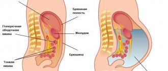
When a growing abdominal aortic aneurysm begins to exert mechanical pressure on the stomach and duodenum, this leads to the development of dyspeptic syndrome, which is characterized by:
- nausea;
- vomit;
- belching air;
- flatulence;
- tendency to chronic constipation.
In some cases, the aneurysmal sac displaces the kidney and compresses the ureter, thereby leading to the formation of a urological syndrome, which is clinically manifested by dysuric disorders (frequent, painful, difficult urination) and hematuria (blood in the urine).
If an abdominal aortic aneurysm compresses the testicular vessels (arteries and veins), the patient experiences pain in the testicular area and also develops varicocele.
Compression of the spinal roots by the increasing protrusion of the abdominal aorta is accompanied by the formation of an ischioradicular symptom complex, which is characterized by persistent pain in the lumbar region, as well as motor and sensory disorders in the lower extremities.
When an aneurysm compresses arteries and veins, a man develops a varicocele.
An abdominal aortic aneurysm can cause chronic disruption of blood supply in the lower extremities, which leads to trophic disorders and intermittent claudication.
When an abdominal aortic aneurysm ruptures, the patient experiences massive bleeding, which can lead to death in a few seconds. Clinical symptoms of this condition are:
- sudden intense pain (so-called dagger pain) in the abdomen and/or lower back;
- a sharp drop in blood pressure, up to the development of collapse;
- feeling of strong pulsation in the abdominal cavity.
Features of the clinical picture of a ruptured abdominal aortic aneurysm are determined by the direction of bleeding (bladder, duodenum, inferior vena cava, free abdominal cavity, retroperitoneal space). Retroperitoneal bleeding is characterized by persistent pain. If the hematoma increases towards the pelvis, then the pain radiates to the perineum, groin, genitals, and thigh. High localization of the hematoma often manifests itself under the guise of a heart attack.
Intraperitoneal rupture of an abdominal aortic aneurysm leads to the rapid development of massive hemoperitoneum, severe pain and bloating are noted. The Shchetkin-Blumberg symptom is positive in all areas. Percussion determines the presence of free fluid in the abdominal cavity.
Simultaneously with the symptoms of an acute abdomen when an aortic aneurysm ruptures, symptoms of hemorrhagic shock arise and quickly intensify:
- sharp pallor of the mucous membranes and skin;
- severe weakness;
- cold clammy sweat;
- lethargy;
- thread-like pulse (frequent, low filling);
- pronounced decrease in blood pressure;
- decrease in diuresis (amount of urine excreted).
With intraperitoneal rupture of an abdominal aortic aneurysm, death occurs very quickly.
If a breakthrough of the aneurysmal sac into the lumen of the inferior vena cava occurs, this is accompanied by the formation of an arteriovenous fistula, the symptoms of which are:
- pain localized in the abdomen and lower back;
- formation of a pulsating tumor in the abdominal cavity, over which systole-diastolic murmurs can be heard well;
- swelling of the lower extremities;
- tachycardia;
- increasing shortness of breath;
- significant general weakness.
Heart failure gradually increases, which becomes the cause of death.
Rupture of an abdominal aortic aneurysm into the duodenum leads to sudden massive gastrointestinal bleeding. The patient's blood pressure drops sharply, bloody vomiting occurs, weakness and indifference to the environment increases. Bleeding with this type of rupture is difficult to diagnose from gastrointestinal bleeding due to other causes, for example, peptic ulcer of the stomach and duodenum.
Branches of the aorta
The location of the BA allows it to supply blood to almost all organs and tissues located in its area of influence. Thus, the inferior artery of the diaphragm is the largest blood branch, which has its own branching. It nourishes the diaphragm and adrenal glands. The branches of the aorta in the lumbar region are presented in the form of four arteries, in turn connected to the epigastric vessels. They supply blood to the spinal cord, abdominal and back muscles, as well as the adjacent skin and subcutaneous tissue.
The internal organs feed the vessels of the abdominal aorta, which are divided into paired and unpaired. The paired ones include: the renal artery and the middle adrenal artery. In women, the ovarian artery, which supplies the ovaries, is also isolated. The unpaired ones are represented by two mesenteric arteries and the celiac trunk. The first of the mesenteric arteries passes over the wall of the duodenum and supplies the pancreas, as well as the cecum, jejunum, ileum and colon. The second supplies blood to the colon and rectum.
Particular attention should be paid to the celiac trunk, which is divided into three arteries: hepatic, left pancreatic and splenic. This branch of the BA supplies blood to such organs as:
- liver;
- gallbladder;
- pancreas;
- stomach (in this case, only its walls and greater omentum);
- spleen
Thus, the abdominal aorta plays a major role in the digestive process, supplying nutrients to most of the digestive organs. Moreover, the celiac trunk itself is a miniature object (its length barely reaches two centimeters). It is located in the area of the twelfth thoracic vertebra.
Diagnostics
In 40% of cases, abdominal aortic aneurysms represent an incidental diagnostic finding during a clinical or radiological examination for another reason.
The presence of the disease can be assumed based on data obtained from collecting anamnesis (indication of family cases of the disease), general examination of the patient, auscultation and palpation of the abdomen. In thin patients, it is sometimes possible to palpate a pulsating, painless formation in the abdominal cavity that has a densely elastic consistency. During auscultation over the area of this formation, a systolic murmur can be heard.
The most accessible and cheapest method for diagnosing an abdominal aortic aneurysm is plain radiography of the abdominal cavity. The X-ray image shows the shadow of the aneurysm, and in 60% of cases, calcification of its walls is noted.
Ultrasound examination and computed tomography make it possible to accurately determine the size and location of pathological expansion. In addition, according to computed tomography data, the doctor can assess the relative position of the abdominal aortic aneurysm and other visceral blood vessels and identify possible anomalies of the vascular bed.
To diagnose an abdominal aortic aneurysm, radiography, ultrasound, and CT are required.
Angiography is indicated for patients with arterial hypertension with severe or unstable angina, significant stenosis of the renal arteries, patients with suspected mesenteric ischemia, as well as patients with symptoms of occlusion (blockage) of the distal arteries.
If indicated, other instrumental diagnostic methods can be used, for example, laparoscopy, intravenous urography.
Thoracic aortic aneurysm
Stenting of a thoracic aortic aneurysm
The main danger of a thoracic aortic aneurysm is the practically zero survival rate from rupture, so diagnosis and treatment must begin in a timely manner. It is especially important to know the symptoms of the disease, although until serious complications occur, it is asymptomatic.
Symptoms
Speaking about the symptoms of a thoracic aortic aneurysm, we can note, first of all, a pain syndrome associated with compression of adjacent structures. Sometimes patients report pain in the lower back. Also, due to compression of the larynx area, hoarseness occurs; characteristic signs of the disease are often coughing and wheezing. When a rupture occurs, if there is no death, pain appears in the chest and lower back, arterial hypertension and shock occur. Any symptom can be a reason to contact a specialist.
Diagnosis and treatment
The first suspicion of a thoracic aortic aneurysm may arise during an X-ray examination. If in doubt, CT and MRA are prescribed. A transesophageal echo (TEE) can determine the size of the AA, as well as the presence of blood leaks. If the disease is diagnosed, surgery with the installation of a synthetic prosthesis can come to the rescue. Endovascular surgery (EVAR) deserves special attention, when an artificial prosthesis is installed into the aorta through a small incision on the thigh, eliminating blood flow through the AA. This operation is recommended for weakened patients, as it is safer, although after some time it is possible to repeat the procedure to completely eliminate blood leakage.
Open intervention with resection of the aneurysm is indicated for patients with ideal indicators and completely eliminates the problem. Meanwhile, direct surgical intervention is not recommended for obese people and weakened patients with serious contraindications. As for the survival rate, deaths occur in only 5% of cases of early treatment.
Treatment of abdominal aortic aneurysm
The presence of an abdominal aortic aneurysm in a patient is an indication for surgical treatment, especially if the size of the protrusion increases by more than 0.4 cm per year.
The main operation for an abdominal aortic aneurysm is aneurysmectomy (excision of the aneurysmal sac) followed by plastic surgery of the removed portion of the blood vessel with a prosthesis made of Dacron or other synthetic material. Surgical intervention is performed through laparotomy (an incision in the abdominal wall). If the iliac arteries are also involved in the pathological process, then bifurcation aorto-iliac prosthesis is performed. Before, during and on the first day after surgery, the pressure in the cardiac cavities and the magnitude of cardiac output are monitored using a Swan-Gantz catheter.
Surgery to remove an abdominal aortic aneurysm is called an aneurysmectomy.
Contraindications to performing elective surgery for abdominal aortic aneurysm are:
- acute cerebrovascular accidents;
- fresh myocardial infarction;
- end-stage chronic renal failure;
- severe degree of cardiac and respiratory failure;
- widespread occlusion of the iliac and femoral arteries (partial or complete blockage of blood flow through them).
If an abdominal aortic aneurysm ruptures, the operation is performed for life-saving reasons on an emergency basis.
Abdominal aortic aneurysm ranks 15th on the list of diseases leading to death.
Currently, vascular surgeons prefer minimally invasive methods for treating abdominal aortic aneurysm. One of them is endovascular prosthetics of the area of pathological expansion using an implantable stent graft (a special metal structure). The stent is installed so that it completely covers the entire length of the aneurysmal sac. This leads to the fact that the blood stops putting pressure on the walls of the aneurysm, thereby preventing the risk of its further enlargement, as well as rupture. This operation for abdominal aortic aneurysm is characterized by minimal trauma, a low risk of complications in the postoperative period, and a short rehabilitation period.
Characteristic symptoms
The robot performed two kidney transplants
Classic triad of symptoms:
- Short-term hypertension, turning into persistent hypotension;
- Acute pain;
- Visible pulsation in the umbilical area.
The onset is acute and is characterized by an attack of sudden, cramping pain in the back. The pain can be tearing, burning, it quickly spreads to the abdomen or pelvis and becomes encircling in nature.
Other symptoms:
- Anxiety, fear of death;
- Shallow rapid breathing;
- Cardiopalmus;
- Dizziness leading to fainting;
- “Marble” pallor of the skin;
- Cold sweat;
- Intestinal bleeding;
- Loss of sensation in the legs.
Typically, patients cannot remain in an upright or sitting position. Internal blood loss leads to collapse - acute vascular insufficiency, which is characterized by a sharp drop in pressure (systolic value can reach 70-50 mm Hg).
The further picture consists of the phenomena of shock - acute liver, kidney and cardiopulmonary failure against the background of cerebrovascular accident:
- Lack of breathing;
- Absence of pulse in peripheral arteries;
- Disorders of consciousness.
Prevention
If there is a genetic predisposition or the presence of other factors that provoke vascular diseases, the patient is recommended to adhere to the following rules of prevention:
- Maintain a healthy lifestyle. Give up bad habits and eat right. The diet should contain a large amount of plant foods, fresh fruits, and fiber.
- It is necessary to regularly monitor blood pressure and cholesterol levels in the blood. If your blood pressure increases, you should contact a cardiologist to prescribe antihypertensive therapy.
- Avoid intense physical activity, avoid physical activity, heavy lifting, and stress.
- Control body weight.
Prognosis for a patient with an aneurysm
This disease is difficult to recognize in a timely manner. Its course is even more difficult to predict. Aortic rupture kills at least 75% of patients. However, about 50% of them do not make it to the hospital.
Nowadays, medicine is making a significant leap in diagnosing and treating complex diseases. A special contribution was made by the endoprosthetic method, which made it possible to reduce the number of deaths in medical practice.
A patient who has undergone surgery should be regularly examined by the attending physician. Timely removal of the aneurysm promises a favorable prognosis. In 70% of cases there is a 5-year survival rate.
As already mentioned, in most cases the cause of the development of an aneurysm is atherosclerosis. This emphasizes the need for constant examination by a doctor, giving up bad habits and foods containing high amounts of cholesterol.
How is surgery to remove an aneurysm performed?
If a patient has an AAA, conservative treatment will not help solve the problem. There are no drugs that would restore the diameter and elasticity of blood vessels, or prevent rupture of the protrusion. But medications are prescribed if it is impossible to perform an operation to remove an aneurysm.
Surgery makes it possible to completely eliminate the pathology by removing a weakened section of the walls or restoring strength. The operation can be emergency or planned. The first option is performed with a high risk of death of the patient (80-90%), since rupture leads to massive bleeding. A planned approach requires preparatory therapy to prevent exacerbations of chronic diseases that complicate the course of surgical intervention.
There are two methods for eliminating an aneurysm:
Cavitary;
Access to the pathology is opened by dissecting the peritoneum. This method of operation allows you to see the entire anatomical structure of tissues and organs, and also allows you to carry out various manipulations in an open space. The cavity type is recommended in emergency cases.
After exposing the vessel, ligation is done in two places. They block the blood flow above and below the intended removal site. The front wall is cut and the tube is fixed inside. The edges are sewn up. Then the clamps are removed and blood circulation is restored. The installed tube does not allow pressure to provoke rupture of the bulging zone.
After the operation, large scars are left because the incision is made from the sternum to the pubic bones. The disadvantages of the abdominal procedure are the possibility of infection of the body and many contraindications. There is also a risk of seams coming apart.
The patient remains in intensive care for 12-24 hours after surgery. If no complications arise, the patient is discharged after hospitalization after 7-14 days. A person becomes able to work only after a month or two. But restrictions remain for an even longer period.
Endovascular;
Requires preparation because it does not require a cut in the abdomen. The morbidity from such an operation is much less. An artificial tube is inserted through the femoral artery, which is secured inside with special anchors. The endovascular method is very effective for saccular protrusion. But it can be carried out only with the preliminary collection of complete information about the condition of the deformed area.
The advantages of surgical intervention are the speed of solving the problem, the absence of deep anesthesia, a low risk of infection, the elimination of suture dehiscence, and a small number of contraindications.
The disadvantages of the endovascular method are the impossibility of treating dissecting and spindle-shaped forms. It is also difficult to perform manipulations in a narrow lumen. If suddenly complications arise during surgery, the abdomen will have to be dissected. The patient is discharged after five days. If there are no negative consequences, then the person returns to work in about 1.5 months.
It is important to note that the choice of surgical method and the speed of repair depend on the diameter and exact location of the AAA
What types are more common?
Single aneurysms predominate in this area.
Saccular and fusiform forms are equally common. True extensions account for 90-95% of cases, false extensions – up to 5%. Small (up to 3 cm) and medium-sized aneurysms (3-5 cm) predominate in size, but due to downward growth they quickly become large (5-7 cm).
The average development time is 2-3 months . In congenital diseases, the pathology is characterized by slow progression (up to 1 mm per year), while in acquired diseases it progresses rapidly (more than 3 mm per year).
Classification
As such, the typification of the defect in question has not been developed, which is due to the rather low prevalence of dissecting aortic aneurysm.
The main classification of violations by localization is carried out according to De Bakey. According to it, three places of separation are distinguished:
- First type. The defect is located in the ascending part of the vessel and can diffusely and expansively cover a group of areas (thoracic and abdominal). This is the most common type. With a massive lesion, the prognosis is initially worse, because the complexity of the proposed operation increases.
- Second type. The deviation is localized strictly isolated, in the ascending aorta. It also presents difficulties for the surgeon; open access is problematic because it is not easy to mobilize the vessel, there is too much tissue.
- Third type. Affects the descending aorta and the abdominal part inclusive.
There is a simplified Stanford classification. According to it, aneurysm dissection is distinguished, originating in the ascending aorta - type (a) and in the descending part, type (b).
It is also possible to subdivide the disorder according to symptoms (acute, chronic), course, and size of the protrusion (an extremely confusing criterion).
There are no universal division methods. Doctors are primarily interested in the diameter of the pathological formation, the rate of its progression and localization.
Finding out the details allows you to reduce risks for the patient through a clear, thorough elaboration of the operational strategy.
How to diagnose pathology?
Although most aneurysms are asymptomatic and do not manifest themselves during examination, the most common complication remains life-threatening rupture with hemorrhage. Most often, the disease is diagnosed “accidentally”, after a chest x-ray or other research methods.
The most common symptom is pain. It can be acute, preceding an impending rupture, or chronic, from compression or sprain. The location of the pain often indicates the area of the aorta, but this is not always the case.
Ascending aortic aneurysms typically cause pain in the anterior chest, whereas arch defects are most likely responsible for pain radiating to the neck. With underlying thoracic aneurysms, localization is found in the back and between the shoulder blades. If the defect is lodged at the level of the diaphragm, pain occurs in the epigastric region.
Large volumes of protrusion affect nearby structures, so typical symptoms of a thoracic aortic aneurysm are:
- inflammation or compression of the superior vena cava manifests itself in the form of swelling of the neck veins;
- aortic insufficiency with increased pulse pressure, diastolic murmur and heart failure;
- stretching of the laryngeal nerves can cause hoarseness;
- with compression of the trachea or bronchus - shortness of breath, stridor, wheezing, cough;
- compression of the esophagus leads to dysphagia;
- erosion into surrounding structures may lead to hemoptysis or bleeding;
- compression of the spinal cord or thrombosis of the spinal arteries can lead to neurological symptoms of paraparesis or paraplegia.
Instrumental diagnostic methods
Chest X-ray
. Carry out using an X-ray machine: it is possible to study the contours and size of the aorta, detect expansion of the mediastinal shadow, compression of surrounding organs, displacement of the trachea and esophagus. In lateral projections, a decrease in the space behind the sternum is observed. Among the disadvantages is the impossibility of diagnosing pathology, which is completely covered by the heart.- Echocardiography. Transthoracic echocardiography is performed with the sensor placed on the chest. It visualizes the aortic valve and proximal aorta, but is less sensitive and specific than transesophageal (the sensor is inserted into the esophagus through the mouth). In this case, detection of the aortic valve, ascending and descending thoracic aorta is possible, but there are limitations in visibility in some other areas. Ischemia can be assessed using dipyridamole-thallium or dobutamine echocardiography.
- Ultrasonography is most often used to demonstrate signs of abdominal aneurysms.
- Aortography. This technique outlines the lumen of the artery using contrast, and this helps determine the extent of the pathological process, the involvement of other structures and their stenosis.
- CT scan. CT with contrast has become the most widely used diagnostic tool. It quickly and accurately evaluates the aorta on either side of the diaphragm to determine the location, extent of pathology, and its relationship to major vessels and surrounding structures.
- Magnetic resonance imaging. Compared with contrast-enhanced CT, magnetic resonance imaging (MRI) and angiography (MRA) have the advantage of avoiding nephrotoxic contrast and ionizing radiation.
For what reasons does an aneurysm develop?
The causes of the development of pathology are varied, they should be studied if you want to know who is at risk and most susceptible to the disease. So, doctors identify several factors that provoke an aneurysm:
- Old age - with the advent of old age, blood vessels weaken, plaques appear in them, and blood circulation is impaired. That is why most of the sick are people over 65 years of age;
- Smoking is a bad habit that negatively affects the functioning of the respiratory and circulatory systems, as well as other organs. In addition to the development of an aneurysm, there is a risk of other abnormalities;
- High blood pressure, therefore, the risk group includes not only hypertensive patients, but also overweight people, since they are more likely to have hypertension;
- Congenital disorders - they develop as a result of hereditary factors, can be congenital, appear at a young age if the patient’s relatives suffered from aortic diseases;
- Chest injuries – with various types of injuries, aneurysms may appear. The same applies to medical intervention in the past, if an inexperienced doctor mistakenly touched the aorta in the thoracic region.
Thoracic aortic aneurysm is a pathology that affects men and women equally. Regardless of gender, age and other factors, the risk of development always exists.
Rehabilitation period after surgery
After the operation, the patient is prohibited from lifting weights or playing sports for the next six months. During the first 12 months after discharge, you must visit the doctor every 30 days, then once every six months and once a year. Before and after the procedure, the patient must strictly adhere to the doctor’s recommendations and be sure to take prescribed medications. To prevent the risk of complications, you need to adhere to the following simple rules:
- Give up bad habits (tobacco, alcohol).
- Eat right and control obesity. It is necessary to completely exclude foods rich in cholesterol, fatty, fried, and spicy from the menu. It is not recommended to eat animal fats, confectionery products, and flour. It is better to cook dishes by steaming, boiling or baking.
- Monitor cholesterol and blood glucose levels with tests, and take statins if necessary.
- Correct the symptoms of concomitant diseases (diabetes, kidney disease, heart disease, etc.). Visit your doctor regularly and take prescribed medications.
Complications after surgery
In general, any operation goes well, and complications during the rehabilitation period are extremely rare . In less than 4% of cases, the following unforeseen situations may occur:
- pulmonary or cerebral edema;
- renal failure;
- divergence of postoperative sutures;
- bleeding;
- blood clotting disorder;
- prosthesis infection;
- removal of the prosthesis;
- vein thrombosis;
- sexual dysfunction;
- postoperative hernia.