Nonspecific ulcerative colitis (UC, modern name “ulcerative colitis”) is a chronic disease that manifests itself as purulent hemorrhagic inflammation of the colon wall. The prevalence of the disease in different countries ranges from 50 to 230 people per 100 thousand population. People aged 20–40 years are most often affected, men and women with equal frequency[1]. Typically, several years pass from the appearance of the first symptoms to an accurate diagnosis. Late diagnosis and the resulting inadequacy of treatment often lead to the development of severe complications and relatively high mortality (in Russia - 17 cases per million population, in Europe - 6 cases per million [2]).
Causes of ulcerative colitis
The exact cause of the disease is still unknown: so far doctors are only talking about provoking factors.
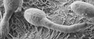
First of all, there is a genetic predisposition: among blood relatives, the incidence of UC is 15 times higher than in the general population[3]. In total, more than a hundred changes in genes associated with ulcerative colitis are known[4]. The altered genetic background disrupts the normal course of immune reactions, causing an excessive inflammatory response to bacterial antigens. Considering that the number of bacteria in the large intestine is 10 times higher than the number of all cells in the human body, against the background of altered immune activity, inflammation becomes almost constant.
Predisposing factors for the development of ulcerative colitis may be:
- intestinal infections;
- nervous stress;
- vitamin D deficiency;
- excess animal protein and lack of dietary fiber in the diet;
- acute viral infection,
Diet and folk remedies
Nutrition for nonspecific ulcerative colitis should be gentle; foods and drinks that have an irritating effect on the intestines should be excluded from the diet. These are raw vegetables, sour fruits, carbonated drinks, alcohol, marinades, spicy foods. But experts believe that there is no evidence that the diet relieves symptoms of UC or somehow increases the effectiveness of treatment. It is recommended to eat liquid or soft pureed food to minimize discomfort during an exacerbation of the disease.
Folk remedies in the treatment of this serious disease are auxiliary and supportive. Before using them, you should consult a doctor, but you cannot replace drug therapy with traditional recipes. Traditionally, herbs and medicinal infusions are used: decoction of chamomile, yarrow, bird cherry leaves, strawberries, parsley decoction, medicinal herb mullein, potato juice, calendula infusion. You must remember that you cannot drink herbal infusions, decoctions, or teas for longer than a month; you need a break. It is wiser to use traditional medicine to treat ulcerative colitis after meals, so as not to aggravate the condition by irritating the intestines.
We recommend: The main symptoms of non-ulcerative intestinal colitis and methods of its treatment
https://youtu.be/d72YXJ4ik8A
Classification of nonspecific ulcerative colitis
According to the nature of the disease:
- acute – the first symptoms appeared less than 6 months before visiting a doctor;
- chronic continuous - breaks between exacerbations of less than six months, despite adequate treatment;
- chronic recurrent: exacerbations are replaced by remissions, each of which lasts 6 months or more.
According to the severity of the attack (current exacerbation):
- light;
- moderate severity;
- heavy.
There are more bacteria in the intestines than cells in the entire body, and this is not always good
| criterion | light | medium-heavy | heavy |
| Frequency of bowel movements with blood | <4 | >=4 | >=6 |
| pulse | within normal limits | <= 90 beats/min | >90 beats/min |
| temperature | <37,5 | >37,5 | |
| hemoglobin | >=105 g/l | <105 g/l | |
| ESR | <=30 mm/h | >30 mm/h | |
| contact vulnerability of the colon mucosa | No | There is | There is |
According to the location of the pathological process:
- distal colitis - proctitis, proctosigmoiditis - inflammation is active in the rectum and sigmoid colon;
- left-sided colitis - inflammation in the rectum, sigmoid, descending colon and partially transverse colon (to the middle);
- total colitis - the entire large intestine is inflamed.
Forms of pathology
Atypical ulcerative colitis has several medical classifications. The forms of the disease and their description are presented in the table.
| Differentiating feature | Type | Description |
| Localization | Distal | Rectum |
| Left-handed | Damage to the remaining parts of the colon up to the splenic flexure | |
| Subtotal | To the hepatic flexure | |
| Total | Ascending colon | |
| Degree of development (according to Truelove and Witts) | Elementary | Up to 4 bowel movements per day inclusive, there is almost no blood, the heart rate and temperature are normal, hemoglobin is more than 110, erythrocyte sedimentation rate is no more than 30, a slight increase in the number of white blood cells, the patient’s weight does not change, the lack of nutrients is not reflected. |
| Average | Up to 6 bowel movements per day inclusive, blood in the stool is noticeable, heart beats - no more than 90, temperature - 37-38 degrees, hemoglobin - up to 100, erythrocyte sedimentation rate - up to 35, a noticeable increase in the number of white blood cells, the patient’s weight decreases, noticeable lack of nutrients. | |
| Heavy | More than 6 bowel movements per day, pronounced blood, heart beats more than 90, temperature - 38-39 degrees, hemoglobin - less than 90, erythrocyte sedimentation rate - more than 35, leukocytosis with a shift in the formula, the patient’s weight is noticeably reduced, lack of nutrients is very noticeable . | |
| Character of the current | Chronic | Stages of exacerbations (up to 2 times per year) and stable remissions. |
| Spicy | Extremely severe course with complications. | |
| Continuous | Diagnosed exacerbations more often than 2 times a year, impossibility of achieving remission. |
The total type is more susceptible to a severe course. The left-sided type is the most common (80 out of 100). The continuous type occurs in 10 cases out of 100.
Necrotizing ulcerative colitis is a separate type of pathology diagnosed in newborns (usually premature infants) who have been exposed to oxygen and nerve starvation in the womb. But it can also occur as a complication in severe colitis in adults. Characterized by cell death (the last advanced stage).
Smoking is considered one of the provoking factors for the development of the disease.
Symptoms of nonspecific ulcerative colitis
The main manifestations of UC can be divided into stool syndrome, hemorrhagic and pain syndromes.
Abnormal bowel movements are manifested by diarrhea: in severe cases, the frequency of bowel movements reaches 20 times a day. The stool is watery, mixed with mucus, pus and blood. Often the urge to defecate turns out to be false (tenesmus) or ends only with the release of bloody mucus (the so-called “rectal spitting”). The more complete the damage to the intestines, the more severe the diarrhea - due to impaired absorption of water and electrolytes against the background of inflammation. In situations where only the sigmoid and rectum are affected, diarrhea may alternate with constipation caused by spasm of the overlying parts of the large intestine.
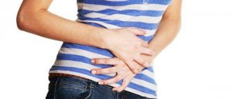
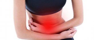
Abdominal pain - aching, cramping, often on the left and below, less often - in the navel area. At the beginning of the disease, they occur 0.5–1.5 hours after eating, but as the inflammatory process progresses, the connection between food intake and pain is lost. Maximum intensity - immediately before defecation, after bowel movement the pain decreases.
Hemorrhagic syndrome is bleeding from inflamed areas of the intestine. If the disease is severe, due to severe inflammatory intoxication, loss of water and electrolytes, general weakness, dizziness appears, the temperature rises, and appetite disappears.
UC - causes, symptoms, treatment, dietary table
Almost half of the patients[5] also have extraintestinal manifestations of UC, which can be caused by autoimmune changes or prolonged inflammation and associated metabolic disorders.
Autoimmune systemic manifestations:
- arthropathy (pain and inflammation in the joints);
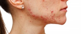
- skin lesions (pyoderma gangrenosum, erythema nodosum);
- aphthous stomatitis (ulcers on the oral mucosa);
- inflammation of the sclera, iris (uveitis, iritis, iridocyclitis, episcleritis).
Systemic manifestations caused by metabolic disorders:
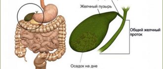
- cholelithiasis (gallbladder stones);
- liver steatosis, steatohepatitis;
- peripheral vein thrombosis;
- pulmonary embolism.
It is precisely because of the variety of systemic manifestations that can be difficult, “drawing” all the attention of both the patient and the doctor, that most diagnostic errors occur. A person with manifestations of, for example, iridocyclitis will consult an ophthalmologist, and with aphthous stomatitis - a dentist. He will not complain to any of these specialists about problems in the intestines.
Possible consequences
Disability from ulcerative colitis and death are the worst complications. You can maintain your ability to work at a mild stage of the disease. Disability group 3 allows for some work.
Inflammation tends to spread and affect other organs (eyes, mouth, bones and joints, skin). For the intestines, the progression of the disease is dangerous due to oncology. There is a risk of fistulas and abscesses.
The most common complications are narrowing, obstruction, persistent bleeding, perforation and dilatation of the intestine. The latter is dangerous due to rupture. Any of these complications require immediate hospitalization.
The first thing to do in the event of an exacerbation of UC is to go to the hospital for qualified help, the purpose of which is to relieve the attack.
Complications of nonspecific ulcerative colitis
- Toxic dilatation of the colon is an acute paralytic dilatation of the intestine with increased intraintestinal pressure. The patient's condition deteriorates sharply: the temperature quickly rises, severe weakness appears, and blood pressure drops.
- Perforation of the colon usually occurs against the background of toxic dilatation. After the intestinal wall is perforated, its contents enter the abdominal cavity, causing peritonitis. The mortality rate for this complication reaches 50%[6].
- Intestinal bleeding.
All these conditions are indications for emergency surgery.
Types of intestinal colitis
Depending on the cause of inflammation, the following types of colitis are distinguished:
- Ulcerative colitis is a type of colitis characterized by ulcers on the walls of the large intestine.
- Acute - a type in which not only the large intestine is affected, but also the small intestine is inflamed, and the stomach is also affected.
- Ischemic – consequences of poor blood circulation in the intestines.
- Chronic is the consequences of acute, incompletely cured colitis.
- Spastic manifests itself with spasms and bloating. It is not considered a severe form.
- Alcoholism occurs when you are dependent on alcohol.
- Erosive - characterized by ulcers over a larger area of the duodenum.
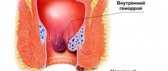
- Atonic is typical for older people. Intestinal activity is reduced, frequent constipation, hemorrhoids subsequently.
- Hemorrhagic is characterized by bloody discharge - diarrhea.
- Radiation colitis occurs after radiation exposure received for cancer.
- Nonspecific ulcerative - similar to chronic with relapses, origin of the immune type.
Diagnosis of nonspecific ulcerative colitis
Abdominal pain and frequent stool mixed with pus, blood and mucus are the main symptoms of UC
The main method for diagnosing UC is colonoscopy with biopsy. During endoscopy, the doctor sees characteristic changes in the intestinal wall, which are confirmed by microscopic examination of the biopsy specimen.
In addition, the condition of the large intestine can be visualized using irrigoscopy, magnetic resonance or computed tomography with contrast.
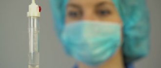
To determine the general condition of the body, general and biochemical blood tests are prescribed. At the first attack of the disease, microscopic and bacteriological examination of stool is recommended to exclude acute intestinal infection.
Detection methods
Diagnosis of UC includes medical history, palpation and examination, and instrumental techniques:
- Classic x-rays allow you to assess the condition of organs and identify complications. The procedure with contrast will show the width of the lumen and mucosal defects.
- Ultrasound locally determines the degree of intestinal damage. It is also carried out as part of treatment monitoring.
Endoscopic diagnostic methods help assess the condition of the gastrointestinal tract
- Computed tomography allows you to differentiate the disease from a similar one - Crohn's. Shows the area of damage in the intestine and the condition of neighboring organs.
- Endoscopic examination is recommended outside of exacerbation. This is the main method. It allows you to clearly assess the condition of the body. Shows ulcers, necrosis, erosion, wall density, condition of blood vessels, presence of blood, granulomas.
- Laboratory tests (general and chemical blood tests, coprogram) will allow you to assess the condition of the body and exclude parasites, antibiotic poisoning, infection, and evaluate blood in the discharge.
- Immunity testing for pANCA and ANCA antibodies. Simultaneous presence in 95% indicates UC.
Symptoms of ulcerative colitis of the intestine in women can be confused with gynecological pathologies, which requires additional consultation with a specialist. Treatment is carried out with hormonal drugs, which also requires consultation with a gynecologist.
If the inflammatory process worsens, it is important to consult a doctor promptly
Treatment of ulcerative colitis
In case of exacerbation, diet Table No. 4 is prescribed; as the symptoms subside, the diet is expanded, focusing on the patient’s reaction. It is advisable to exclude milk and dairy products, since many patients with UC have impaired tolerance to them.
To reduce the activity of inflammation, mesalazine is recommended. The form of application will depend on the extent of the process: for mild to moderate proctitis, rectal suppositories or rectal foam may be sufficient; for more extensive lesions, tablets are prescribed.
To improve the regeneration of the mucous membrane, gastroprotectors based on rebamipide are prescribed. They reduce the activity of inflammation, reduce the permeability of the epithelial barrier and promote the rapid restoration of the normal structure and function of the intestinal wall.
If these measures are not enough, glucocorticosteroids (budesonide, prednisolone), drugs that suppress the immune system (azathioprine), and monoclonal antibodies (infliximab, adalimumab, golimumab or vedolizumab) are prescribed to relieve inflammation. The regimen of use and dosage is determined by the doctor, but you need to tune in to a long course of maintenance therapy: premature cessation of treatment can provoke a relapse.
If conservative therapy is ineffective and complications occur, surgery is necessary to remove the affected area of the colon.
Intestinal bleeding is extremely dangerous
Traditional treatment
Treatment of ulcerative colitis with drugs involves taking corticosteroids (Prednisolone, Budesonide), 5-ASA (Mesalazine, Colazal), antidepressants (Methotrexate) and cytostatic drugs (Infliximab). In severe cases with severe fever and strong signs of inflammation, antibiotics (Metronidazole) are used.
Therapeutic baths are one of the methods of treating ulcerative colitis
To eliminate symptoms, medications that relieve pain and stop diarrhea (Loperamide) are prescribed. If necessary, rehydration is carried out, the body is saturated with iron.
5-ASA is usually prescribed as an anti-inflammatory agent. The use of corticosteroids is indicated only during periods of severe exacerbation of the second and third degrees of severity of the disease and for only a few months.
The goal of treatment at present is to eliminate symptoms, reduce inflammation, and prevent relapses. However, new treatments for ulcerative colitis are being developed regularly. Research is being conducted on the effectiveness of innovative topical drugs based on bioprocesses and gene structures. In Israel, Remicade, an anti-TNF drug (tumor necrosis factor), is actively used in practice.
If a combination of medications, diet and physiotherapy is ineffective, surgical treatment is indicated: resection with anastomosis or segmental resection.
During an exacerbation of the disease, the patient is only allowed to drink water
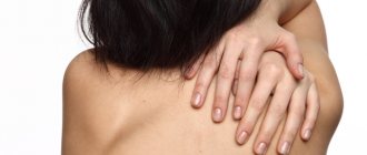
The most common skin diseases in IBD
Erythrema nodosum
Erythema nodosum
Erythema nodosum (erythema nodosum) is an allergic or granulomatous inflammatory process of the blood vessels of the skin and subcutaneous fat. It is characterized by the appearance of painful red nodules on the legs or arms. Despite the fact that the fair half of humanity is more at risk of getting erythema nodosum, in the case of IBD the chances between men and women are equal. In addition to the above nodules, the patient may have a fever and joint pain.
Methods for treating erythrema nodosum include various painkillers, glucorticoids (oral or parenteral), potassium iodide solution, and cold compresses on the affected areas of the skin. Typically, symptoms of the disease disappear within six weeks.
Pyoderma gangrenosum
Pyoderma gangrenosum
Pyoderma gangrenosum is a rapidly spreading rash consisting of red and/or purple blisters. Eventually, these blisters coalesce and form deep, open sores (ulcers) with a blue or purple border.
The prevalence of pyoderma gangrenosum is practically unlimited.
Pyoderma gangrenosum on the legs can make it difficult for the patient to move. Moreover, in some cases the disease can affect internal organs, which puts a person’s life at risk. Sometimes a rash develops around the site of an injury or surgical wound.
Matilda Hagan, MD
Unlike erythema, pyoderma gangrenosum appears even when inflammatory bowel disease is in remission. In some cases, pyoderma gangrenosum is difficult to treat.
Pyoderma gangrenosum can leave terrible scars. The set of measures should include treatment of the underlying disease. those. UC or CD.
Joaquin Brieva, MD, dermatologist at Northwestern Memorial Hospital in Chicago
Medications for the treatment of pyoderma gangrenosum include anti-inflammatory creams and steroid ointments; oral or parenteral glucorticoids, as well as immunosuppressants such as Azathioprine and Methotrexate, known to a wide range of IBD patients.
Aphthous stomatitis
Aphthous stomatitis
Aphthous stomatitis is one of the most unpleasant types of stomatitis, the distinctive feature of which is the presence of aphthous (painful ulcers) in the patient’s oral cavity. Some people experience aphthous stomatitis just before their IBD flares up.
Typically, in people with ulcerative colitis and Crohn's disease, aphthae reach more than 10 mm in diameter. In most cases, the duration of treatment is from 2 to 3 weeks.
Treatment includes antibiotic therapy (particularly tetracycline) and a glucocorticoid-based mouthwash. Among other things, lidocaine can be used to reduce pain.
Pyoderma vegetans
Pyoderma vegetans
Pyoderma vegetans (ulcerative-vegetative pyoderma) is a rare condition characterized by the presence of ulcerated lesions covered with flaccid granulations and vegetations. Sometimes the ulcers become crusty. The distribution is limited to the hands, legs, and skin folds.
Pyoderma vegetans is similar in symptoms to pemphigus vegetans, blastomycosis, verrucous tuberculosis, and bromoderma.
Treatment for this skin condition is usually not required: in the case of IBD, it is enough to treat the underlying disease.
Sweet's syndrome
Sweet's syndrome
Sweet's syndrome (acute febrile neutrophilic dermatosis) is another rare skin complication associated with ulcerative colitis and Crohn's disease. The disease is accompanied by a fever and a rash of many pink or bluish-red bumps or spots. The rash spreads to the arms, legs, torso, face, or neck. Associated problems may include arthritis and eye inflammation.
Treatment for Sweet's syndrome usually includes glucocorticoid ointments and injections or tablets.
Seborrheic dermatitis
Seborrheic dermatitis
Seborrheic dermatitis is a condition that affects those areas of the scalp and body where the sebaceous glands are developed. It appears in the form of certain “scales” and is accompanied, so to speak, by “dandruff” even on, for example, the hands or feet. Seborrheic dermatitis is caused by fungi of the Malassezia species – Malassezia restricta and Malassezia globosa.
The reason, in the case of UC and CD. lies in the malfunction of the immune system, which leads to the loss of the body’s ability to control the growth of Malassezia fungi and keep them in a saprophytic state (bacteria use decomposition products of complex organic substances as a source of nutrition).
A diet excluding fatty and sweet foods, as well as glucocorticoid ointments, will help reduce the symptoms of seborrheic dermatitis.
Introduction
According to a study published in the journal Inflammatory Bowel Diseases in August 2015, up to 15% of people with inflammatory bowel disease experience skin problems.
Controlling your UC or CD is the key to solving your skin problems.
Matilda Hagan, MD
If you notice any skin changes that are unusual for you, it is important to immediately tell your doctor and also consult a dermatologist.