We eat to live! The mechanisms for assimilation of nutrients from food have been refined by evolution. However, if until recently the main problem was hunger, and getting food was difficult, today the variety and availability of food no longer surprises anyone. Abuse of food for pleasure, and not for survival, creates a lot of problems with the gastrointestinal tract. In this article we will look at the phenomenon of creatorrhea - a problem of protein processing.
What are the muscle fibers in feces?
Striation is an indicator that indicates the degree of digestion of muscle fibers. The coprogram form has separate columns indicating the presence or absence of muscle fibers with or without striations.
Muscle fibers with striations are fragments of meat food, which is partially processed in the stomach and intestines.
Under a microscope, striated fibers appear as long cylindrical structures with smooth corners. Stripes are clearly visible across or along them, indicating an insufficient effect of enzymes.
Unstriated muscle fibers are completely digested fibers that look like small lumps under a microscope.
Normally, the striations of muscle fibers should disappear in the stomach during digestion of food.
The striations should finally disappear in the duodenum when bile reaches the chyme.
Microscopic examination
Muscle fibers - with striations (unchanged, undigested) and without striations (changed, digested). A large number of changed and unchanged muscle fibers in the feces (creatorrhoea) indicates a violation of proteolysis (protein digestion):
- in conditions accompanied by achlorhydria (lack of free HCl in gastric juice) and achylia (complete absence of secretion of HCl, pepsin and other components of gastric juice): atrophic pangastritis, a condition after gastric resection;
- with accelerated evacuation of food chyme from the intestines;
- in case of violation of the exocrine function of the pancreas;
- with putrefactive dyspepsia.
Connective tissue (remnants of undigested vessels, ligaments, fascia, cartilage). The presence of connective tissue in the feces indicates a deficiency of proteolytic enzymes of the stomach and is observed with hypo- and achlorhydria, achylia.
Fat is neutral. Fatty acid. Salts of fatty acids (soaps)
The appearance of large amounts of neutral fat, fatty acids and soaps in the stool is called steatorrhea. This happens:
- with exocrine pancreatic insufficiency, a mechanical obstruction to the outflow of pancreatic juice, when steatorrhea is represented by neutral fat;
- if the flow of bile into the duodenum is impaired and if the absorption of fatty acids in the small intestine is impaired, fatty acids or salts of fatty acids (soaps) are found in the feces.
Plant fiber
Digestible - found in the pulp of vegetables, fruits, legumes and grains. Indigestible fiber (the skin of fruits and vegetables, plant hairs, the epidermis of cereals) has no diagnostic value, since there are no enzymes in the human digestive system that break it down
. It is found in large quantities during rapid evacuation of food from the stomach, achlorhydria, achylia, and the syndrome of bacterial overgrowth in the colon.
Starch
The presence of a large amount of starch in the feces is called amilorrhea and is observed more often with increased intestinal motility, fermentative dyspepsia, and less often with exocrine insufficiency of pancreatic digestion.
Iodophilic microflora (clostridia)
With a large amount of carbohydrates, clostridia multiply intensively. A large number of clostridia is regarded as fermentative dysbiosis.
Epithelium
A large amount of columnar epithelium in the feces is observed in acute and chronic colitis of various etiologies.
Leukocytes
A large number of leukocytes (usually neutrophils) are observed in acute and chronic enteritis and colitis of various etiologies, ulcerative necrotic lesions of the intestinal mucosa, intestinal tuberculosis, and dysentery.
Red blood cells
The appearance of slightly changed red blood cells in the stool indicates the presence of bleeding from the colon, mainly from its distal parts (ulceration of the mucous membrane, disintegrating tumor of the rectum and sigmoid colon, anal fissures, hemorrhoids). A large number of red blood cells in combination with leukocytes and columnar epithelium is characteristic of ulcerative colitis, Crohn's disease with damage to the colon, polyposis and malignant neoplasms of the colon.
Worm eggs
Eggs of roundworms, tapeworms, etc. indicate a corresponding helminthic infestation.
Pathogenic protozoa
Cysts of dysenteric amoeba, Giardia, etc. indicate a corresponding invasion by protozoa.
Yeast cells
Found in feces during treatment with antibiotics and corticosteroids. Identification of the fungus Candida albicans is carried out by culturing on special media (Sabouraud's medium, Microstix Candida) and indicates a fungal infection of the intestine.
Calcium oxalate (oxalic lime crystals)
Detection of crystals is a sign of achlorhydria.
Triple phosphate crystals (ammonium phosphate-magnesia)
Triple phosphate crystals found in feces (pH 8.5-10.0) immediately after defecation indicate increased protein putrefaction in the colon.
Causes and possible diseases
In a healthy body, when food passes through the gastrointestinal tract, the striations of muscle fibers disappear under the action of enzymes, gastric juice and bile. The end products of protein breakdown are water, uric acid, and ammonia.
Important! Normally, there should be no muscle fibers in the stool.
There are physiological reasons and external factors for the appearance of muscle fibers without striations in the stool (creatorrhoea) in an adult:
- impossibility of final digestion due to insufficient heat treatment of food;
- insufficient chewing of foods;
- excessive consumption of food.
Pathological disorders of various etiologies:
- Changes in the stomach: insufficient secretion or absence of hydrochloric acid for gastric digestion, gastric resection, gastritis with decreased function of digesting protein foods.
- Pancreatic disorders: cancer, inflammation (pancreatitis) with reduced secretion of enzymes.
- Intestinal diseases: diseases associated with accelerated excretion of intestinal contents, infections (proteins, when decomposing, create conditions for the proliferation of pathogenic microorganisms).
The appearance of striated muscle fibers in stool analysis cannot be a physiological condition; it is a sign of gastrointestinal disease.
general description
Feces are formed in the large intestine. It consists of water, remnants of food taken and secretions of the gastrointestinal tract, products of the transformation of bile pigments, bacteria, etc. For the diagnosis of diseases associated with the digestive organs, stool examination in some cases can be crucial
. A general stool analysis (coprogram) includes macroscopic, chemical and microscopic examination.
Possible complications of creatorrhea
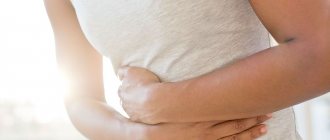
The presence of muscle fibers in excrement does not pose a danger to the human body. A complication is caused by a disease that causes creatorrhea. If the disease is not treated in time, complications arise:
- the appearance of ulcers, inflammation of the appendix due to chronic gastritis;
- constant inflammation of the pancreas causes thinning of the walls of the stomach and small intestine, this leads to peritonitis (inflammation of the peritoneum);
- infection leads to sepsis (bacteria entering the blood), which can lead to death;
- the appearance of blood in the stool due to bleeding of the stomach and intestines due to erosions and ulcers leads to anemia (reduction of red blood cells and hemoglobin in the blood);
- the occurrence of malignant neoplasms.
Norms
Macroscopic examination
| Parameter | Norm |
| Quantity | A healthy person produces an average of 100-200 g of feces per day. Normally, feces contain about 80% water and 20% dry matter . With a vegetarian diet, the amount of feces can reach 400-500 g per day; when using easily digestible food, the amount of feces decreases. |
| Consistency | Normally, formed feces have a dense consistency. Pasty feces can occur normally and are caused by the intake of predominantly plant foods. |
| Form | Normally cylindrical. |
| Smell | Normally, stool has a mild odor, which is called fecal (ordinary). It may intensify with the predominance of meat products in the diet, with putrefactive dyspepsia and weaken with a dairy-vegetable diet, constipation. |
| Color | Normally, stool is brown in color. When eating dairy foods, stool turns yellowish-brown, and meat stool turns dark brown . Ingestion of plant foods and some medications can change the color of stool (beets - reddish; blueberries, blackcurrants, blackberries, coffee, cocoa - dark brown; bismuth, iron color stool black). |
| Slime | Normally absent (or in scanty quantities). |
| Blood | Normally absent. |
| Pus | Normally absent. |
| Leftover undigested food (lientorrhea) | Normally none. |
Chemical research
| Parameter | Norm |
| Fecal reaction | Normally neutral, less often slightly alkaline or slightly acidic. Protein nutrition causes a shift in the reaction towards the alkaline side, while carbohydrate nutrition causes the reaction to shift towards the acidic side. |
| Reaction to blood (Gregersen reaction) | Normally negative |
| Reaction to stercobilin | Normally positive. |
| Reaction to bilirubin | Normally negative. |
| Vishnyakov-Triboulet reaction (for soluble protein) | Normally negative. |
Microscopic examination
| Parameter | Norm |
| Muscle fibers | Normally absent or single in the field of view. |
| Connective tissue (remnants of undigested vessels, ligaments, fascia, cartilage) | Normally absent. |
| Fat is neutral. Fatty acid . Salts of fatty acids (soaps). | Normally there are no or scanty amounts of fatty acid salts. |
| Plant fiber | Normally, there are single cells in the p/z. |
| Starch | Normally absent (or single starch cells). |
| Iodophilic microflora (clostridia) | Normally, single in rare areas (normally, iodophilic flora lives in the ileocecal region of the large intestine). |
| Epithelium | Normally, there are no or single columnar epithelial cells in the p/z. |
| Leukocytes | Normally, there are no or single neutrophils in the p/z. |
| Red blood cells | Normally none. |
| Worm eggs | Normally none. |
| Pathogenic protozoa | Normally none. |
| Yeast cells | Normally none. |
| Calcium oxalate (oxalic lime crystals) | Normally none. |
| Triple phosphate crystals (ammonium phosphate-magnesia) | Normally none. |
- 1. Chronic hemorrhoids
- 2. Anal fissure
- 3. Crohn's disease
- 4. Colon diverticulosis
- 5. Duodenal ulcer
- 6. Stomach ulcer
- 7. Chronic pancreatitis
- 8. Hemolytic anemia
- 9. Benign neoplasms of the colon
- 10. Intestinal helminthiases
- 11. Liver cirrhosis
- 12. Ulcerative colitis
- 13. Constipation
- 14. Malignant neoplasm of the colon
- 15. Irritable bowel syndrome, chronic colitis
- 16. Cholera
- 17. Amoebiasis
- 18. Typhoid fever
- 19. Peptic ulcer of the stomach and duodenum
- 20. Dyspepsia
- 21. Giardiasis
- 22. Salmonellosis
- 23. Food allergies
Chronic hemorrhoids
With hemorrhoids, the feces have the form of small lumps (“sheep feces” indicates a spastic condition of the intestines). Due to bleeding, the stool has a pronounced red color; it may contain scarlet blood and slightly changed red blood cells.
Anal fissure
With an anal fissure, feces may take the form of small lumps (“sheep feces” indicates a spastic condition of the intestines). Due to bleeding, scarlet blood and slightly changed red blood cells may be present in the stool.
Crohn's disease
With Crohn's disease, you may find blood in your stool. The Vishnyakov-Triboulet reaction reveals soluble protein in it
. Crohn's disease affecting the colon is characterized by the presence in the feces of a large number of red blood cells in combination with leukocytes and columnar epithelium.
Colon diverticulosis
With diverticular disease, due to the long stay of stool in the colon, it takes the form of “large lumps”.
Duodenal ulcer
With a duodenal ulcer, the feces have the form of small lumps (“sheep feces” indicates a spastic condition of the intestines).
Stomach ulcer
With a stomach ulcer, the feces have the form of small lumps (“sheep feces” indicates a spastic condition of the intestines).
Chronic pancreatitis
In chronic pancreatitis with exocrine insufficiency, stool may have a pasty consistency.
Hemolytic anemia
With hemolytic jaundice (anemia), due to massive hemolysis of red blood cells, the amount of stercobilin in the feces increases.
Benign neoplasms of the colon
With a tumor accompanied by bleeding from the distal colon, the stool may have a pronounced red color. With disintegrating colon tumors, blood can be detected in the stool
. Pus on the surface of the stool occurs when there is severe inflammation and ulceration of the colon mucosa (disintegration of an intestinal tumor), often along with blood and mucus. When a colon tumor is in the stage of decay due to bleeding, the reaction to blood (Gregersen reaction) is positive.
Intestinal helminthiases
With helminthic infestation, the feces contain eggs of roundworms, tapeworms, etc.
Cirrhosis of the liver
In liver failure, including liver cirrhosis, the stool is grayish-white, clayey (acholic).
Ulcerative colitis
With colitis, there is mucus covering the outside of the stool in the form of thin lumps. With ulcerative colitis, blood may be found in the stool; pus on the surface of the stool, often along with blood and mucus; soluble protein in the Vishnyakov-Triboulet reaction; a large number of leukocytes (usually neutrophils); a large number of red blood cells in combination with leukocytes and columnar epithelium.
Constipation
With prolonged constipation caused by chronic colitis, peptic ulcers and other conditions associated with increased absorption of fluid in the intestine, the amount of feces decreases. With constant constipation due to excessive absorption of water, the consistency of stool is dense
. With constipation, there may be mucus covering the outside of the stool in the form of thin lumps.
Malignant neoplasm of the colon
The form of feces in the form of “large lumps” - when feces remain in the colon for a long time - is noted in colon cancer. Pronounced red color of stool - with a tumor accompanied by bleeding from the distal parts of the colon and rectum. Blood in the stool can be found in disintegrating colon tumors
.
Pus on the surface of the stool occurs when there is severe inflammation and ulceration of the colon mucosa (disintegration of an intestinal tumor), often along with blood and mucus. A positive reaction to blood (Gregersen reaction) indicates bleeding with a tumor of the colon in the stage of decay
. A large number of red blood cells in combination with leukocytes and columnar epithelium is characteristic of malignant neoplasms of the colon.Irritable bowel syndrome, chronic colitis
With colitis with diarrhea, the amount of feces increases. The amount of feces decreases with prolonged constipation caused by chronic colitis. Mucus covering the outside of formed feces in the form of thin lumps is found in colitis. An alkaline reaction (pH 8.0-10.0) occurs in colitis with constipation. A large number of leukocytes (usually neutrophils) are observed in colitis of various etiologies.
Cholera
In cholera, stool looks like a gray inflammatory exudate with fibrin flakes and pieces of the colon mucosa (“rice water”).
Amoebiasis
With amebiasis, the stool is jelly-like, deep pink or red.
Typhoid fever
In typhoid fever, the stool looks like “pea soup.”
Peptic ulcer of the stomach and duodenum
With prolonged constipation caused by peptic ulcers, the amount of feces decreases. With an ulcer of the duodenum and stomach, the feces have the form of small lumps (“sheep feces” indicates a spastic state of the intestines).
To diagnose the condition of the gastrointestinal tract, a coprogram is performed. If an adult has muscle fibers without striations in the stool (creatorrhoea), this indicates a violation of the digestive process
. The small amount of fiber in feces may be physiological due to a protein-rich diet. If a lot of them are found, you should consult a doctor.
Diagnosis of creative rhea
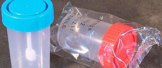
Muscle fibers in stool are detected by microscopic examination of stool samples. To do this, the patient donates feces for coprogram. They are placed in a sterile container purchased at a pharmacy and handed over to a laboratory assistant in the morning.
The study is carried out for a preventive examination and if a disease is suspected.
A coprogram is a stool analysis that evaluates the enzymatic function of the gastrointestinal tract (digestion of food with the help of enzymes) and the formation of hydrochloric acid in the stomach. The study allows you to determine dysfunction of the stomach, intestines, liver, and pancreas.
Features of stool analysis for muscle fibers (coprograms)
There are rules that the patient must follow when preparing for a stool test for creatorrhea in order for the diagnosis of the disease to be reliable:
- The material is submitted for analysis in the morning. The intestines should be emptied no longer than 8 hours before donating feces. The material is stored on the refrigerator door.
- A week before the study, medications are discontinued. You cannot use enemas or laxatives.
- If it is impossible to discontinue medications due to health conditions, the patient provides a list of medications used to the attending physician.
- Defecation should be independent.
- A week before taking a stool test for muscle fiber, you need to follow a protein-free diet. This is done to ensure that the result is not false positive.
- Women should not allow vaginal secretions to get into the biomaterial. To do this, use a hygienic tampon.
General stool analysis data
The coprogram determines all stool indicators that help make a diagnosis. Gastrointestinal diseases entail changes in many parameters of biological materials. Stool test results show:
- fats, fatty acids in feces;
- connective tissue fibers;
- presence of muscle fibers in feces;
- erythrocytes, leukocytes;
- starch;
- bacteria, parasites, helminths.
Based on the color of feces, the analysis determines which part of the gastrointestinal tract is disturbed:
- Red. Shows bleeding in the intestines: inflammation and ulceration of the mucous membrane of the large or small intestine. Its focus is determined using endoscopy.
- Transparent. Disorders of the gallbladder and bile duct.
- Black. Bleeding of the stomach and intestines.
- Green. Additionally, a foul odor occurs. The reason is fermentation in the intestines, the presence of pathogens.
- White. The appearance of white areas in the stool is a sign of liver pathology.
Deviations from the norm are identified by the shape and quantity
Small amounts: constipation, ulcers, fasting, chronic colitis.
Large: accelerated evacuation, reduced intestinal nutrient absorption function.
- Dense feces - lack of water in the body.
- The shape of small lumps (sheep feces) indicates a violation of the intestinal mucosa (cracks, ulcers).
- Ribbon-shaped feces indicate narrowing of the small intestine.
- Diarrhea occurs due to infections, impaired digestion and absorption.
Additional Research
After creatorrhea (the presence of muscle fibers in the stool) is confirmed, additional studies are carried out. This is done in order to determine the source of damage causing the disorder. Carry out:
- gastroscopy (examination of the gastrointestinal tract using a camera inserted through the pharynx);
- colonoscopy (endoscopic method of examining the intestinal tract);
- blood and urine tests;
- Ultrasound of the peritoneal organs.
Important! The examination is prescribed depending on the severity of the patient’s condition and the location of the pain.
Macroscopic examination
Quantity
In pathology, the amount of feces decreases with prolonged constipation caused by chronic colitis, peptic ulcers and other conditions associated with increased absorption of fluid in the intestines. With inflammatory processes in the intestines, colitis with diarrhea, and accelerated evacuation from the intestines, the amount of feces increases.
Consistency
Thick consistency - with constant constipation due to excessive absorption of water. Liquid or mushy consistency of stool - with increased peristalsis (due to insufficient absorption of water) or with abundant secretion of inflammatory exudate and mucus by the intestinal wall. Ointment-like consistency - for chronic pancreatitis with exocrine insufficiency
. Foamy consistency - with enhanced fermentation processes in the large intestine and the formation of a large amount of carbon dioxide.
Form
The form of feces in the form of “large lumps” - when feces remain in the colon for a long time (hypomotor dysfunction of the colon in people with a sedentary lifestyle or who do not eat rough food, as well as in cases of colon cancer, diverticular disease). The form in the form of small lumps - “sheep feces” indicates a spastic state of the intestines, during fasting, stomach and duodenal ulcers, a reflex nature after appendectomy, with hemorrhoids, anal fissure
.
Ribbon or “pencil” shape - for diseases accompanied by stenosis or severe and prolonged spasm of the rectum, for rectal tumors
. Unformed feces are a sign of maldigestion and malabsorption syndrome.
Color
If staining of stool by food or drugs is excluded, then color changes are most likely due to pathological changes. Grayish-white, clayey (acholic feces) occurs with obstruction of the biliary tract (stone, tumor, spasm or stenosis of the sphincter of Oddi) or with liver failure (acute hepatitis, cirrhosis of the liver). Black (tarry) stool—bleeding from the stomach, esophagus, and small intestine
.
Pronounced red color - with bleeding from the distal parts of the colon and rectum (tumor, ulcers, hemorrhoids). Gray inflammatory exudate with fibrin flakes and pieces of the colon mucosa (“rice water”) - with cholera
.
The jelly-like character is deep pink or red in amoebiasis
.
In typhoid fever, the stool looks like “pea soup.”
With putrefactive processes in the intestines, the feces are dark in color, with fermentative dyspepsia - light yellow.
Slime
When the distal colon (especially the rectum) is affected, the mucus occurs in the form of lumps, strands, ribbons, or a glassy mass. With enteritis, the mucus is soft, viscous, mixing with feces, giving it a jelly-like appearance. Mucus, covering the outside of formed feces in the form of thin lumps, occurs with constipation and inflammation of the large intestine (colitis).
Blood
When bleeding from the distal parts of the colon, the blood is located in the form of streaks, shreds and clots on formed stool. Scarlet blood occurs when bleeding from the lower parts of the sigmoid and rectum (hemorrhoids, fissures, ulcers, tumors). Black stool (melena) occurs when there is bleeding from the upper digestive system (esophagus, stomach, duodenum)
. Blood in the stool can be found in infectious diseases (dysentery), ulcerative colitis, Crohn's disease, disintegrating colon tumors.
Pus
Pus on the surface of the stool occurs with severe inflammation and ulceration of the mucous membrane of the colon (ulcerative colitis, dysentery, disintegration of an intestinal tumor, intestinal tuberculosis), often together with blood and mucus. Large amounts of pus without mucus are observed when opening paraintestinal abscesses.
Leftover undigested food (lientorrhea)
The release of undigested food residues occurs with severe insufficiency of gastric and pancreatic digestion.
What to do if muscle fibers are found in stool
If muscle fibers without striations are absent or are found singly, this indicates increased protein in the food.
Creatorrhea is a sign of an underlying disease. Therefore, treatment is carried out after laboratory and instrumental tests and diagnosis by the attending physician.
Treatment of creatorrhoea is carried out depending on the diagnosis:
- Nutritional creatorrhea (due to increased protein content in the diet) is treated with diet. Reduce food protein to minimum values.
- The infection is treated with antibacterial drugs. They are prescribed after an analysis to determine the sensitivity of bacteria to antibiotics.
- If muscle fibers are found with insufficient pancreatic function, a reduced amount of gastric enzymes is prescribed enzymes that break down organic substances.
- Surgical intervention. It is used for massive blood loss as a result of mechanical damage to the mucosal wall and peptic ulcer.
- Symptomatic treatment (relieves symptoms, but does not cure the disease): painkillers, antipyretics.
A single change in the function and operation of the gastrointestinal tract leads to a failure of the entire system. A persistent disorder of the digestive tract that does not go away over time is a signal to contact a specialist. If symptoms of malaise, pain in the abdominal area, or a change in the normal appearance of feces appear, you must consult a physician and gastroenterologist and take a stool test for muscle fibers. Timely treatment will help avoid complications. The prognosis of the disease is positive if a person adheres to the doctor’s recommendations, diet, and uses medication.
Chemical research
Fecal reaction
An acidic reaction (pH 5.0-6.5) is observed when iodophilic flora is activated, producing carbon dioxide and organic acids (fermentative dyspepsia). An alkaline reaction (pH 8.0-10.0) occurs with insufficient digestion of food, with colitis with constipation, sharply alkaline with putrefactive and fermentative dyspepsia.
Reaction to blood (Gregersen reaction)
A positive reaction to blood indicates bleeding in any part of the gastrointestinal tract (bleeding from the gums, rupture of varicose veins of the esophagus, erosive and ulcerative lesions of the gastrointestinal tract, tumors of any part of the gastrointestinal tract in the stage of decay).
Reaction to stercobilin
The absence or sharp decrease in the amount of stercobilin in the feces (the reaction to stercobilin is negative) indicates obstruction of the common bile duct with a stone, compression by a tumor, stricture, stenosis of the common bile duct, or a sharp decrease in liver function (for example, in acute viral hepatitis). An increase in the amount of stercobilin in feces occurs with massive hemolysis of red blood cells (hemolytic jaundice) or increased bile secretion.
Reaction to bilirubin
The detection of unchanged bilirubin in the stool of an adult indicates a disruption in the process of bilirubin recovery in the intestine under the influence of microbial flora. Bilirubin can appear during rapid evacuation of food (sharp increase in intestinal motility), severe dysbiosis (syndrome of bacterial overgrowth in the colon) after taking antibacterial drugs.
Vishnyakov-Triboulet reaction (for soluble protein)
The Vishnyakov-Triboulet reaction is used to identify a hidden inflammatory process. The detection of soluble protein in stool indicates inflammation of the intestinal mucosa (ulcerative colitis, Crohn's disease).
Nutritional Features
The patient is advised to follow a diet. High-fat foods, fried foods, pickles, marinades, smoked foods, that is, all heavy foods, are removed from the diet. During treatment it is necessary to exclude alcoholic drinks. Processed meats and low-quality foods also contribute to the formation of steatorrhea.
The diet should contain proteins in the form of lean poultry, turkey, and rabbit. Steamed sea fish, cottage cheese, and low-fat kefir are allowed. It is allowed to use a small amount of butter - no more than 50 g per day.
If you have fatty stools, it is not recommended to eat legumes - peanuts, beans, peas. A person should consume no more than 50-60 grams of oil and fat per day.
The diet is followed until the symptoms of steatorrhea disappear completely. It is important to enrich your diet with fortified foods.
To improve digestive processes the following are prescribed:
- enzymes - Pancreatin, Mezim, Creon;
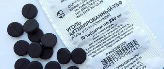
- sorbents - Enterosgel, Smecta;
- antacids (they enhance the action of enzymes and neutralize excess hydrochloric acid) - Phosphalugel, Omez;
- vitamin complexes containing fat-soluble vitamins and B vitamins.