Unreasonable headaches, poor sleep, fatigue, irritability - all this can be a consequence of poor blood circulation in the brain or abnormalities in the nervous system. For timely diagnosis of negative disorders in blood vessels, EEG - electroencephalogram of the brain is used. This is the most informative and accessible examination method, which does not harm the patient and can be safely used in childhood.
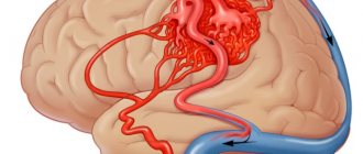
An electroencephalogram is used to examine the blood vessels of the brain.
EEG of the brain - what is it?
An encephalogram of the head is a study of a vital organ by exposing its cells to electrical impulses.
The method determines the bioelectrical activity of the brain, is very informative and the most accurate, as it shows the complete clinical picture:
- level and distribution of inflammatory processes;
- the presence of pathological changes in blood vessels;
- early signs of epilepsy;
- tumor processes;
- the degree of impairment of brain functioning due to pathologies of the nervous system;
- consequences of a stroke or surgery.
EEG helps identify signs of epilepsy
EEG helps monitor changes in the brain, both structural and reversible. This allows you to monitor the activity of a vital organ during therapy and adjust the treatment of identified diseases.
Echoencephalography as a method for studying brain structures
EchoEG is an ultrasound examination that allows you to study the anatomical structures of the brain. It is based on the reflection of an ultrasonic signal at the boundaries of media of different densities: brain matter, dura maters and cerebrospinal fluid (CSF). Allows you to diagnose so-called “space-occupying formations”: abscesses, cystic formations, hemorrhages, foreign bodies.
During the examination, the patient is in a supine position.
It can be used to diagnose: Parkinsonism, VSD, ICP, bruises, heart attacks, strokes, cerebrovascular accidents, concussions and much more.
Where can the examination be done and the price?
Electroencephalography can be done at any specialized medical center. Institutions can be either public or private. Depending on the form of ownership, the level of qualifications of the clinic, as well as the equipment used, prices for the procedure vary significantly.
In addition, the following factors influence the cost of an encephalogram:
- duration of the diagnostic procedure;
- carrying out functional tests;
- use of special programs (for mapping, studying epileptic impulses, comparing zones of symmetrical brain zones).
The average cost for an electroencephalogram is 2,680 rubles. Prices in Russian clinics start at 630 rubles.
Types of human brain activity recorded during EEG recording
The main types of activity that are recorded during the procedure and subsequently subjected to interpretation and further study are wave frequency, amplitude and phase.
Frequency
The indicator is estimated by the number of wave oscillations per second, recorded in numbers, and expressed in a unit of measurement - hertz (Hz). The description indicates the average frequency of the activity being studied. As a rule, 4-5 recording sections with a duration of 1 s are taken, and the number of waves in each time interval is calculated.
Amplitude
This indicator is the range of wave oscillations of the eclectic potential. It is measured by the distance between the peaks of waves in opposite phases and is expressed in microvolts (µV). A calibration signal is used to measure the amplitude. If, for example, a calibration signal at a voltage of 50 µV is determined on a record with a height of 10 mm, then 1 mm will correspond to 5 µV. In deciphering the results, interpretations are given to the most common meanings, completely excluding rare ones.
Phase
The value of this indicator evaluates the current state of the process and determines its vector changes. On the electroencephalogram, some phenomena are assessed by the number of phases they contain. Oscillations are divided into monophasic, biphasic and polyphasic (containing more than two phases).
Rhythms of brain activity
The concept of “rhythm” in the electroencephalogram is considered to be a type of electrical activity related to a certain state of the brain, coordinated by appropriate mechanisms. When deciphering the EEG rhythm indicators of the brain, its frequency corresponding to the state of the brain region, amplitude, and its characteristic changes during functional changes in activity are entered.
Indications for an electroencephalogram
Before prescribing an encephalography to a patient, a specialist examines the person and analyzes his complaints.
The following conditions may be the reason for an EEG:
- sleep problems - insomnia, frequent awakenings, sleepwalking;
- regular dizziness, fainting;
- fatigue and constant feeling of tiredness;
- causeless headaches.
If you have frequent headaches, you need to undergo an EEG
Insignificant, at first glance, changes in well-being may be the result of irreversible processes in the brain.
Therefore, doctors may prescribe an encephalogram if they detect or suspect pathologies such as:
- vascular diseases of the neck and head;
- vegetative-vascular dystonia, disruptions in cardiac activity;
- condition after a stroke;
- speech delay, stuttering, autism;
- inflammatory processes (meningitis, encephalitis);
- endocrine disorders or suspected tumor foci.
An EEG study is considered mandatory for people who have suffered head trauma, neurosurgical surgery, or suffering from epileptic seizures.
CT, ultrasound, MRI, EchoEG and EEG, or big confusion
People without medical education get confused trying to figure out the numerous abbreviations. In fact, it's very simple: CT and MRI scans show the internal structure of organs and the brain, exactly as it would be seen during an autopsy.
Ultrasound also shows processes visible to the eye: contraction of the heart, movement of blood through the vessels. EchoEG indicates the symmetry of signal reflection inside the brain, from which a conclusion is made about structural anomalies.
And only EEG records the invisible , that is, it registers the biological currents of the brain, which are the “echoes” of its secret and obvious work. If you take an encephalogram of the brain, you can see chaotic lines. Therefore, in addition to recording, a qualified decoding of the encephalogram of the brain is required. It is performed by a neurophysiologist, and then transmits the results and their interpretation to the doctor. Usually this study is ordered by neurologists. How does the research work and what do its results say?
How to prepare for research
Monitoring electrical activity in the brain requires little preparation. For the reliability of the results, it is important to follow the basic recommendations of the doctor.
- Do not use anticonvulsants, sedatives, or tranquilizers 3 days before the procedure.
- 24 hours before the test, do not drink any carbonated drinks, tea, coffee or energy drinks. Avoid chocolate. No smoking.
- On the eve of the procedure, thoroughly wash your scalp. Avoid the use of cosmetics (gels, varnishes, foams, mousse).
- Before starting the study, you need to remove all metal jewelry (earrings, chain, clips, hairpins)
- Hair should be loose – all kinds of weaves need to be undone.
- You need to remain calm before the procedure (avoid stress and nervous breakdowns for 2-3 days) and during it (do not be afraid of noises and flashes of light).
An hour before the examination you need to eat well - the examination is not carried out on an empty stomach.
The day before the examination you should not eat chocolate.
The essence of the method
EEG is used to determine the health and activity of the brain function after suspicion or detection of a lesion in the central nervous system, for example, poliomyelitis, encephalitis, meningitis. Based on the results of the procedure, it is possible to identify the level of damage to the brain and the location of the lesion.
An EEG can be performed both in a state of wakefulness and sleep, but special tests are required. Standard tests for EEG are:
- Stimulation with bright and intense flashes of light, while the eyes are closed;
- Alternate blinking of eyes;
- Achieving saturation of the brain with oxygen through deep breathing for 3-5 minutes.
Among the auxiliary methods there are:
- Stay in the dark for 40 minutes;
- Use of medications;
- Sleep deprivation.
How is an electroencephalogram performed?
The electrical activity of brain cells is assessed using an encephalograph. It consists of sensors (electrodes) that resemble a swimming pool cap, a block and a monitor, where the monitoring results are transmitted. The study is carried out in a small room that is isolated from light and sound.
The EEG method takes little time and includes several stages:
- Preparation. The patient takes a comfortable position - sits on a chair or lies down on the couch. Then the electrodes are applied. The specialist puts a “cap” with sensors on the person’s head, the wiring of which is connected to the device, which records the bioelectric impulses of the brain.
- Study. After turning on the encephalograph, the device begins to read information, transmitting it to the monitor in the form of a graph. At this time, the power of electric fields and its distribution in different parts of the brain can be recorded.
- Use of functional tests. This is performing simple exercises - blinking, looking at flashes of light, breathing rarely or deeply, listening to sharp sounds.
- Completion of the procedure. The specialist removes the electrodes and prints out the results.
During the EEG, the patient take a comfortable position and relax
If the study requires a more in-depth study (day monitoring), breaks in the procedure are possible. The sensors are disconnected from the wires, and the patient can go to the toilet, have a snack, and communicate with relatives.
Features of EEG in children
Monitoring brain activity in children has its own nuances. If the child is under one year old, then the study is carried out in a state of sleep. To do this, the baby should be fed and then rocked to sleep. After a year, children are examined while awake.
For the procedure to be successful, it is important to prepare the child:
- On the eve of the examination, it is recommended to talk with the child and tell him about the upcoming procedure. You can come up with a game to make your baby adapt faster by calling him a superhero or an astronaut.
- Take your favorite toys with you. This will help distract the fidget and calm him down at the right time.
- Feed the child before starting the study.
- Discuss with the doctor the time of manipulation and choose convenient hours when the child is awake and does not feel sleepy.
- On the eve of the examination, wash your baby’s hair well. If it is a girl, unwind her hair and remove all jewelry (immediately before monitoring).
If your baby takes certain medications on an ongoing basis, you should not give them up. It is enough to inform your doctor about this.
Carrying out electroencephalography in children
EEG in children is performed in the first year of life during sleep. The procedure helps to assess the state of the brain and analyze its development at the cellular level. At the same time, abnormalities are identified that can be treated before they begin to progress into serious diseases.
Electroencephalography for children is prescribed for:
- Epilepsy attacks, convulsions or similar attacks. An EEG is also done to adjust treatment.
- Sleep disorders.
- The need to assess brain development, its formation and biological activity.
- The presence of ischemic disorders of the central nervous system.
- To assess the severity of the disease.
EEG can detect developmental abnormalities already in the first year of life. For older children, preparation for the procedure includes playful moments. Since babies are more active, it is difficult for them to sit still. The child should gradually learn not to move for several minutes and overcome the fear of putting on a helmet with suction cups. The rest of the requirements are standard.
How long does the procedure take?
A regular encephalogram is a routine EEG or diagnosis of a paroxysmal state. The duration of this method depends on the area being studied and the application in monitoring functional tests. On average, the procedure does not take more than 20–30 minutes.
During this time, the specialist manages to:
- rhythmic photostimulation of different frequencies;
- hyperventilation (breaths are deep and rare);
- load in the form of slow blinking (open and close your eyes at the right moments);
- detect a number of functional changes of a hidden nature.
If the information received is insufficient, specialists may resort to a more in-depth examination.
There are several options:
- Encephalogram of night sleep. A long-term period is studied - wakefulness before bedtime, dozing, going to bed and waking up in the morning.
- EEG with deprivation. The method involves depriving the patient of sleep at night. He should wake up 2-3 hours earlier than usual and stay awake the next night.
- Continuous electroencephalogram. Monitoring of bioelectrical activity of the brain occurs during daytime sleep. The method is very effective in cases of suspected paroxysm (seizure) or identifying the causes of sleep disturbances.
Based on the EEG method, the duration of such a study can vary from 20 minutes to 8–15 hours.
The procedure for taking an electroencephalogram
The patient's position is sitting/reclining or lying down. Electrodes are attached to the head, usually using a special cap. A gel is applied between the skin and the electrode; for long-term recordings, the electrodes are secured with a special compound.
The subject must remain motionless, because motor activity causes “noise” when recording an EEG. This requirement causes some difficulty when taking an encephalogram in children. There is no way to stop kids from moving; you can only attract attention with toys. In this case, there will be “noises”. This is taken into account when decrypting.
Infants cannot hyperventilate because... To do this, the child must be able to follow instructions about the rhythm and depth of inhalation/exhalation. This makes it impossible to see some pathologies that are noticeable only under such a load. And they are not diagnosed alone.
The duration of the examination depends on its type. The procedure is not dangerous and not painful.
Decoding EEG indicators
A qualified diagnostician interprets the results of the encephalogram.
When deciphering, the patient’s clinical symptoms and basic EEG indicators are taken into account:
- state of rhythms;
- symmetry of the hemispheres;
- changes in gray matter when using functional tests.
The results obtained are compared with established standards, and deviations (dysrhythmia) are recorded in the conclusion.
Table “EEG interpretation”
| Indicators | Norm | Deviations | Possible pathological processes | |
| In adults | The child has | |||
| Alpha rhythm | 8–15 Hz – the rhythm is regular, observed at rest or with eyes closed. Maximum concentration of impulses in the area of the back of the skull and crown | The appearance of alpha waves in the frontal part of the brain. The rhythm becomes paroxysmal. Violation of the stability of frequency and symmetry of the hemispheres (above 30%) | Development of tumor processes, appearance of cysts. Condition of stroke or heart attack. Presence of serious skull injuries | Neuroses of varying degrees Psychopathy Delayed psychomotor development – neurophysiological immaturity of brain cells |
| Beta rhythm | 12–30 Hz – reflects excitement, anxiety, nervousness and depression. Sensitive to sedatives. Localized in the suprafrontal lobes | Diffuse beta waves Amplitude increase Violations of hemispheric symmetry Paroxysmal discharges | Concussion Encephalitis | |
| Delta rhythm | 0.5–3 Hz – records the state of natural sleep. Does not exceed 15% of all rhythms. Amplitude not higher than 40 µV | High amplitude The appearance of delta and theta waves outside sleep, localization in all parts of the brain High frequency rhythms | Irritation of the structural centers of gray matter (irritation) Neuroses Dementia | |
| Theta rhythm | 3.5–8 Hz – reflects the normal state during sleep in adults. In children, this indicator is dominant | |||
Based on the study of rhythms, a conclusion is made about the bioelectrical activity of the brain. In a normal state, it should be without attacks (paroxysms), have regular rhythm and synchronicity. Diffuse (moderate) changes are acceptable if other pathological disorders are not identified (irritation of parts of the brain, dysfunction of regulatory systems, disorganization of rhythms). In this case, the specialist can prescribe corrective treatment and monitor the patients.
Important!
It is important to consider that moderate changes in rhythms (delta and theta), the appearance of paroxysmal discharges and epileptic activity on the EEG in children and people under 21 years of age are the norm and do not relate to deviations in the structures of a vital organ.
The need to use EEG
It is difficult to overestimate the relevance of what the electroencephalogram of the brain shows. This information allows you to identify fairly complex diseases and abnormalities that may not have visible symptoms. It is worth noting that at the moment more modern diagnostic techniques are available to patients. We are talking about magnetic resonance and computed tomography.
Nevertheless, EEG is still actively used in Russian clinics, allowing doctors to make fairly accurate diagnoses. The use of this technique is especially important when working with patients suffering from epilepsy. This is explained by the fact that EEG makes it possible to record important details and features of the patient’s condition at any time.
No need to be afraid
If you have to do an encephalogram of the child’s brain , then you need to warn that there is no need to be afraid. It won't hurt, but it will be very interesting. They will put a beautiful wig on your head, sit you on a comfortable chair, like an astronaut, and ask you to breathe, and then the light will blink. And in general, this is all very similar to flying into space.
How is this research actually conducted?
- First, a rubber “harness” is put on the patient’s head to secure the electrodes;
- then the scalp is lubricated with a conductive gel, and, according to a special scheme, electrodes are attached to rubber rims so that they fit tightly to the scalp, and the wires are connected.
This is where the preparation ends, the patient is seated in a chair, the lights are turned off, and they are asked to relax. The laboratory assistant - neurophysiologist goes into another room. Sometimes she will ask you to breathe deeply and forcefully, and then suddenly a bright light will flash into your eyes.
Electroencephalography results
Example of EEG results
Bioelectrical current activity caused by neurons in the brain is read by electrodes and represented as wave-like oscillations. In the recorded vibration waves, it is customary to distinguish alpha, beta, delta, theta and mu waves.
The results of the encephalogram, recorded on paper or presented on the screen, are analyzed by two doctors at once - a neurophysiologist (who also performs the study itself) and a neuropathologist.
What is the interpretation of EEG? First of all, the doctor evaluates the symmetry of the activity of neurons in the left and right hemispheres. The doctor is also interested in the brain’s reaction to the tests performed during the study.
Based on what the encephalogram shows, the doctor can make a conclusion about the correct functioning of the human brain. If a pathology is detected, the necessary treatment is selected by a neurologist.
Contraindications
Electroencephalography has no contraindications, but in young children it is best performed during sleep. In case of exacerbation of mental illness, you need to wait for the condition to normalize. The study should be carried out with caution in people with injuries and wounds to the head.
Electroencephalography is a safe, informative research method that allows you to reliably diagnose some brain diseases. An EEG of the brain shows what underlies loss of consciousness, fatigue, insomnia, and headaches. It is used in children and adults in any condition.
Basic rhythms of electrical activity of the brain
Before deciphering the result of brain activity, all factors are taken into account:
- anatomy, physiology, genotype and age of the patient;
- general condition of the body, presence of head bruises;
- presence of disorders or pathology;
- the presence of tremor, convulsive phenomena, weakness in the limbs;
- time of last attack or fit;
- many other manifestations.
QEEG - a computer electroencephalogram consists of rhythms that come from the bioelectric activity of the brain at certain periods of time or situations.
The main biorhythms are alpha, beta, theta and delta. In addition to them, other impulses are observed:
- high frequency gamma;
- kappa, found in the temporal zone;
- mu rhythms, which are associated with mental stress.
The study is carried out not only during waking hours, but also during sleep. Often, the doctor prescribes a referral for EEG monitoring at night, since such an examination is considered to be the most productive. When recording brain activity, the main rhythmic parameters are read.
Alpha rhythm
Such impulses are recorded in the normal state of a person, when the patient is awake. The frequency of the waves ranges from 8-14 Hz, the amplitude reaches up to 100 μV.
The a-rhythms are most clearly expressed in the parietal and occipital zones. When brain activity changes, low-amplitude irregularity in brain activity occurs, which is reflected in the desynchronous movement of neurons.
With a bright flash of light this happens quite sharply. If there is no emotiogenic stimulus, then a-rhythms are restored instantly. Doctors call this phenomenal phenomenon the “desynchronization reaction.”
Beta rhythm
It is clearly seen in the central part of the brain, but is also common in the peripheral posterior and frontal lobes.
The frequency ranges from 15 to 41 Hz, the amplitude does not exceed 25 μV. The norm is expressed weakly, within 15 microvolts.
B-rhythms are associated with the sensory somatic system and motor functions of the cerebral cortex, which leads to a reaction of extinction of tactile stimulation (decrease in the pain threshold, etc.).
Theta and delta rhythms
Theta activity occurs at a frequency of 4 to 7 Hz with an amplitude of no more than 40 μV. In pathologies it can reach more than 300 microvolts.
T-rhythms are considered key for children 3-6 years old. A clear manifestation can be seen on EEG readings in the central region of the brain even in the first 28 days of life.
D-rhythms are detected at a frequency of up to 3 Hz, the amplitude is equivalent to theta. These fluctuations take up about 15% of the time of the entire procedure.
The delta rhythm can indicate the presence of a tumor, neoplasm, brain contusion, or narcotic state.
Theta and delta rhythms are recorded in the patient during sleep.
Methodology for performing EEG during pregnancy
The pregnant woman sits in a comfortable chair. To study brain activity during an EEG, electrodes are used that are fixed on the head. Their inner surface is lubricated with water-soluble gel. An indifferent electrode is attached to the earlobe. The read brain potentials are amplified by a special device, transmitted to the monitor and recorded on the hard drive. The electroencephalogram is then transferred to mobile media and printed on paper tape. A pregnant woman can receive an EEG transcript in her hands on the second day.
What are they studying?
The biocurrents of neurons in the cerebral cortex are very weak. If a person rolls his eyes closed and wrinkles his forehead, then these signals will drown out the activity of the cortex, and the doctor will not understand anything, so you need to sit and relax.
An encephalogram can show disorders that another research method cannot see: a predisposition to convulsive manifestations and epileptic seizures. Therefore, the exclusion of epilepsy, or any epileptiform activity, is the main indication for an encephalogram, or EEG for short.
Therefore, it is usually prescribed:
- specialists – epileptologists in the treatment and management of patients;
- during medical examinations of pilots, railway workers, drivers;
- upon conscription for active military service and examination of the health of the conscript;
- during forensic medical and forensic psychiatric examination;
- just to check the functioning of the cerebral cortex, when patients complain of fainting, loss of consciousness, and other symptoms under the guise of which epilepsy may be hiding.
An encephalogram of the child’s brain can be prescribed for autism, or vice versa, motor hyperreactivity, psychomotor agitation, night terrors and other complaints.
Brief introduction to EEG
This type of examination allows, through scanning, to track the activity of each part of the brain, neural impulses and, most importantly, the EEG clearly records the level of coordination of the actions of all components of the brain part (or the property of rhythm). If a person’s central nervous system is attacked by one or another ailment, diagnostics will reveal a disruption in the functioning of the organ.
Carrying out the test is not particularly difficult. The patient sits comfortably on an equipped couch in a reclining position. Certain points of the head are treated with a gel that ensures the conduction of impulses, and small electrodes are attached to the surface. When making a diagnosis, a person must relax his muscles and close his eyelids.
Next, a computer program is launched, which, using sensors, records brain activity and provides the necessary information in the form of several graphs. The procedure can last 1 hour, and in some cases the whole night under the close supervision of a doctor. The decrypted data will be received after a few days.
Despite the painlessness of EEG, a small child may become alarmed by what is happening, so the mother should be nearby and, if necessary, calm the child
Using EEG it is possible to detect:
- foci of epileptic activity;
- possible causes of fainting and panic attacks;
- various pathologies in the functioning of the brain and central nervous system as a whole;
- the effect of the prescribed complex of drugs on the body;
- violation of relevant functions, etc.
The procedure is considered safe and completely painless, so it is performed on both adults and children.