201
Despite the amazing abilities (intellectual and extrasensory) of some people, the human brain does not work at 100%, but only at 5-7%. Thanks to this, brain tissue has unlimited reserve capacity, which allows restoration of normal function even after major strokes. This also creates a whole direction for scientific research that seeks to make the human brain work at its full capacity. I wonder what will a person be able to do then?
The brain is the main organ of the human central nervous system; it regulates all human life processes. The brain is located in the cranial cavity, where it is reliably protected from external negative influences and mechanical damage. As the brain develops, it takes on the shape of a skull. In appearance it resembles a yellowish gelatinous mass, since the brain tissue contains a large amount of specific lipids.
The brain has always been and remains an extraordinary mystery for scientists, which they have been trying to solve for thousands of years and, probably, will do so for just as long. This is a perfect mechanism created by nature, which allows a person to be called homo sapiens, or a reasonable person. Our brain is the work of millions of years of evolution.
A look into history and modernity
Initially, the heart was considered the organ of thoughts and feelings. However, with the development of mankind, a connection between behavior and GM was determined (in accordance with traces of trepanation on the found turtles). This neurosurgery was probably used to treat headaches, skull fractures, and mental illness.
From the point of view of historical understanding, the brain comes into focus in ancient Greek philosophy, when Pythagoras, and later Plato and Galen, understood it as the organ of the soul. Significant advances in determining brain functions were provided by the conclusions of doctors who, based on autopsies, examined the anatomy of the organ.
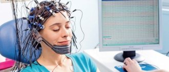
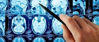
Today, to study the brain and its activity, doctors use EEG, a device that records brain activity through electrodes. The method is also used to diagnose cerebral tumors.
To eliminate a tumor, modern medicine offers a non-invasive method (without an incision) - stereosurgery. But its use does not exclude the use of chemical therapy.
Diencephalon
This section is located between the midbrain and the corpus callosum. There are visual hillocks here that have a number of important functions, in particular the processing of centripetal impulses coming from the outside world and their transmission to the brain. In addition, they are responsible for such parameters of emotional behavior as pulse, breathing, blood pressure, facial expressions, etc.
Embryonic development
The GM develops during embryonic development from the anterior part of the neural tube emerging in the 3rd week (days 20-27 of development). At the head end of the neural tube, 3 primary cerebral vesicles are formed - anterior, middle, posterior. At the same time, the occipital and frontal regions are created.
During the 5th week of child development, secondary brain vesicles form, forming the main parts of the adult brain. The frontal brain is divided into the intermediate and terminal brain, the posterior - into the Varoliev bridge, the cerebellum.
Cerebrospinal fluid is formed in the chambers.
hindbrain
The structure of the human brain provides for the presence of a hindbrain. This section includes two main parts - the pons and the cerebellum. The pons is a component of the brainstem that is located between the midbrain and medulla oblongata. The main functions of this department include reflex and conduction.
The pons, which from an anatomical point of view is considered a structure of the hindbrain, is presented in the form of a thickened cushion. At the bottom of the bridge there is an oblong section, at the top there is a middle section.
The pons contains centers that control the functioning of the masticatory, facial, and some oculomotor muscles. Nerve impulses from the receptors of the sensory organs, skin, and inner ear go to the bridge; thanks to this zone, we can feel taste, maintain balance, and have hearing sensitivity.
Anatomy
The GM, as the energy, control, and organizational center of the nervous system, is stored in the neurocranium. In adult humans, its volume (weight) is about 1500 g. However, specialized literature shows great variability in the mass of the GM (both in humans and in animals, for example, in monkeys). The lowest weights - 241 g and 369 g, as well as the highest weight - 2850 g were found in representatives of the population with severe mental retardation. The volume between the floors also differs. The male brain weighs about 100 g more than the female brain.
The location of the brain in the head is visible in the section.
The brain, along with the spinal cord, forms the central nervous system. The brain is located in the skull, protected from damage by the fluid that fills the cranial cavity, cerebrospinal fluid. The structure of the human brain is very complex - it includes a cortex, divided into 2 hemispheres, which are functionally different.
The function of the right hemisphere is to solve creative problems. It is responsible for the expression of emotions, perception of images, colors, music, facial recognition, sensitivity, and is the source of intuition. When a person first encounters a task, a problem, it is this hemisphere that begins to work.
The left hemisphere dominates in tasks that a person has already learned to cope with. Metaphorically, the left hemisphere can be called scientific, since it includes logical, analytical, critical thinking, calculation and use of language skills, intelligence.
The brain contains 2 substances – gray and white. The gray matter on the surface of the brain produces the cortex. The white matter consists of a large number of axons with myelin sheaths. It is located under the gray matter. The bundles of white matter passing through the central nervous system are called nerve tracts. These tracts provide signal transmission to other structures of the central nervous system. Depending on the function, the pathways are divided into afferent and efferent:
- afferent pathways bring signals to the gray matter from another group of neurons;
- efferent pathways form the axons of neurons, leading signals to other cells of the central nervous system.
Brain protection
The protection of the brain includes the skull, meninges, and cerebrospinal fluid. In addition to tissue, the nerve cells of the central nervous system are also protected from the effects of harmful substances from the blood by the blood-brain barrier (BBB). The BBB is an adjacent layer of endothelial cells, closely interconnected, preventing the passage of substances through the intercellular spaces. In pathological conditions such as inflammation (meningitis), the integrity of the BBB is compromised.
Shells
The brain and spinal cord are covered by 3 layers of membranes - hard, arachnoid, soft. The constituent components of the membranes are the connective tissues of the brain. Their general function is to protect the central nervous system, the blood vessels supplying the central nervous system, and the collection of cerebrospinal fluid.
Midbrain
The midbrain has a relatively simple structure, small in size, and includes two main parts: the roof (the centers of hearing and vision are located in the subcortical part); legs (contain conducting pathways). It is also customary to include black matter and red nuclei in the structure of the cloth.
The subcortical centers, which are part of this department, work to maintain the normal functioning of the hearing and vision centers. Also located here are the nerve nuclei that ensure the functioning of the eye muscles, the temporal lobes that process various auditory sensations, turning them into sound images familiar to humans, and the temporoparietal node.
The following functions of the brain are also distinguished: controlling (together with the medulla oblongata) the reflexes that arise when exposed to a stimulus, helping with orientation in space, forming an appropriate reaction to stimuli, turning the body in the desired direction.
The gray matter in this part is a high concentration of nerve cells that form the nerve nuclei inside the skull.
The brain actively develops between the ages of two and eleven years. The most effective method of improving your intellectual abilities is to engage in unfamiliar activities.
The main parts of the brain and their functions
The GM is divided into several parts - departments that perform different functions, but work together to form the main body. How many departments are there in the brain and which brain is responsible for certain abilities of the body?
What does the human brain consist of - sections:
- The hindbrain contains a continuation of the spinal cord - the medulla oblongata and 2 other parts - the pons and the cerebellum. The pons and cerebellum together form the hindbrain in the narrow sense.
- Average.
- The forebrain contains the diencephalon and telencephalon.
The combination of the medulla oblongata, midbrain, and pons forms the brain stem. This is the oldest part of the human brain.
Medulla
The medulla oblongata is a continuation of the spinal cord. It is located at the back of the skull.
Functions:
- entrance and exit of cranial nerves;
- transmission of signals to the centers of the brain, the course of descending and ascending neural pathways;
- the place of the reticular formation is the coordination of the activity of the heart, the content of the vasomotor center, the center of unconditioned reflexes (hiccups, salivation, swallowing, coughing, sneezing, vomiting);
- when the function is impaired, reflexes and cardiac activity are disrupted (tachycardia and other problems, including stroke).
Cerebellum
The cerebellum forms 11% of the total lobe of the brain.
Functions:
- center for coordination of movements, control of physical activity - coordination component of proprioceptive innervation (guidance of muscle tone, accuracy and coordination of muscle movements);
- balance and posture support;
- When cerebellar function is impaired (depending on the degree of the disorder), muscle hypotonia, slowness in walking, inability to maintain balance, and speech impairment occur.
By monitoring movement activity, the cerebellum evaluates information received from the statokinetic apparatus (inner ear) and proprioceptors in the tendons associated with the body's current position and movement. The cerebellum also receives information about planned movements from the motor cortex of the brain, compares it with current body movements, and ultimately sends signals to the cortex. She then directs the movements as they were planned. Using this feedback, the cortex can restore commands and send them directly to the spinal cord. As a result, a person can do well-coordinated actions.
Pons
It forms a transverse wave over the medulla oblongata and is connected to the cerebellum.
Functions:
- the area of exit of the nerves of the head and the deposition of their nuclei;
- transmission of signals to higher and lower centers of the central nervous system.
Midbrain
This is the smallest part of the brain, the phylogenetically oldest brain center, part of the brain stem. The upper part of the midbrain is formed by the quadrigeminalis.
Functions of the quadrigeminal region:
- the superior colliculi are involved in the visual pathways, work as a visual center, and participate in visual reflexes;
- the lower colliculi are involved in auditory reflexes - they provide reflexive reactions to sounds, volume, and reflexive appeal to sound.
Diencephalon
The diencephalon is largely closed by the telencephalon. It is one of the 4 main brain parts. Consists of 3 pairs of structures - thalamus, hypothalamus, epithalamus. Separate parts limit the third ventricle. The pituitary gland is connected to the hypothalamus through the infundibulum.
Thalamus function
The thalamus makes up 80% of the diencephalon and is the basis for the lateral walls of the ventricle. The thalamic nuclei redirect sensory information from the body (spinal cord) - pain, touch, visual or auditory signals - to specific brain areas. Any information sent to the cerebral cortex must be redirected to the thalamus, the gateway to the cerebral cortex. Information in the thalamus is actively processed and changes - it increases or decreases signals intended for the cortex. Some of the thalamic nuclei are motor.
Function of the hypothalamus
This is the lower part of the diencephalon, on the lower side of which there are the intersections of the optic nerves (chiasma opticum), downward is the pituitary gland, which secretes a large number of hormones. The hypothalamus stores a large number of gray matter nuclei; functionally, it is the main center for controlling the organs of the body:
- control of the autonomic nervous system (parasympaticus and sympaticus);
- control of emotional responses - part of the limbic system includes an area for fear, anger, sexual energy, joy;
- regulation of body temperature;
- regulation of hunger, thirst - areas of concentration of nutrient perception;
- behavior management – control of motivation for food consumption, determination of the amount of food eaten;
- sleep-wake cycle control – responsible for the timing of the sleep cycle;
- monitoring of the endocrine system (hypothalamic-pituitary system);
- memory formation – receiving information from the hippocampus, participating in the creation of memory.
Function of the epithalamus
This is the most posterior part of the diencephalon, consisting of the pineal gland - the pineal gland. Secretes the hormone melatonin. Melatonin signals the body to prepare for the sleep cycle, affects the biological clock, the onset of puberty, etc.
Pituitary gland function
Endocrine gland, adenohypophysis – production of hormones (GH, ACTH, TSH, LH, FSH, prolactin); neurohypophysis - secretion of hormones produced in the hypothalamus: ADH, oxytocin.
Finite brain
This element of the brain structure is the largest part of the human central nervous system. Its surface consists of gray bark. Below this is the white matter and the basal ganglia.
Hemispheres:
- the telencephalon consists of hemispheres constituting 83% of the total brain mass;
- between the 2 hemispheres there is a deep longitudinally oriented groove (fissura longitudinalis cerebri), extending to the brain muscle (corpus callosum), connecting the hemispheres and mediating cooperation between them;
- There are grooves and convolutions on the surface.
Cerebral cortex:
- control of the nervous system - the seat of human consciousness;
- formed by gray matter - formed from the bodies of neurons, their dendrites and axons; does not contain nerve pathways;
- has a thickness of 2-4 mm;
- makes up 40% of the total GM volume.
General information about the brain
The brain is made up of more than 100 billion nerve cells. The structure of the organ is anatomically divided into the cerebrum, which consists of the right and left hemispheres, the cerebellum and the brain stem. The brain is covered with 3 membranes and occupies up to 95% of the capacity of the cranium.
Infographics: structure of the human brain
The mass of brain tissue in healthy people varies and on average is in the range of 1100-1800 grams. No connection has been established between human abilities and brain weight. In women, as a rule, the central organ of the NS weighs 200 grams less than in men.
The brain is covered with gray matter - the main functional sphere, where the bodies of almost all neurons are located, which form the cerebral cortex. Inside there is white matter, which consists of processes of neurons and represents the pathways along which information enters the cortex for analysis and, after this, commands are transmitted down.
Not only are there control centers in the cerebral cortex, which are called screen centers, they are also present deep in the brain, surrounded by white matter. Such centers are called nuclear or subcortical (clusters of nerve cell bodies in the form of nuclei).
Inside the brain there is a hollow system, which consists of 4 ventricles and several ducts. It connects to the spinal cord canal. Within this system, cerebrospinal fluid, or cerebrospinal fluid, circulates, which performs a protective function.
Cortical areas
On the surface of the hemispheres there are permanent grooves dividing them into 5 lobes. The frontal lobe (lobus frontalis) lies in front of the central sulcus (sulcus centralis). The occipital lobe extends from the central to the parieto-occipital sulcus (sulcus parietooccipitalis).
Frontal lobe regions
The main motor area is located in front of the central sulcus, where there are pyramidal cells, the axons of which form the pyramidal (cortical) path. These paths provide precise and comfortable movements of the body, especially the forearms, fingers, and facial muscles.
Premotor cortex. This area is located in front of the primary motor area and controls more complex free activity movements that depend on sensory feedback - grasping objects, moving over obstacles.
Broca's speech center is located in the lower part, usually the left or dominant hemisphere. Broca's center in the left hemisphere (if it is dominant) controls speech, in the right hemisphere it supports the emotional coloring of the spoken word; this area is also involved in short-term memory for words and speech. Broca's center is associated with the preferential use of one hand for work - left or right.
The visual area is the motor part that controls the required rapid eye movements when viewing a moving target.
The olfactory region is located at the base of the frontal lobes and is responsible for the perception of smell. The olfactory cortex connects to the olfactory areas in the lower centers of the limbic system.
The prefrontal cortex is a large area of the frontal lobe responsible for cognitive functions: thinking, perception, conscious storage of information, abstract thinking, self-awareness, self-control, perseverance.
Areas of the parietal lobe
The sensitive area of the cortex is located just behind the central sulcus. Responsible for the perception of general bodily sensations - the perception of skin (touch, warmth, cold, pain), taste. This center is capable of localizing spatial perception.
Somatosensitive area - located behind the sensitive one. Participates in recognizing objects based on their shape based on previous experience.
Occipital lobe regions
The main visual area is located at the end of the occipital lobe. It receives visual information from the retina and processes information from both eyes together. Here the orientation of objects is perceived.
Associative visual area - located in front of the main one, cooperates with it to determine the color, shape, and movement of objects. It also communicates with other parts of the brain through the anterior and posterior pathways. The anterior tract runs along the lower edge of the hemispheres and is involved in word recognition during reading and face recognition. The posterior pathway passes into the parietal lobe and is involved in spatial connections between objects.
Temporal lobe areas
The hearing zone and vestibular region are located in the temporal lobe. There is a distinction between the main and associative areas. The main one perceives volume, pitch, rhythm. Associative – based on memorizing sounds and music.
Speech area
The speech area is a broad area associated with speech. The left hemisphere is dominant (in right-handed people). To date, 5 areas have been identified:
- Broca's area (speech formation);
- Wernicke's area (speech understanding);
- lateral prefrontal cortex in front of and below Broca's area (speech analysis);
- area of the temporal lobe (coordination of auditory and visual aspects of speech);
- internal share – articulation, recognition of rhythm, voiced word.
The right hemisphere is not involved in the speech process in right-handed people, but works on the interpretation of words and their emotional connotation.
Brain functions
The brain has a very complex structure, which corresponds to the functions it performs. It is very difficult to list them, since this includes the entire sphere of activity of the human body. Let's dwell on the basic functions of life:
- Physical activity . All movements of the body are associated with the activity of the area of the cerebral cortex, which is located in the parietal lobe in the central anterior gyrus. The activity of all skeletal muscle groups is under the direction of this part of the brain.
- Sensitivity. The central posterior gyrus in the parietal lobe of the cerebral cortex is responsible for this function. In addition to skin sensitivity (tactile, pain, temperature, baroreceptor), there is also a center of proprioceptive sensitivity, which controls the sense of the position of the body and its individual parts in space.
- Rumor . The area of the brain that is responsible for hearing is located in the temporal lobes of the cortex.
- Vision. The visual center is localized in the occipital region of the cortex.
- Taste and smell . The center that is responsible for these functions can be found on the border of the frontal and temporal lobes, in the gyri.
- Human speech , both motor function and sensory (pronunciation of words and their understanding) are located in the Broca's and Wernicke's centers of the cerebral hemispheres.
- In the medulla oblongata there are all centers important for life - breathing, heartbeat, regulation of the lumen of blood vessels, food reflexes, for example, swallowing, all protective reflexes (coughing, sneezing, gagging, lacrimation, etc.), regulation of the state of smooth muscle fibers of internal organs.
- The posterior part of the organ regulates the maintenance of balance and coordination of motor activity, in addition, there are many pathways that carry information to the higher and lower centers of the brain.
- The midbrain contains subcortical centers that regulate visual, auditory and motor functions at the lower level.
- Diencephalon : the thalamus regulates all types of sensitivity, and the hypothalamus converts nerve signals into endocrine signals (the central organ of the human endocrine system), and also regulates the activity of the autonomic nervous system.
These are the main centers of the brain that provide a person with life, but there are many others, for example, the center of writing, counting, music, centers of human character, irritability, color differences, appetite, etc.
Main functional centers of the brain
Laterality of the hemispheres
There are differences in the functioning of the left and right hemispheres. Both hemispheres coordinate opposite parts of the body and have different cognitive functions. For most people (90-95%), the left hemisphere controls, in particular, language skills, mathematics, and logic. In contrast, the right hemisphere controls visual spatial abilities, facial expressions, intuition, emotions, artistic and musical abilities. The right hemisphere works with large images, and the left hemisphere works with small details, which it then logically explains. In the rest of the population (5-10%), the functions of both hemispheres are opposite, or both hemispheres have the same degree of cognitive function. Functional differences between the hemispheres tend to be greater in men than in women.
Medulla
An important part of the central nervous system, which is called the bulbus in various medical descriptions. It is located between the cerebellum, pons, and spinal region. The bulbus, being part of the central nervous system trunk, is responsible for the functioning of the respiratory system and regulation of blood pressure, which is vital for humans.
In this regard, if this department is damaged in any way (mechanical damage, pathologies, strokes, etc.), then the probability of human death is high.
The most important functions of the oblongata are:
- Working together with the cerebellum to ensure balance and coordination of the human body.
- The department includes the vagus nerve with autonomic fibers, which helps ensure the functioning of the digestive and cardiovascular systems and blood circulation.
- Ensuring the swallowing of food and liquids.
- Presence of coughing and sneezing reflexes.
- Regulation of the respiratory system and blood supply to individual organs.
The medulla oblongata, the structure and functions of which differ from the spinal cord, has many common structures with it.
The brain contains about 50-55% fat and in terms of this indicator it is far ahead of other organs of the human body.
Basal ganglia
The basal ganglia are located deep in the white matter. They work as a complex neural structure that cooperates with the cortex to control movement. They start, stop, regulate the intensity of free movements, are controlled by the cerebral cortex, can select the appropriate muscles or movements for a particular task, and inhibit opposing muscles. When their function is impaired, Parkinson's disease and Huntington's disease develop.
Cortex
The structure of the cortex is very complex and represents a multi-level system. Moreover, the structure is not the same in all areas. In some there are only 3 layers of cells (old cortex), and in some all 6 (new cortex). If the bark is straightened, its area will be approximately 220 thousand square millimeters.
The entire cerebral cortex is functionally divided into separate fields or centers (Brodmann fields), which are responsible for one or another function in the body. This is a kind of map of what a person can do and where these skills are hidden in the brain.
Localization of body functions in the cerebral cortex
Despite the amazing abilities (intellectual and extrasensory) of some people, the human brain does not work at 100%, but only at 5-7%. Thanks to this, brain tissue has unlimited reserve capacity, which allows restoration of normal function even after major strokes. This also creates a whole direction for scientific research that seeks to make the human brain work at its full capacity. I wonder what will a person be able to do then?
inf
The brain's memory capacity is practically unlimited
Tatiana Ayazo / rd.com
It is impossible to know too much or get so much new information that there is nowhere else to put it (although that is exactly what it seems after long meetings). In our brain, unlike computers and phones, space never runs out. Although, for example, lack of sleep can negatively affect the ability to remember data.
Subcortical structures.
Below the cortex lies a number of important brain structures, or nuclei, which are collections of neurons. These include the thalamus, basal ganglia and hypothalamus. The thalamus is the main sensory transmitting nucleus; it receives information from the senses and, in turn, forwards it to the appropriate parts of the sensory cortex. It also contains nonspecific zones that are connected to almost the entire cortex and probably provide the processes of its activation and maintenance of wakefulness and attention. The basal ganglia are a collection of nuclei (the so-called putamen, globus pallidus and caudate nucleus) that are involved in the regulation of coordinated movements (starting and stopping them).
The hypothalamus is a small region at the base of the brain that lies beneath the thalamus. Richly supplied with blood, the hypothalamus is an important center that controls the homeostatic functions of the body. It produces substances that regulate the synthesis and release of pituitary hormones (see also pituitary gland). The hypothalamus contains many nuclei that perform specific functions, such as regulation of water metabolism, distribution of stored fat, body temperature, sexual behavior, sleep and wakefulness.
The brain, like the muscles, is subject to the “use it or lose it” rule.
Tatiana Ayazo / rd.com
We can expand our cognitive reserve, or the brain's innate ability to repair, through different types of learning and new experiences. People with greater cognitive reserve have been shown to cope better with the unexpected. But if the brain is not used, this reserve will decrease.
Functionality of the telencephalon
Higher nervous activity is recognized as the most important task of the telencephalon. It is aimed at uniting and managing all organs and systems of the body. The basic mechanism of the cerebral cortex of this part of the organ, consisting of neurons, closely interacts with conditioned types of reflexes. They, in turn, are responsible for human mental behavior. The main components of this organ are both hemispheres of the cerebrum and the olfactory brain, including the corpus callosum and striatum.
The left hemisphere is entrusted with the role of abstract thinking and the formation of correct speech. This segment is also designed to handle logical and analytical functions. In this part, the most complex actions are formed (for example, motor acts), and abstract, invariant and generalized recognition is formed.
Here, too, analytical perception is born, mathematical calculations are constructed, and the organs of the right half of the human body are controlled, similarities are established, and other tasks are performed.
If only the left hemisphere is damaged, cognitive functions are impaired and apraxia, agraphia, and alexia may develop. In addition, in such cases there is a risk of violation of logic and analysis.
Other systems.
The limbic system is a broad network of interconnected areas of the brain that regulate emotional states and support learning and memory. The nuclei that form the limbic system include the amygdala and hippocampus (part of the temporal lobe), as well as the hypothalamus and the so-called nuclei. transparent septum (located in the subcortical regions of the brain).
The reticular formation is a network of neurons that extends through the entire brainstem to the thalamus and is further connected to large areas of the cortex. It is involved in the regulation of sleep and wakefulness, maintains the active state of the cortex and promotes focusing attention on certain objects.
Nerve cells require oxygen and glucose to survive
Tatiana Ayazo / rd.com
These two substances are essential for the functioning and survival of the human brain. If it does not receive enough oxygen or glucose within 3-5 minutes, irreversible damage occurs. It is curious that death is almost never instantaneous. Even if a person is decapitated, the brain does not die for several minutes, as long as there is oxygen and glucose in its cells.
The brain produces enough electricity to light a small light bulb
Tatiana Ayazo / rd.com
According to scientists from Stanford University, a robot with a processor comparable in intelligence to the human brain will need at least 10 megawatts of electricity to fully operate. And our neurons themselves produce enough energy throughout the day to light a light bulb. In addition, the brain works much faster than the smartest computers.
Intermediate department
If we consider the structural features of the brain without characteristics of the diencephalon, structure and its functions, the picture would be incomplete. The intermediate department consists of:
- thalamic (visual);
- third ventricle;
- hypothalamus.
The entire structure is located under the corpus callosum.
The functions of the diencephalon include the regulation and distribution of signals received by it to other parts. The thalamus plays the main role in this process, acting as an intermediary between the stimulus and the cerebral hemispheres. Thanks to the visual thalamus, the body easily adapts to changes in the environment.
The main functions of the system include:
- extrapyramidal sensitivity wire;
- control over the motor system;
- regulation of the autonomic system.
The intermediate department has another important function. This is giving sensations an emotional coloring of any character.
With a detailed examination of the parts of the brain and their functions, we can safely say that this organ is a block of programming, control and regulation of all human activity.
Our well-being will depend on its condition. It is the main regulator of all processes of a living organism, as well as one of the significant elements of the central nervous system.
The brain is 60% fat
Tatiana Ayazo / rd.com
The brain is the organ that contains the most fat. Therefore, a diet rich in healthy fats (omega-3 and omega-6) is so necessary for his health. They strengthen the walls of brain cells and also transport and store fat-soluble vitamins. Additionally, fats reduce inflammation and help the immune system function properly.
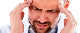
The brain doesn't feel pain
Tatiana Ayazo / rd.com
Have you ever wondered how neurosurgeons perform brain surgery without anesthesia? There are simply no pain receptors in the brain. But they are present in the meninges and blood vessels. Therefore, when we experience a headache, it is not the brain itself that hurts, but the tissues surrounding it.
The brain is more active when we sleep
Tatiana Ayazo / rd.com
As the brain works, it creates electrical fields that can be measured on the surface of the scalp using electroencephalography (EEG). It seems to us that during sleep the brain is turned off, but in fact it works even more actively than during the day. During wakefulness, it produces alpha and beta waves, and during sleep, especially in its early stages, theta waves. Their amplitude is greater than that of other waves.