Characteristics of the disease
Hydrocephalus can develop at any age. In 70% of cases it is found in young children. The frequency of detection of hidden or obvious pathology in infants is 1:500.
Less commonly, the anomaly develops in adults. At older ages, the disease is more severe and requires particularly intensive treatment.
Hydrocephalus is characterized by constant progression and persistently increased intracranial pressure. Dropsy of the brain, the symptoms and treatment of which are the sphere of activity of a neurologist, requires a competent approach. In the absence of quality therapy, the disease can gradually lead to disability.
Classification of pathology by form
Hydrocephalic syndrome is classified according to localization, pathogenesis, fluid pressure level, and flow rate.
The localization of the outbreak comes in three varieties:
- internal - cerebrospinal fluid accumulates in excess in the lateral ventricles;
- external - overconcentration of cerebrospinal fluid is determined in the subarachnoid space;
- mixed - simultaneous accumulation of cerebrospinal fluid in the ventricles and subarachnoid space.
Based on the pathogenesis, GM hydrocephalus can be:
- occlusive (closed) – the most dangerous form, resulting from blockage (occlusion) of the cerebrospinal fluid passages by a tumor, hematoma, or post-inflammatory adhesions;
- communicating (open) - with this pathogenesis, there is a disruption of resorption processes due to damage to the structures involved in the absorption of cerebrospinal fluid into the venous system.
Based on the CBF pressure indicator, they are distinguished:
- hypertensive hydrocephalus – the level of intracranial pressure is increased;
- normotensive – ICP remains within normal limits;
- hypotensive – the pressure inside the skull is reduced.
Hydrocephalus is diagnosed based on the rate of progression:
- acute - from the appearance of the first signs to the phase of severe clinical decompensation it takes no more than 72 hours;
- subacute – develops within 30 days;
- chronic - formation occurs at a slow pace, over months and even years (more common in open forms).
In ICD-10, hydrocephalus is assigned a general code of G91. Each form has its own alphanumeric symbol: communicating - G91.0; occlusal – G91.1; normotensive – G91.2; post-traumatic unspecified – G91.3; another type – G91.8; hydrocephalus of unspecified origin – G91.9.
Reasons for the development of pathology
If dropsy of the brain has developed, the causes of the pathology may be as follows:
- infection of the fetus during intrauterine development;
- congenital malformations of the central nervous system;
- atrophy of the gray or white matter that forms the brain.
During the gestation period, the unborn child may be exposed to various infections. Diseases such as herpes, rubella, syphilis, toxoplasmosis, encephalitis, and mumps can be transmitted from the mother.
Congenital defects of the central nervous system, due to which hydrocele of the brain can form, include Adams, Chiari, and Dandy-Walker syndromes. Against the background of these pathologies, cerebrospinal fluid loses its ability to circulate freely and accumulates in certain intracranial areas. Congenital hydrocephalus also develops against the background of basilar compression and aneurysm of the great cerebral vein.
Atrophy of the medulla occurs due to degenerative processes occurring in the central nervous system. Pathology is often preceded by vascular, toxic encephalopathy, Creutzfeldt-Jakob disease.
The disorder may be associated with traumatic brain or birth injuries. Sometimes dropsy occurs due to damage to the body by parasites, the presence of tumors, metastases, and cystic formations in the brain.
Definition
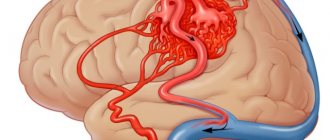
This phenomenon is understood as excessive accumulation of cerebrospinal fluid in the cavities of the central nervous system, which are intended for its circulation. These are slits, spaces, ventricles and cisterns. It would be a big mistake to assume that in this case the brain matter is saturated with cerebrospinal fluid and can be “squeezed into a bucket.” The brain, unlike other organs, is a very dense structure. And if during pulmonary edema it is filled with fluid, then with hydrocephalus we are not talking about “wateriness of the brain”, but about the accumulation of this fluid in various spaces that are not occupied by dense tissue and communicate with each other.
A little about the normal anatomy and physiology of liquor circulation in the central nervous system
The volume of the cranium, or cranial cavity, is constant in an adult. Normally it is distributed like this:
- brain tissue – 80%;
- cerebrospinal fluid – 10%;
- venous and arterial blood located in the cranial cavity – 10%.
In absolute figures, the volume of cerebrospinal fluid in the cranial cavity occupies about 150 ml, and a little more is accounted for by the structures of the spinal cord and the terminal cistern. Normal cerebrospinal fluid pressure (when measured while lying down during a lumbar puncture) is about 150–180 mm, but not the mercury column, but the water column. And given that mercury is 13.6 times heavier than water, we can assume that intracranial pressure is approximately 13 times lower than arterial pressure.
In the cranial cavity there is not a single ventricle, aqueduct or tank that does not communicate with the other cavities containing cerebrospinal fluid. Constant circulation of cerebrospinal fluid is one of the necessary conditions for normal brain function. Therefore, the following structures in which it circulates communicate with each other:
- unpaired (axial) ventricles of the brain (third, fourth);
- lateral ventricles;
- brain cisterns at its base;
- a narrow slit of the subarachnoid space on the surface of the convex, or cerebral hemispheres.
Of course, cerebrospinal fluid can easily “flow” into the subarachnoid space of the spinal cord, right up to the terminal cistern at the level of 3–4 lumbar vertebrae.
The production of cerebrospinal fluid occurs in the vascular, or choroidal, plexuses in the lateral ventricles, and absorption occurs in special formations (pachyonic granulations) near the sagittal (sagittal) sinus.
The daily production of cerebrospinal fluid in adults is about 600 ml, and the same amount is absorbed. Judge for yourself how finely balanced this mechanism is. But in some cases it is disrupted, which leads to either excess production (hyperproductive form), or insufficient absorption of cerebrospinal fluid (aresorptive, which occurs in 80% of all cases), or a combination of both. What reasons lead to increased intracranial pressure (ICP) and why does hydrocephalus of the brain develop in adults?
Variants of the course of hydrocephalus
There are several types of hydrocephalus. Regardless of the patient’s age, dropsy can be open, non-occlusive, aresorptive, biventricular, or atrophic.
Open hydrocephalus
This form of the disease occurs due to the slow absorption of cerebrospinal fluid into the blood. Such a violation leads to unbalanced production and resorption of cerebrospinal fluid. The body tries to eliminate the pathology by increasing intracranial pressure.
Non-occlusive
The disease occurs with overflow of the cerebral ventricles with cerebrospinal fluid. The substance accumulates in the ventricular system, filling the subdural and subarachnoid space.
Aresorptive hydrocephalus
The deviation is more common in older patients. With this disorder, the venous sinuses are unable to absorb cerebrospinal fluid normally.
Biventricular and atrophic
The first type of disease is provoked by the closure of the holes located in the 3rd ventricle. Atrophic dropsy of the brain results from a decrease in brain volume. This pathology is mainly diagnosed in old age - in people suffering from atherosclerosis, arterial hypertension, and diabetic angiopathy.
There is also a mixed type of hydrocephalus, in which the accumulation of cerebrospinal fluid occurs simultaneously in several areas.
What types of external hydrocephalus of the brain are there?
External hydrocephalus of the brain refers to the accumulation of cerebrospinal fluid (cerebrospinal or cerebrospinal fluid) outside the cerebral hemispheres - in the subarachnoid space. Due to the large accumulation of fluid, the subarachnoid fissures widen, which causes increased pressure on the cerebral cortex and the resulting negative consequences.
The nature and level of complexity of the disease directly depend on the specific type of dropsy. Several criteria are used for classification. The most common ones are:
- intensity of manifestation (pronounced - accumulation of a large amount of cerebrospinal fluid, causing neurological symptoms; moderate - minimal amount of fluid, no signs);
- degree of impact on brain structures (compensated - cerebrospinal fluid does not affect the brain; decompensated - there is a deterioration in the functioning of the nervous system and brain);
- causes of occurrence (replacement - more often diagnosed in older people and is accompanied by the death of brain cells; acquired - occurs due to the spread of infections and mechanical traumatic brain injuries);
- nature of the course (chronic form - a gradual increase in neurological disorders; acute form - a sharp deterioration in the patient’s well-being).
Employees of the Department of Neurosurgery of the City Clinical Hospital named after. Eramishantsev will first determine the type of external hydrocephalus and only then begin treatment. Particular attention is paid to diagnostic data and a thorough study of the symptoms of the identified disease.
Symptoms of hydrocephalus
In the acute course of the disease, the symptoms of hydrocephalus take the form:
- Pressing, bursting headaches radiating to the eye area (most often they appear in the morning and weaken during the day).
- Enlargement of the head.
- The appearance of a venous network.
- Increased intracranial pressure.
- Nausea, vomiting (these symptoms also predominate in the morning and are not associated with food intake).
- Cloudiness in the field of vision, burning sensation in the eyeballs.
- Oculomotor dysfunction.
- Difficulty breathing.
- Drowsiness.
- Epileptic seizures.
In severe forms of dropsy, depression of consciousness is observed, which can lead to coma.
Newborn babies with hydrocephalus have an enlarged forehead that hangs over the eyes. Such babies often develop strabismus and are underweight. Infants with dropsy are excessively tearful, irritable, and experience nervous overexcitation.
The chronic form of the disease occurs with less severe symptoms. Adult patients experience periodic confusion, problems falling asleep, deterioration of memory and mental activity. A sick person loses self-care skills and may suffer from loss of balance and unsteady gait.
If the patient does not receive appropriate treatment, dropsy of the brain progresses, leading to uncontrolled urination and defecation, significant weakening or complete absence of thinking, and decreased muscle tone. The disease gives rise to constant lethargy and apathy.
Diagnostics
In addition to the typical complaints described above, evidence and objective data are needed. Therefore, a neurologist, in order to make the correct diagnosis:
- Examines the condition of the fundus (presence of congestion). It is known that the veins through which blood flows from the optic nerves have a connection with the cavernous sinus. Therefore, congestion and swelling of the optic nerve head is an early symptom of the development of hydrocephalus and increased ICP.
- Refers for a regular radiograph of the skull - craniogram. If the disease is long-term and chronic, then changes in bone tissue occur - osteoporosis and deformation of the sella turcica, and the anterior edge of the foramen magnum becomes thinner. The phenomenon of “finger impressions” is observed - the bones of the cranial vault are thinned, and from the inside the fingertips appear to be imprinted on them in the form of depressions, the channels in the diploe - the spongy substance of the skull bones - are expanded.
- Recommends CT angiography to show late filling of the venous system, sinuses, and slowing of cerebral blood flow in the post-capillary phase. Normally, the contrast from the veins disappears 6 hours after administration, and with an increase in ICP this time is extended by 1.5–2 times.
- MRI both with and without contrast. Shows the type and nature of hydrocephalus and very often allows you to find the cause (adhesions, space-occupying formations).
- In some cases, lumbar puncture. Currently, the indications for it in ICP are extremely limited, since with a sharp decrease in pressure caused by a decrease in its volume, compensatory cerebral edema occurs, and such manipulation can end in failure.
A little about the actual numbers
It is important for all patients to know that if they have not undergone the above studies and are not bothered by the above complaints, then a diagnosis of hydrocephalus (except for Hakim-Adams syndrome) is unlikely. The level of intracranial pressure can be directly determined only by “screwing” a pressure gauge into the skull and bringing its sensor into the ventricular cavity or subarachnoid space.
This is what is done in neurosurgical intensive care units. The level can be indirectly determined using lumbar puncture data. Therefore, if a doctor says that “you have intracranial”, then it doesn’t hurt to ask him what criteria he was guided by in the absence of complaints, data from instrumental research methods and direct measurements?
Currently, there is a very intensive search abroad for non-invasive methods of measuring ICP (using computer online analysis of pressure in the eyeball adjusted for ICP and scleral curvature), but so far the error remains high, and such methods have not found widespread use in clinical and outpatient settings. practice.
Diagnosis of cerebral hydrocele
The initial stage of diagnosing hydrocephalus is examining the patient. In addition to a neurologist, the patient may need consultations with a neurosurgeon and ophthalmologist.
To clarify the degree and form of dropsy, the patient is prescribed:
- ultrasound examination of the brain (ultrasound);
- radiography;
- cisternography;
- magnetic resonance imaging (MRI);
- computed tomography (CT);
- lumbar puncture.
After an ultrasound, it is possible to clarify the degree of intracranial hypertension - one of the main signs of hydrocephalus. When undergoing radiography, the specialist finds out in what condition the vessels and vascular walls are.
Cisternography is necessary to clarify the form of the pathology and clarify the direction in which the cerebrospinal fluid moves. Magnetic resonance imaging determines the cause of the disease and visualizes the accumulation of fluid in the brain. CT scan clarifies the form of the disease and its severity.
A lumbar puncture is a procedure to remove cerebrospinal fluid and then determine intracranial pressure. The method is aimed at studying the composition of the cerebrospinal fluid and detecting the inflammatory process.
Pathology often provokes eye problems. To clarify visual function, ophthalmoscopy is performed.
Liquor shunt interventions
CSF shunting is the installation of elastic silicone implant systems to remove cerebrospinal fluid outside the central nervous system. The systems are equipped with catheters in the form of flexible hollow tubes, as well as valves with an anti-siphon (reverse) mechanism and with fixed or adjustable opening pressure.
The operation can be performed in various ways. But surgeons consider ventriculoperitoneal (the most commonly used method) or ventriculoatrial shunting to be the most successful in terms of safety. Interventions are performed under endotracheal anesthesia, manipulations are controlled by intraoperative fluoroscopy, CT, and ECG.
- Ventriculo-peritoneal shunt. The principle of the procedure is based on the implantation of silicone catheters, through which an excess of CBF goes into the intra-abdominal cavity, where it is resorbed between the intestinal loops.
- The procedure begins by making an incision on the scalp, after which a small burr hole is created in the skull. The dura mater is opened sparingly.
- A ventricular catheter is introduced through the created access, its end is placed in the lateral ventricle of the brain.
- The valve element is implanted in the area of the auricle (at the back or slightly above). The ventricular (ventricular) and distal catheter (DC) are fixed to it.
- Next, the neurosurgeon places a distal catheter into the abdominal cavity through a specially formed subcutaneous channel.
- Upon reaching the desired abdominal area, the specialist makes a small incision (no more than 10 mm) and inserts the end of the DC into the abdominal cavity.
- The procedure ends with thorough disinfection of the surgical field, followed by closing the wound areas with antiseptic dressings (sutures are applied if necessary).
- Ventriculoatrial shunting. The essence of this operation is to divert cerebrospinal fluid through installed shunts from the ventricle of the brain into the right atrium.
- Dissection of tissue in the neck along the anterior sternocleidomastoid muscle is performed to open the common facial or internal jugular vein.
- The atrial catheter is removed into one of the indicated veins, fixing it with specially designed ligatures.
- The shunt is directed through the catheterized vein to the right atrium. The end of the atrial shunt is generally located in the superior vena cava.
- For the installation area of the distal end of the vascular catheter, yes, the superior vena cava is often preferred. Here the blood flow is turbulent, and this reduces the likelihood of thrombosis of the drainage system by blood clots.
- The cranial part of the intervention, when the ventricular element of the system is implanted, a valve and two catheters are connected to it, is identical to VP shunting.
For adults, shunts are permanently implanted. In childhood, they are periodically replaced with elongated models. We emphasize that patients after surgery with shunt implantation are shunt-dependent people.
How is hydrocephalus treated?
Dropsy of the brain is mainly treated with surgical methods. Manipulations are performed under general anesthesia.
The main types of operations for hydrocephalus:
- palliative;
- radical.
During palliative intervention, a specialist performs a spinal or ventricular puncture. Elimination of excess fluid in the youngest patients is carried out through the orbit. The radical effect consists of complex procedures - Küttner Wenglovsky's surgery, ventriculocisternostomy.
Treatment for hydrocephalus often involves the installation of a shunt (special tube), which can take up to several months to wear. All this time, the patient remains under constant medical supervision (this is necessary to prevent infectious processes and monitor possible complications).
In addition to surgery, medicinal treatment of cerebral hydrocele is carried out. A course of medications provides improvement in patients with small fluid collections and moderately high intracranial pressure.
If hydrocephalus develops, drugs and their dosages are determined individually. Therapy in children and adults is carried out with the prescription of diuretics:
- Furosemide;
- Diacarba;
- Mannitol.
In addition to the listed drugs, antibiotics, vitamins, and absorbent agents are indicated. A frequently prescribed medication is Asparkam, which saturates the body with important microelements (potassium and magnesium).
After the subsidence of acute symptoms, physiotherapeutic procedures and exercise therapy under the supervision of a specialist are indicated.
Surgical and medical treatments are often combined. If the doctor considers it necessary, folk remedies for hydrocephalus can be used. With timely treatment, most patients recover, and the likelihood of relapse is minimized.
Treatment
In the initial stages, hydrocephalus can be treated with medications; in acute or advanced forms, surgical treatment is indicated. Folk remedies relieve only symptoms.
Conservative therapy
Drug treatment is aimed at reducing cerebrospinal fluid pressure, restoring blood supply and nutrition to the brain. It includes:
- Diuretics. These medications reduce fluid levels in the body. They use Diacarb, Furosemide.
- Preparations containing potassium. They are prescribed to replenish potassium levels in the body. In particular, Asparkam is used.
- Painkillers. Aimed at reducing the intensity of headaches. This could be Nurofen, Ketorolac.
- Nootropic. Medicines of this class help restore blood supply. Prescribed Pantogam, Noofen.
- Hormonal. Prednisolone and Dexamethasone relieve swelling and inflammation.
Operations
If it is necessary to take urgent measures and conservative therapy is ineffective, surgery is performed.
Bypass surgery involves the installation of a special catheter with a valve and tubes. When intracranial pressure increases, the opening of the valve facilitates the release of cerebrospinal fluid into the abdominal cavity. In children, due to constant growth, the operation is performed repeatedly.
Endoscopic surgery is performed if bypass surgery has caused complications or after injury. A hole is made in the bottom of the ventricle and connected to the cistern. This allows the outflow of cerebrospinal fluid to be restored.
Folk remedies
Lingonberry or cranberry fruit drinks are used as a means of improving the removal of fluids from the body. To prepare them, take 2 tablespoons of berries and 30 g of sugar per glass of water. The mixture is boiled, cooled and drunk.
Rosehip decoction can be prepared from 100 g of the fruits of this plant and a liter of water. Take 100 g three times a day.
A dill decoction will help not only get rid of excess fluid, but also lower blood pressure. Boil a teaspoon of seeds and the same amount of greens in half a liter of water. The seeds can be taken dry, half a teaspoon.
A decoction of nettle leaves is prepared in a water bath. To do this, take 1 liter of water and 4 tablespoons of the plant.
Complications of cerebral hydrops
With the development of pathology, it is important to understand why hydrocephalus is dangerous. In case of untimely treatment of dropsy in children there is a high risk of:
- late mental, physical development;
- the appearance of speech defects;
- inability to concentrate;
- memory problems.
In adults, complications include convulsive seizures, impaired coordination of movements, deterioration of vision or hearing (up to the onset of blindness or deafness), lack of control over urination and defecation.
Poorly treated hydrocephalus can make a person disabled. To avoid such complications, treatment should be prescribed at the initial stage of the disease.
Endoscopic operations
Endoscopic surgical techniques are used in the treatment of occlusive hydrocephalus, among them:
- ventriculocisternostomy;
- ventricular cystocisternostomy;
- ventriculoplasty of the aqueduct of Sylvius;
- septostomy;
- etiotropic endoscopy (getting rid of the causative factor - removal of a tumor, cyst, hematoma, etc.).
In 90% of cases, the method of endoscopic ventriculocisternostomy is used. The purpose of this operation is perforation of the bottom of the third ventricle of the brain under endoscopic control through a miniature trepanation window. The anastomosis created during the endoscopic procedure allows you to restore the natural path of outflow of cerebrospinal fluid between the third ventricle and the basal cisterns of the brain.
Endoscopy of any kind is a more gentle tactic of neurosurgery; it does not require the implantation of foreign bodies into the body and is less likely to cause postoperative consequences. Despite the promising characteristics of endoscopic methods, in some cases it is impossible to do without shunting or the use of open microsurgery.
Modern neurosurgery technologies have been honed to perfection in the Czech Republic; brain surgery in this country is a leading field of medicine. Neurosurgical care in clinics in the Czech Republic is no worse than in Germany and Israel, but the price is much lower (about 2 times). People with this diagnosis are operated on here at the most exemplary level, and upon completion of the full course of rehabilitation they are discharged with excellent and good results.
Prevention of hydrocephalus in children and adults
To exclude cerebral hydrops in newborns during pregnancy, it is necessary to take special vitamin complexes, including folic acid. During pregnancy, you need timely treatment for infectious diseases, completely abandon bad habits, and uncontrolled use of various medications.
Hydrocephalus in adults can be prevented by preventing sexually transmitted and other dangerous diseases. It is important to avoid severe stress, overwork, and lead a healthy lifestyle. If you have a family history of hydrocele, you should undergo regular medical examinations.
Share:
A little anatomy and physiology
Hydrocephalus (dropsy of the brain) (from the gr. “hydro” - water, “cephalon” - head) is a condition accompanied by an increase in the volume of the ventricles of the brain. The human brain has several interconnected cavities filled with cerebrospinal fluid (CSF). These cavities are called ventricles. The ventricular system consists of two lateral ventricles, which connect to the slit-like third ventricle (III ventricle), which, in turn, through a thin canal (Aqueduct of Sylvius) connects to the fourth ventricle (IV ventricle). Liquor is produced in the choroid plexuses of the ventricles and moves freely from the lateral to the fourth ventricle, and from there into the subarachnoid space of the brain and spinal cord (the subarachnoid space is the space between the brain and the dura mater), where it washes the outer surface of the brain. There it is reabsorbed into the bloodstream. Liquor is a clear, colorless liquid, very similar in appearance to water, containing a small amount of cells, protein and salts. In an infant, the amount of cerebrospinal fluid is about 50 ml, in a teenager and an adult - up to 120-150 ml. Liquor is constantly produced and absorbed; up to 500 ml of cerebrospinal fluid are produced per day by the choroid plexuses of the brain.
Any disturbance in the system of liquor production, liquor circulation and absorption of liquor leads to its excessive accumulation in the cavities of the brain, which is called hydrocephalus, or dropsy of the brain.
Back to contents
Rehabilitation and drug treatment
Medicines are prescribed for stable or regressive forms of the disease, for contraindications to surgery or during the recovery period after it. The choice of drugs depends on the patient’s condition and the severity of the disease.
To improve the outflow of fluid, Mannitol or Diacarb is used, they remove it into the bloodstream. In addition to medications, diuretics of the thiazide group are taken, for example, Furosemide. It prevents the increase in blood pressure while taking potassium-sparing diuretics.
To improve the nutrition of nervous tissue and its blood supply, Detralex, Glycine and Piracetam are prescribed. Anticonvulsants, anti-inflammatory and sedative medications can be used as symptomatic drugs. In the early stages, treatment at home is possible, but if the disease progresses, it is necessary to go to the hospital. The doctor will decide whether a course of medications is enough or surgical intervention is necessary.
Rehabilitation measures help the patient live with the consequences of the disease or shunt after surgery. He needs to limit physical activity and follow a diet with strict control of fluid and salt. You should also be regularly examined by a neurologist and attend treatment procedures.
Modern medicine does not deny the use of some unconventional methods of treatment. The most effective among them is hirudotherapy: leeches reduce blood and intracranial pressure. The use of various herbs to improve well-being is permissible only with the consent of the attending physician. The effectiveness of homeopathy and similar remedies has not been proven in clinical trials.
Risk factors for hydrocephalus
Babies who are born prematurely have a high risk of severe bleeding into the ventricles of the brain, which can result in hydrocephalus. Also, the risk of this disease in a child increases the presence of infection in the uterus. Sometimes a child has congenital developmental defects that are not noticed at birth. Subsequently, they can also provoke the manifestation of hydrocephalus in children even at an older age. Other risk factors include tumors or other changes in the brain and spinal cord, central nervous system infections, and bleeding in the brain.
Prognosis, complications
With occlusive hydrocephalus, intracranial pressure progresses, so without neurosurgical treatment the prognosis is unfavorable. The risk of severe complications and death is high.
The outcome of the operation depends on the primary disease. The prognosis is least favorable for malignant neoplasms and severe brain abnormalities.
The performed shunting causes shunt dependence - if the functioning of the shunt is disrupted, the patient’s condition sharply worsens, and it is necessary to restore the drainage system.
One of the severe complications of the pathology is occlusive-hydrocephalic crisis. In this case, the symptoms of the disease sharply increase and consciousness is depressed.
The most severe complication is mass effect. In this case, the brain tissue is displaced, the medulla oblongata is compressed, which leads to disruption of the cardiovascular and respiratory activity it regulates. This can be fatal.
Classification
Occlusive hydrocephalus is one of the forms of cerebral hydrocephalus and, in turn, differs in several ways.
Depending on the time of development of such a pathological condition, it can be congenital or acquired . In the first case, the pathology is formed during intrauterine development, and its signs appear immediately from the birth of the child. Acquired dropsy implies the possibility of formation at any stage of life. The cause usually lies in injury or disease of the central nervous system. In this case, the clinical picture is more extensive.
Anatomical and topographical classification implies that cerebral hydrocele can be mono-, bi-, tri- or tetraventricular . This division is based on the number of brain ventricles affected by pathology:
- With monoventricular hydrocele, dilatation of only one lateral ventricle is observed. Pathology develops due to blockage of the foramen of Monroe.
- In the case of the biventricular form, the two lateral ventricles are affected. Their volume increases, and occlusion occurs at the level of the third ventricle.
- With triventricular hydrops, three ventricles are involved, the outflow of cerebrospinal fluid is disrupted against the background of the Sylvian canal.
- In the case of tetraventricular pathology, all ventricles of the brain are affected. Disruption of the outflow of cerebrospinal fluid occurs at the exit from the fourth ventricle.
Surgery for dropsy of the brain
Conservative treatment of dropsy of the brain in acute form or severe symptoms against the background of the mid-late stage of the chronic process does not give a therapeutic result.
Surgery for hydrocephalus in such cases is considered as a real way to combat not only the symptoms of the disease, but also a means of eliminating its cause. After professionally performed intervention, patients are completely cured.
To assess the stage of the disease and establish indications for surgical treatment, the so-called ventriculocranial coefficients are often calculated, showing the degree of expansion of the ventricles and a decrease in their volume after the intervention.
To more accurately predict the outcome of surgical treatment, a patient with chronic cerebral hydrops is injected with a small volume of CSF into the spinal cord canal (TAP test), and an improvement in the condition is observed for a short time. If the FNA test is positive, then there is a high probability of full recovery after surgery. But a negative result does not exclude a favorable outcome, which appears a certain time after the operation.
There are two main techniques for surgical treatment of hydrocephalus (dropsy) of the brain.
Shunt method for hydrocephalus
Brain shunting for hydrocephalus is an surgical technique in which excess cerebrospinal fluid from the spaces of the brain is diverted to areas where it can be easily absorbed into the vascular bed. For this method, a special cerebrospinal fluid shunt system is implanted into the brain through a small hole in the skull. In this case, exclusively high-quality silicone materials are used, which are not rejected by their own tissues.
This system includes:
- shunt - a tubular structure that provides CSF circulation bypassing blocked liquor channels. In this case, the valve installed on the shunt regulates the outflow of cerebrospinal fluid, triggering when its pressure increases;
- a ventricular catheter installed in the ventricles of the brain, and a peripheral catheter leading into the cavity where cerebrospinal fluid is absorbed into the venous system.
During the VPS operation (ventriculo-peritoneal shunt), the cerebrospinal fluid is diverted into the abdominal cavity, during the ventriculo-thoracic shunt - into the pleural cavity, and the VAS technique or ventriculo-atrial shunt involves the removal of fluid into the right atrium.
In some cases, other types of surgical interventions are used according to indications:
- Lumboperitoneal shunting, in which a connection is established between the spinal canal and the peritoneum.
- Torkildsen's operation - diversion of cerebrospinal fluid from the lateral ventricle into the cerebellocerebral (occipital) cistern - ventriculocisternostomy.
Shunt surgery for hydrocephalus lasts approximately 2 hours. After 3 days (if there are no complications), the patient goes home.
The bypass method for hydrocele of the brain is effective, but has quite unpleasant consequences and associated complications:
- hypo- and hyperdrainage syndrome (intracranial increase or decrease in pressure due to insufficient or overly active excretion of cerebrospinal fluid);
- breakdown or blockage of the shunt system, which requires repeated surgery;
- inflammatory processes, infection with pathogens.
The earlier surgical treatment for hydrocephalus is performed, the lower the risk of any complications after shunt surgery.
Endoscopic operations
Surgical techniques using an endoscope are considered a priority in neurosurgery due to:
- low trauma, since there is no need to install a catheter in the brain tissue;
- high precision;
- reducing the risk of overdrainage and related complications;
- shortened recovery period.
Under the control of a computer video surveillance system, the neurosurgeon inserts endoscopic instruments with a tiny video camera into the cavity of the skull through a mini-hole. When forming an outflow channel for cerebrospinal fluid, the specialist has full control over his manipulations and surveys the surgical field.
Often, with hydrocele of the brain, endoscopic ventriculocisternostomy is performed, which restores the physiological outflow of cerebrospinal fluid from the ventricles into the basal cisterns. Within 20–30 minutes after endoscopy, the absorption of CSF is normalized and its pressure decreases to physiological levels.
The concept of hydrocephalus: types, mechanisms of development, causes
Hydrocephalus is understood as a progressive multifactorial (having many provoking factors) condition associated with a pathological increase in the volume of cerebrospinal fluid in the ventricles of the brain due to a violation of its movement from places of secretion (i.e., secretion) to places of absorption (i.e., absorption) into the bloodstream . Or while maintaining normal circulation due to an imbalance between the amount of newly formed CSF and that which has already entered the blood (subjected to resorption, i.e., reabsorption). In the first case, hydrocephalus is considered occlusive, in the second - aresorptive.
This pathology can occur at any age, although more often it is congenital and manifests itself as an independent nosological form, that is, the initial disease, and as a complication of some other damage to the body.
Classification
Aresorptive, open, communicating or non-occlusive hydrocephalus is a form in which expansion of all cerebrospinal fluid spaces of the brain occurs. In this case, there is no obstacle to the flow of fluid through the central nervous system. The main mechanism for the development of this type of hydrocephalus is a discrepancy between the resorption of cerebrospinal fluid and its secretion, as a result of which CSF accumulates in quantities much higher than normal limits. As a rule, the disease occurs after meningitis. The course is mild, and with timely treatment, the prognosis for life is favorable.
Closed or occlusive hydrocephalus is a more serious pathology that occurs in the central nervous system due to blockage of the passages through which the cerebrospinal fluid outflows. Clinically, the following types are distinguished:
- Monoventricular - expansion of only one ventricle of the brain;
- Biventricular – two ventricles;
- Tri- and tetraventricular - respectively, expansion of the three and four cerebrospinal fluid spaces of the brain.
Causes of occlusive hydrocephalus
Occlusion, or blockage of the cerebrospinal fluid passages, can occur for a variety of reasons. It can be:
- Anomalies in the development of the ventricular system of the brain;
- Adhesive processes in the CSF outflow system;
- Tumors of the brain;
- The presence of massive blood clots in the central nervous system;
Provoking factors that lead to the appearance of the above conditions in an adult are: traumatic brain injuries, cerebral infarctions, hemorrhages, infectious processes in the nervous system.
Classification and characteristic symptoms
Depending on the time of occurrence, hydrocephalus is divided into:
- congenital - diagnosis is possible during the period of intrauterine development;
- acquired - occurs as a result of traumatic brain injuries or inflammatory processes that directly affect the brain.
According to the developmental features, the pathology has the following types:
- open - some parts of the brain are destroyed, which leads to incomplete release of cerebrospinal fluid into the bloodstream;
- closed - characterized by the fact that the liquid begins to spread incorrectly due to obstacles formed as a result of defective changes, which leads to the formation of blood clots or even tumors;
- atrophic – replacement of substances with spinal cord fluid occurs;
- hypersecretory – excessive volume of cerebrospiral fluid is produced.
Depending on the location of the cerebrospinal fluid, cerebral hydrocele is divided into:
- moderate internal - fluid formation occurs in the ventricular space;
- moderately expressed - can occur as an independent disease or act as a symptom of some other pathology;
- moderate external - localization of excess cerebrospinal fluid is in the subarachnoid space;
- mixed replacement - the formation of cerebrospinal fluid is observed not only in the space of the ventricle, but also in the area under the meninges;
- replacement external – found in newborns, localized in the frontal lobe;
- external expressed - one of the types of dropsy of external form.
Based on the degree of development of the disease, it is classified into the following forms:
- regressing;
- progressive;
- stabilized.
It should be noted that the development of hydrocephalus can be acute or chronic. This determines the main symptoms of the pathology.
Acute dropsy has all the standard signs that accompany high intracranial pressure:
- severe headaches in the morning;
- congestion of the optic discs, which is associated with impaired current in the nerves, causing increased pressure, leading to visual impairment;
- axial dislocation of the brain - expressed by loss of consciousness, the head acquires a forced position, oculomotor disorders occur; cases of suppressed functioning of the heart and respiratory system, which leads to death, cannot be excluded;
- attacks of nausea and vomiting in the morning, after which his headaches subside;
- drowsiness is one of the dangerous signs indicating the possible development of a more serious neurological disorder.
In the chronic form of hydrocephalus, the symptoms differ significantly from the acute form and are expressed in the following manifestations:
- Apraxia of walking. This syndrome is characterized by a condition where the patient, while lying down, can easily show how to walk or ride a bicycle correctly, but when trying to walk, he cannot do it normally. When moving, the legs are spread wide apart, swaying and shuffling occurs;
- Dementia. In most cases, the first signs of the disease begin to appear 15-20 days after injury, meningitis or some other disease occurs: the patient experiences confusion between day and night; activity decreases; there is indifference, lack of initiative to everything that happens; memory deteriorates, primarily numerical, when the patient cannot even tell his age; a later stage has a likelihood of developing mnestic-intellectual disorders;
- Urinary incontinence. This symptom may not appear in all cases.
No changes in the fundus are observed.
What should be done to prevent the disease?
Prevention of childhood hydrocephalus:
- Timely registration of a pregnant woman is necessary;
- Preventing the occurrence of infectious diseases in a pregnant woman;
- Take medications with maximum caution during pregnancy;
- A control ultrasound of the pregnant woman and newborn baby is mandatory for early diagnosis of hydrocephalus.
Prevention for adults includes:
- Timely treatment of pathological conditions that can lead to the development of factors contributing to impaired outflow of cerebrospinal fluid, therapy for hypertension, atherosclerosis, etc.;
- Avoiding head injuries;
- Early diagnosis of tumor processes in the brain;
- Strengthening the immune system, reducing the risk of infection with ARVI and other viral diseases;
- Maintaining a proper lifestyle, diet and sleep.
Surgery
Surgery is the most effective way to get rid of dropsy. Several types of intervention are used for treatment:
- removal of the cause of the disease in a closed form (thrombus, tumor, septum);
- shunting in a communicating (open) or hypersecretory form , if it is impossible to eliminate the cause of the pathology;
- combined operations for mixed type.
In most cases, the doctor decides to bypass the affected ventricle of the brain. The essence of the operation is that the patient is implanted with a tube (shunt), which drains excess fluid from the source of the disease. The free end of the implant is directed into the abdominal cavity, less often into the atrium. Artificial communication allows you to get rid of unnecessary cerebrospinal fluid and remove it to a place where absorption into the blood is not impaired. The result is the restoration of normal liquor circulation and the cessation of its accumulation in the cranial cavity. If the operation is performed on a child, the shunt is installed “for growth” and is replaced every few years. To prevent reverse flow of liquid due to the pressure difference, it is equipped with a valve, the design of which allows the movement of the cerebrospinal fluid only in the desired direction.
If the disease can be cured, the causative factors are eliminated: blood clots and blood clots, tumors, adhesions. Such operations can prolong the patient’s life. If the measures taken are not enough and the fluid continues to accumulate, an additional shunt may be performed.
There are a number of contraindications to the operation:
- active inflammatory process in nervous tissue - encephalitis, meningitis, ventriculitis;
- stable or regressing form of dropsy;
- irreversible consequences of the disease - blindness, neurological disorders, atrophy of the cerebral hemispheres;
- exhaustion, severe pathologies of the heart, blood vessels, respiratory system.
Symptoms of the disease
The clinic can be divided into deterioration of general condition, cerebral symptoms and focal phenomena. In any case, the main damaging factors are the accumulation of fluid and increased intracranial pressure, hypotrophic and atrophic changes in the adjacent nervous tissue.
Deterioration of the general condition is a rather uncharacteristic and unexpressed symptomatology, which does not allow making an unambiguous diagnosis. It is manifested by moderate headaches, nausea, and, less commonly, discomfort in the eyes, which is paroxysmal in nature. These signs are most pronounced with internal hydrocephalus, and with external hydrocephalus they are insignificant.
General cerebral symptoms are associated with a malnutrition of the brain; in the initial stages they accompany general symptoms and are not of great value for diagnosis. Such manifestations include convulsive syndrome, photophobia, tinnitus, memory impairment, attention, and coordination of movements.
Of higher value for diagnosis are focal phenomena that are associated with the expansion of the cerebral ventricles. They quite accurately indicate the affected area. Most often they manifest themselves in the form of impaired sensitivity of the limbs, paralysis, partial convulsive seizures (when one area is affected, for example, an arm or leg), and can be both asymmetrical and symmetrical in nature.
In children of the first year of life, hydrocephalic syndrome is formed - an increase in the volume of the head, a lag in physical and intellectual development, up to severe mental retardation. With a mild degree of the disease, partial restoration of functions is possible, and with timely surgical intervention, hydrocephalus becomes curable. If the genesis of the disease is caused by intrauterine causes, the death of a small hydrocephalus is possible during childbirth or during the first year of life.
Methods of modern diagnosis of hydrocephalus
The characteristic manifestations of the disease in question enable the doctor to assume and confirm its presence already at the first appointment.
In order to find out what diseases caused dropsy of the brain, how far this disease has gone in its development, a number of hardware studies :
- X-ray of the head. Needed to determine the type, subtype, and degree of hydrocephalus. The image will show the divergence of the bone walls on the outer side of the skull, the presence of small rounded dents in the inner part. A more popular method of studying this disease is CT and MRI of the brain. They help to visualize in more detail the structure of the ventricles, subarachnoid area, and skull, and to more clearly understand the cause of cerebral hydrocele.
- Echoencephalography is an ultrasound study of intracranial pressure fluctuations.
- Ultrasonography. Relevant for newborn babies. Through their fontanelle, this diagnostic method can be used to study the state of the brain.
- Ophthalmoscopy. Needed to determine the degree of malfunction of the eye organs and check visual acuity. For these purposes, you need to contact an ophthalmologist, who, in addition to ophthalmoscopy, may prescribe another medical examination (perimetry).
- Lumbar puncture. Its implementation is feasible in the absence of contraindications for the patient. It is needed to take a sample of the liquid for further examination.
Rate
—
Complications of the disease
Hydrocephalus is a dangerous enough disease to ignore the symptoms of its manifestation. Ignoring treatment of pathology can cause disability or even a threat to life.
If treatment is not treated in a timely manner, a person’s ability to work and importance are lost. Impaired mental functioning, problems with movement and urination, blurred vision, epileptic seizures - all these are possible complications that result from delays in seeking medical help.
The greatest danger, perhaps, is the development of cerebral edema, characterized by a gradual loss of consciousness, which is accompanied by drowsiness, unilateral dilation of the pupil, fever and pyramidal insufficiency.
Chronic hydrocephalus in adults has a more favorable prognosis with appropriate and timely treatment.
Lymphostasis of the lower extremities: treatment at home.
Effective drugs for migraines.
Folk remedies for high blood pressure are listed in this article.