Removing a specific section of the intestine that is damaged by disease is called digestive organ resection. Bowel resection is a dangerous and traumatic operation. The procedure is different from many others using anastomosis. After excision of part of the digestive organ, its ends are connected to each other. Therefore, a person should be aware of the indications for performing the procedure and what complications may arise.
- 5.1 Complications and pain after resection
Anatomy of the intestine
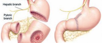
As an anatomical formation, the intestinal tube begins with the duodenum , which is located retroperitoneally, i.e. covered with parietal peritoneum (a thin film lining the inside of the abdominal wall). Thus, it is isolated from the free abdominal cavity and tightly fixed in the retroperitoneal space. The duodenum has a horseshoe shape, its final part passes into the jejunum. In this case, the transition site is tightened by the ligament of Treitz and forms an acute angle, which normally prevents the backflow of intestinal contents into the duodenum. The small intestine begins from the ligament of Treitz . It is located intraperitoneally and has a fairly long mesentery, which ensures its significant mobility. The length of the small intestine varies from three to seven meters. It is divided into two sections: the jejunum and the ileum . The border is arbitrary because it has no anatomical landmarks. The ileum continues into the large intestine.
The blood supply to the small intestine comes from the superior mesenteric artery, which arises from the aorta. From it, small intestinal arteries (from 12 to 16 branches) pass through the mesentery, which are divided into jejunal and ileal branches. By anastomosing with each other they form arcades of the first, second and higher orders, the caliber of which decreases as they approach the intestine. At the wall itself, they form a continuous vessel (marginal), from which straight short vessels extend to the intestinal wall.
Venous outflow is carried out in the reverse order through straight veins, which form wide arcades, from which the veins of the jejunum and ileum are collected. All veins merge to form the superior mesenteric vein. The latter runs parallel to the artery of the same name; it collects blood from the small, blind, ascending colon, part of the transverse colon.
Thus, due to the arcade, a sufficiently rich blood supply to the small intestine is ensured, which creates favorable conditions for resection and anastomosis.
The lymphatic system is also widely developed. The main collectors of the lymph nodes are located in three rows. The first row of lymph nodes is located along the mesenteric edge of the intestine, the second runs at the level of the vascular arcades, and the third along the main branches of the superior mesenteric artery. However, some lymphatic vessels pass into the thoracic duct, bypassing the listed barriers from the lymph nodes. This explains the cases of rapid metastasis of the malignant process.
The large intestine is the final section of the digestive tract and is divided into sections: the cecum, colon and rectum.
The cecum is the initial section where intestinal chyme enters from the ileum. At the junction of the ileum and the cecum there is a special valve (Bauhinian valve), which prevents the backflow (reflux) of the contents of the large intestine into the small intestine. Thus, complete isolation of the small and colon occurs. The peculiarities of the initial section of the large intestine are that it is located below the entry point of the small intestine and is located in the right iliac region in the form of a bag, which is why it is called “blind”. In addition, there is also a rudimentary continuation of the cecum - the vermiform appendix ( appendix ).
The next section of the large intestine is the colon . It in turn is divided into ascending, transverse and descending parts. Already from the names it is clear that the colon surrounds the loops of the small intestine in a U-shape. The ascending colon is a continuation of the cecum, starts from the level of the confluence of the small intestine, is located in the right lateral half of the abdomen and, at the level of the liver, after bending, passes into the transverse colon. The transverse colon is located under the liver, below the stomach, runs horizontally to the left flexure, where it becomes the descending colon. All sections are covered with peritoneum and are limited in mobility. The sigmoid colon, being a continuation of the descending colon, on the contrary, is very mobile. It often has an S-shape, which is why it gets its name “sigma”. It is located in the left iliac region and is the final section of the colon. The place where the sigmoid colon enters the rectum is called the rectosigmoid region by experts.
The large intestine is supplied with blood from the systems of the superior and inferior mesenteric arteries, which arise from the aorta. The right half is supplied by the superior mesenteric artery. The ileocolic, right and middle colic arteries extend to the corresponding parts of the intestine (blind, ascending and part of the transverse colon). The left half of the colon, the sigmoid and partially the rectum are supplied, respectively, by the sections of the left colon, sigmoid and superior rectal arteries, which arise from the inferior mesenteric artery.
The radially extending branches of the superior and inferior mesenteric arteries form an arterial arcade (Arch of Riolan) in the mesentery of the transverse colon, which provides communication between the superior and inferior arterial basins. From the arch directly to the intestinal wall, the straight vessels approach.
The venous and lymphatic systems accompany the arterial one, and the peculiarity is that the first lymphatic barrier is located in close proximity to the intestine in the peripheral vascular branches, and the second and third are around the great vessels.
The preservation of the Riolan arch is very important when performing resection of various parts of the intestine.
The rectum is located in the small pelvis, adjacent to the sacrum, and is completely retroperitoneal. The length of the intestine is about 15 cm. It is shaped like an ampoule and has three bends. According to these features, the upper ampullary, middle ampullary and lower ampullary sections are distinguished. This division is important when choosing the level of rectal resection. The rectum ends with the anal canal. A complex sphincter apparatus, consisting of external and internal sphincters, provides the closing function of the anal canal.
The rectum has three main sources of blood supply: the superior rectal artery (a continuation of the inferior mesenteric artery) and the paired middle and inferior rectal arteries, which enter the bloodstream of the internal iliac artery.
Venous outflow is carried out through the veins of the same name. At the same time, they form venous plexuses in the intestinal wall.
The outflow of lymph from the upper part of the ampulla occurs through the superior rectal artery into the lymph nodes of the inferior mesenteric artery, which should be taken into account when performing resection. Lymph flows from the midgut to the lymph nodes of the iliac arteries. From the lower section - to the inguinal lymph nodes. In addition, the process involves the lymph nodes of the mesorectum, the so-called. mesorectum. Through them, metastases can spread along the intestine.
This is very important knowledge, since its use in practice makes it possible to perform extended resections in compliance with the principles of oncological safety, as will be discussed below.
Forms of benign tumors
In medicine, there are several types of polyps and leiomyomas, which have their own characteristics and characteristics. Gastric polyps are oval or spherical neoplasms on a stalk with a dense or soft consistency. Types of polyps:
- single;
- multiple;
- polyposis (formation of a large number of polyps that cannot be accurately counted) - adenomatous polyposis (arises from the glandular epithelium, can degenerate into cancer), hyperplastic polyposis (tumor-like polyps with a low risk of malignancy), Metrier's disease (precancerous condition of polyposis);
- inflammatory fibrous polyposis (contain a large number of blood cells).
Among other forms of benign tumors it is worth noting:
- fibroma - a tumor of connective tissue;
- angioma is a tumor consisting of blood vessels;
- neuroma is a tumor consisting of nerve tissue;
- lipoma – a tumor consisting of submucosal tissue;
- Leiomyoma is a tumor of muscle tissue.
Indications for small bowel resection
In emergency surgery, the following conditions are indicated for small bowel resection:
- Massive injury to the intestine and mesentery . In cases where simple suturing of the wound is technically impossible and unreliable, economical resection of the damaged area is performed and continuity is restored using an intestinal suture.
- Mesenteric thrombosis is a severe situation associated with thrombosis of the mesenteric (mesenteric) vessels supplying the intestine. In this case, necrosis of a significant part of the intestinal tube occurs, the blood supply of which was provided by the pool of “blocked” mesenteric vessels. In this situation, it is very difficult to determine the demarcation boundary between the already “dead” section of the intestine and the remaining living part, the blood supply of which remains intact. Resection is performed in emergency conditions in unprepared patients, most often elderly and senile patients, burdened by concomitant diseases. Surgeons have to perform an extended resection of the small intestine, presumably within the preserved areas of the intestine. In equipped clinics, the operation is complemented by vascular intervention to remove the blood clot and attempts to restore blood supply to ischemic areas. It all depends on the severity of the lesion and the stage of the process. These factors ensure the outcome of the operation. If the anastomosis is performed between the viable ends of the intestine, and the progression of thrombosis can be stopped, there is hope that the anastomosis will be successful. Thus, the doctor has a chance to defeat the disease, and the patient has a chance to survive.
- Strangulated hernia of the abdominal wall with necrosis of the strangulated intestinal loop . A loop of the small intestine strangulated in the hernial orifice suffers not only from the strangulation of its wall, but also from the cessation of blood supply as a result of compression of the vessels supplying the loop. If the situation is not resolved within 2-3 hours, intestinal necrosis occurs. The afferent part of the intestine suffers more than the efferent part. As a rule, its lumen is expanded, atonic, microcirculation is impaired. In this case, an extended resection of the small intestine is performed. The afferent section is intersected at a distance of at least 40 cm from the place of infringement, the efferent section is intersected at a distance of 15 cm. Taking into account the different diameters of the adductor and efferent sections, it is rational to restore the patency of the intestine with a side-to-side anastomosis.
- Crohn's disease is a chronic inflammatory bowel disease of an autoimmune nature. When the disease worsens and an acute abdomen appears, patients undergo emergency surgery. The scope of the intervention includes economical resection of the affected area of the intestine with anastomosis.
- Volvulus occurs more often in children and elderly patients. The predisposing factor is anatomical features: elongation of the intestine and its mesentery. The resulting excessive mobility of the loops can lead to twisting of a section of the intestine around its axis with the formation of a volvulus. If the blood supply is disrupted, necrosis of the intestinal loop involved in the process occurs. In this case, bowel resection within healthy tissue is required.
- Inflammation of Meckel's diverticulum . Meckel's diverticulum is a rare pathology of the ileum, occurring in 3% of the population. This is the residual part of the umbilical-mesenteric duct, looks like a sac-like protrusion at a distance of 60-80 cm from the cecum. When the diverticulum becomes inflamed, a clinical picture of an acute abdomen occurs, similar to that of acute appendicitis. During surgery, once the diagnosis has been established, the surgeon performs a sectoral resection of the intestine. In this case, the sector of the intestine with the lesion is removed without completely crossing it. Next, the intestinal defect is sutured.
Routinely, intestinal resection is indicated in the presence of small intestinal neoplasms.
Benign formations, depending on their size, can be removed using either sectoral or economical resection with appropriate restoration of patency. For malignant neoplasms, an extended resection is performed with a wedge-shaped resection of the mesentery and removal of the arterial-lymphatic complex associated with the affected area of the intestine.
Possible complications
A thorough examination, preparation for surgery, trained medical staff, and high professionalism of the doctor, unfortunately, do not guarantee the absence of complications. Having studied the medical statistics, we can say that one or another difficulty awaits 90% of those operated on for this reason.
Complications that are most common:
- Infection (most often suppuration of sutures, peritonitis). High temperature, increased pain. Redness and swelling are the first signs of the development of this condition.
- Hernia development. Even correctly placed sutures and excellent tissue fusion do not guarantee the same strength of the abdominal wall as it was before the operation. Therefore, the incision site is often transformed into a hernial orifice.
- Adhesions and scars. They can not only cause pain and pulling sensations, but also disrupt intestinal patency and cause other unpleasant consequences; In this condition, the temperature may be elevated.
- Bleeding. It is also a common occurrence after intestinal resection, which can even cause death if assistance is not provided in a timely manner.
After the operation, at least a week, and most often 10 days, which the patient spends in a hospital setting. This allows doctors to keep the patient’s condition under constant control and, if necessary, adjust treatment.
Indications for colon resection
Resection for emergency indications.
The lower section of the intestinal tube, which includes the colon and rectum, has a number of features. First of all, this is a segmental blood supply, not as abundant as that of the small intestine, as well as a very aggressive intestinal flora of the lumen of the lower sections. Under these conditions, emergency resection cannot always be completed with anastomosis. The danger of insolvency is too great. In the vast majority of cases, a so-called obstructive bowel resection is performed, in which, after removing the section of bowel with the lesion, an anastomosis is not performed. The lower (outgoing) end of the intestine is sutured tightly. The upper (adductor) is brought out in the form of a colostomy on the anterior abdominal wall.
The operation is safe because there is no weak link - “anastomosis”. It is performed for complicated diseases of the colon: diverticulum ruptures with the formation of an inflammatory infiltrate or abscess, massive intestinal wounds, acute obstructive obstruction, and other situations accompanied by inflammation and necrosis of the intestinal wall.
Restoration of intestinal integrity and anastomosis are performed routinely no earlier than four or more months after obstructive resection.
Planned operations on the colon.
It should be noted that elective surgery of the colon and rectum is one of the main areas of surgical activity of our Center. Planned operations are performed mainly for:
- intestinal neoplasms (for benign and malignant tumors);
- diverticular disease;
- dolichosigma.
For benign diseases, the affected areas are removed using the method of economical resection with end-to-end anastomosis.
Colon cancer requires more severe extended resections. The purpose of the operation is not only to remove the cancerous tumor, but also to prevent recurrence of the disease in the long term. Therefore, resection must adhere to certain oncological principles. Depending on the location of the cancerous tumor, these resections are supplemented by the removal of anatomical structures through which the spread of cancer cells passes.
The principles of the operation include wide mobilization of the mesentery while maintaining the integrity of the fascia, central intersection of the great vessels supplying the resected segment of the intestine, with removal of all lymphatic drainage collectors closely associated with the vascular complex.
The length of the resected section of the intestine must be sufficient; the operation must include the removal of all regional lymph nodes through which cancer cells may spread.
Causes
To begin with, it is worth noting that the nature and causes of oncology still remain a mystery to doctors. This means that doctors make certain assumptions, but it is impossible to say that it is a specific factor that causes cancer. The primary, that is, the main causes are unknown even to science, but we can dwell in more detail on the secondary causes of cancer, that is, talk about risk factors. They can contribute to the development of duodenal cancer.
Types of operations for colon cancer
For uncomplicated forms of cancer of the right half of the colon, a so-called right hemicolectomy - removal of the entire right flank of the colon, including 20 cm of the ileum and a third of the transverse colon. This extent of resection is explained by the fact that lymphogenous metastasis occurs along all three colonic arteries, which supply blood to this particular segment of the intestine. They must be ligated at the point of origin with the obligatory removal of surrounding fatty tissue. The operation is completed by applying an end-to-side anastomosis between the small and transverse colon.
If the transverse part of the colon , resection of the transverse colon is performed along with the hepatic and splenic flexures. At the end of the operation, an end-to-end anastomosis is formed between the ascending and descending colon. Depending on the extent of the process and the location of the tumor (closer to the hepatic or splenic angles), the extent of resection may vary.
For cancer of the left third of the transverse colon, left flexure and descending colon, a left-sided hemicolectomy is performed - removal of a third of the transverse colon with the splenic flexure, the entire descending colon to the initial part of the sigmoid colon.
As mentioned above, such a massive volume of resection is associated with the characteristics of the blood supply and lymphatic drainage. Mobilization of the intestine begins with the intersection of the inferior mesenteric artery at its origin from the aorta, which is the prevention of relapse of the disease.
When the sigmoid colon , the volume of intervention depends on the location of the malignancy. For tumors of the middle third of the intestine, its resection is performed. Depending on the stage and extent of the process, it is possible to expand the resection with ligation and intersection of the inferior mesenteric artery. After removing the resected portion of the intestine, an anastomosis is performed between the descending colon and the ampulla of the rectum.
For rectal cancer, the choice of resection method depends on the location of the cancer. When the tumor is located in the rectosigmoid region or ampulla of the rectum, a so-called anterior rectal resection , which not only allows for radical removal of the tumor, but is also accompanied by good functional results. During the operation, the upper parts of the rectum and the lower part of the sigmoid are removed. An anastomosis is performed between the sigmoid and the remaining part of the rectum.
If the middle and lower part of the rectum is affected, low anterior resection . The operation involves removing almost the entire rectum, its mesorectum (mesorectum) down to the anal sphincter muscles. Total mesorectumectomy is currently the standard treatment for tumors of the rectum that are located in the lower rectum. With this method of surgical treatment, the frequency of disease return (relapse) is minimal. After part of the rectum is removed, an anastomosis is created between the reduced end of the colon and the remaining lowermost part of the rectum or anal canal. This operation is considered sphincter-sparing, however, the formation of a temporary ilestomy may be required to unload the anastomosis and therefore protect it.
Diagnostics
To cure the disease, it is very important to start treatment on time, and for this it is necessary to detect the cancer. Today, diagnosis is carried out using laboratory and instrumental methods. Testing for tumor markers for stomach and duodenal cancer will help identify cancer cells in time. It is worth noting that tumor markers are substances that promote the production of cancer cells. Diagnostics consists of three stages:
- To begin with, an analysis of pathologies and collection of anamnesis is carried out. The doctor examines the patient and palpates the abdomen. The person’s clinical complaints, such as vomiting, pain, loss of appetite, are taken into account.
- At the second stage, a laboratory test of the patient is carried out, consisting of a general blood test, urine and stool tests, a biochemical blood test and a tumor marker.
- The third stage consists of an instrumental examination.
The last stage is the most important in diagnosing duodenal cancer. The first symptoms may be erroneous or indicate another disease, but instrumental examination allows an accurate diagnosis. The doctor performs an esophagogastroduodenoscopy, during which the esophagus, stomach, bile duct and 12-fingered colon are examined. During the analysis, tissues are taken for biochemical analysis.
After this, the doctor sends the patient for a chest x-ray and contrast x-ray of the duodenum and stomach. The doctor may also prescribe ultrasound, magnetic resonance and computed tomography. At the same time, the patient undergoes examination by all specialists.
Types of intestinal anastomoses
The method of restoring the integrity of the intestinal tube after resection depends on the anatomical conditions and condition of the intestine. Three types of anastomoses are considered.
- End-to-end anastomosis (similar to the restoration of gas pipeline pipes) is the most physiological and is most often used to restore the integrity of the intestinal tube.
- A side-to-side anastomosis is performed in cases of discrepancy in the width of the lumen of the connected ends of the intestine. In this situation, the lumens of the intestinal tubes are sutured tightly, and the lumen of the adjacent afferent and efferent loops is opened from the side. A lateral anastomosis is performed.
- An end-to-side anastomosis is performed under certain conditions to ensure the physiological flow of intestinal contents. Parts of the small intestine may be anastomated, for example, during Roux-en-Y gastrectomy. Or an anastomosis is created between the small and large intestines, for example, when removing the right flank of the colon (right hemicolectomy).
According to the methods of execution, there are manual and hardware seams . The development and introduction of stapling devices into surgical practice made a revolutionary breakthrough in gastrointestinal surgery. The devices provide reliable, precise suture placement. The dependence of the quality of anastomosis on the qualifications of the surgeon is excluded. Hardware anastomosis is reliable, first of all, due to a three-row staple suture, which creates a hermetically sealed tissue suturing.
The use of staplers has significantly accelerated and facilitated the application of reliable anastomoses in hard-to-reach places that are inconvenient for manual suture and has greatly facilitated the performance of complex operations, in particular, with low resections of the descending colon and rectum.
It should be noted that the Auto Suture™ company was the first in the world to use titanium staples, which do not produce artifacts during magnetic resonance imaging, which subsequently became the world standard for staplers.
Stapling devices are divided into three groups depending on their functional purpose:
- devices for stitching the intestine, closing the intestinal defect (a staple suture is applied, tissue is cut off with a scalpel);
- linear staplers for the formation of interintestinal anastomoses (simultaneous dissection and suturing of tissue);
- circular devices cut off and stitch together intestinal tissue, ensuring the formation of a complete end-to-end interintestinal anastomosis.
General information about the diet
Main indications for intestinal surgery:
- Detection of rectal fistula.
- Crohn's disease.
- Intestinal ischemia.
- Paraproctitis and diverticulitis.
- Injuries of the rectum and sigmoid colon.
- Oncology.
- Acute inflammatory processes.
- Hemorrhoids, fissures.
The operation is not prescribed in every case: for example, if the intestines begin to whistle, drug therapy is first prescribed. If medications are not effective, resection is used. The specifics of nutrition and care after surgery are determined by its complexity and the patient’s health status.
Principles of diet after surgery:
- You should avoid foods that cause flatulence, diarrhea or constipation.
- During the first three days, the patient is fed intravenously. If the resection was extensive, parenteral nutrition will last several weeks.
- Natural nutrition begins with treatment table No. 0, gradually moving to table No. 1a, 1b. After 1.5-2 months to table No. 15.
- For the first 15 days after surgery, you should not eat fresh vegetables, eggs, milk or legumes.
- From the diet we exclude canned food, pickles, marinades, spices, fatty meats, rich broths, and sausages. You also need to give up foods with flavor enhancers, fast food, processed foods, mushrooms, baked goods, sweets, sauces and mayonnaise.
- At first, we grind all the products in a blender or grate them - this way the food is better absorbed.
- The diet provides for fractional meals: 5-6 times a day.
- Food can be steamed, boiled or baked (but not until golden brown).
- To prevent constipation, you need fiber: these are vegetables (pumpkin, carrots, zucchini, cauliflower, etc.) and fruits (apple, banana, pear). Citrus fruits and berries with rough skin should be avoided for a while.
- The preferred meats are turkey, veal, chicken, and rabbit.
- To avoid increased gas formation, cabbage, legumes, white bread and pastries, and carbonated drinks are excluded from the diet.
- If a person suffers from constipation, it is worth giving up hard-boiled eggs, white rice, strong tea, pomegranates, and lingonberries.
- Keep your body hydrated: you need to drink at least 2 liters of water per day, in small portions. Fruit drinks, compotes, herbal teas, and vegetable juices diluted with water are also useful.
- Train yourself not to drink food. You can have a tea party only 1.5-2 hours after eating.
- Monitor the temperature of your food: very cold and hot food is harmful to the gastrointestinal tract.
- You can't go hungry. If your body requires it, you can make a healthy snack: vegetable puree, fruit juice, a glass of kefir or acidophilus milk.
Recommendations during the recovery period
Features of the body's recovery are as follows:
- The diet after rectal surgery is primarily aimed at restoring the balance of enzymes.
- Restoration of peristalsis.
- Improving the functions of the gastric mucosa.
- Prevention of complications.
- If necessary, care for the seam.
- Providing necessary and feasible physical activity.
We would like to immediately make a reservation that proper nutrition after rectal surgery is not enough. The doctor must prescribe medications that can prevent possible complications. Neuroleptics, immunomodulators, biological products, enzymes, painkillers, antibiotics and others can only be taken after consultation with a specialist. Physical rehabilitation is no less important: it helps improve blood circulation, tone of the muscle group and abdominal tissue. Some exercises can be performed as early as 2-3 weeks after surgery. In particular, breathing exercises help improve peristalsis and reduce intra-abdominal pressure.
Do not forget about timely examinations: x-rays, colonoscopy, ultrasound. It is mandatory to take tests in order to notice a relapse in time. There may be problems with stool for some time, but if you follow the diet, the situation will stabilize. The intestine has a good feature: in the absence of a specific section, the functions it performs are distributed among neighboring ones.
Indicative menu for the week
Ideally, the diet should be prescribed by the attending physician (photo: www.zhenskiy.club)
| Day of the week | Diet |
| 1st | For breakfast: 1 soft-boiled egg. For second breakfast: low-fat cottage cheese (120g). For lunch: vegetable puree soup (180g), boiled chicken (50g). Afternoon snack: banana. For dinner: pumpkin puree (80g), steamed meatballs (80g) |
| 2nd | For breakfast: oatmeal (100g). For second breakfast: baked apples (80g). For lunch: rice soup (180g), chicken cutlet (80g). For an afternoon snack: rosehip decoction with crackers. For dinner: mashed potato soup with broth (150g) |
| 3rd | For breakfast: 2 egg omelet. For second breakfast: applesauce (80g). For lunch: cauliflower soup (180g), boiled beef (80g). Afternoon snack: banana. For dinner: rice porridge (80g), chicken (80g). |
| 4th | For breakfast: buckwheat porridge (120g). For second breakfast: a piece of hard mild cheese (40g), biscuits. For lunch: fish soup (200g), a slice of whole grain bread (40g). For afternoon snack: baked apples (100g). For dinner: mashed potatoes (100g), boiled fish (80g) |
| 5th | For breakfast: chicken soufflé (120g). For second breakfast: banana. For lunch: puree soup with meatballs (200g). For afternoon snack: carrot puree with apple (120g). For dinner: zucchini puree with cheese (140g). |
| 6th | For breakfast: viscous barley porridge (200g). For second breakfast: fruit compote with crackers. For lunch: broccoli puree (80g), boiled fish (100g). Afternoon snack: yogurt. For dinner: chicken cutlet (80g), mashed potatoes (80g) |
| 7th | For breakfast: semolina porridge (140g). For second breakfast: grated apple (80g). For lunch: noodle soup (200g), boiled meat of your choice (80g). For an afternoon snack: cocoa, a piece of bread (40g). For dinner: rice porridge (100g), meatballs (80g) |
Often after rectal surgery, patients experience constipation. This is an unpleasant phenomenon and very harmful for a fragile intestine. What products help combat this problem? Watch the helpful video below.