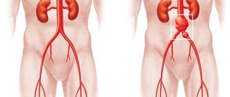
Ascites is a pathological accumulation of fluid in the abdominal cavity.
Ascites can develop rapidly (over several days) or over a long period (weeks or months). Clinically, the presence of free fluid in the abdominal cavity is manifested when a fairly large volume is reached - from 1.5 liters.
The amount of fluid in the abdominal cavity sometimes reaches significant figures - 20 liters or more. By origin, ascitic fluid can be of an inflammatory nature (exudate) or non-inflammatory, resulting from a violation of hydrostatic or colloid-osmotic pressure in pathologies of the circulatory or lymphatic system (transudate).
In more than 80% of cases, ascites is caused by decompensation of a chronic disease or acute inflammatory process in the liver
What is abdominal ascites
A symptomatic phenomenon in which transudate or exudate collects in the peritoneum is called ascites. The abdominal cavity contains part of the intestines, stomach, liver, gall bladder, and spleen. It is limited by the peritoneum - a membrane that consists of an internal (adjacent to the organs) and an external (attached to the walls) layer. The task of the translucent serous membrane is to fix the internal organs and participate in metabolism. The peritoneum is abundantly supplied with vessels that ensure metabolism through lymph and blood.
Between the two layers of the peritoneum in a healthy person there is a certain volume of fluid, which is gradually absorbed into the lymph nodes to free up space for new fluid to enter. If for some reason the rate of water formation increases or its absorption into the lymph slows down, then transudate begins to accumulate in the peritoneum. This process can occur due to multiple pathologies, which will be discussed below.
Causes of abdominal dropsy
The presence of serious diseases in women or men leads to complications, including dropsy of the abdomen. The abdomen swells gradually and the reason for this can only be found out through diagnosis.
Dropsy can occur not only with cirrhosis of the liver, but also be caused by heart failure or oncology.
The main causes of ascites include:
- kidney disease;
- Budd-Chiari syndrome, neoplasms;
- heart and vascular diseases;
- liver pathologies;
- gastrointestinal diseases;
- diets, poor nutrition;
- tumor of the stomach and abdominal organs;
- Rhesus conflict between fetus and mother.
There are cases of congenital dropsy, the cause of which is hidden bleeding or hemolytic diseases.
Causes of fluid accumulation in the abdominal cavity
Ascites of the abdominal cavity often occurs in oncology and many other diseases, when the barrier and secretory function of the peritoneal layers is disrupted. This leads to the filling of the entire free space of the abdomen with fluid. The constantly increasing exudate can reach up to 25 liters. As already mentioned, the main reason for damage to the abdominal cavity is its close contact with the organs in which a malignant tumor forms. The tight fit of the peritoneal folds to each other ensures the rapid capture of nearby tissues by cancer cells.
The main causes of abdominal ascites:
- peritonitis;
- peritoneal mesothelioma;
- peritoneal carcinosis;
- cancer of internal organs;
- polyserositis;
- portal hypertension;
- cirrhosis of the liver;
- sarcoidosis;
- hepatosis;
- hepatic vein thrombosis;
- venous congestion in right ventricular failure;
- heart failure;
- myxedema;
- gastrointestinal diseases;
- introduction of atypical cells into the peritoneum.
Among women
Fluid inside the abdominal cavity in the female population is not always a pathological process. It can be collected during ejaculation, which occurs monthly in women of reproductive age. This liquid dissolves on its own without posing a health hazard. In addition, the cause of water is often purely female diseases that require immediate treatment - inflammation of the reproductive system or ectopic pregnancy.
The development of ascites is provoked by intra-abdominal tumors or internal bleeding, for example, after surgery, due to injury or cesarean section. When the endometrium lining the uterine cavity grows uncontrollably, which is why it extends beyond the female organ, water also collects in the peritoneum. Endometriosis often develops after viral or fungal infections of the reproductive system.
In men
In all cases of dropsy in representatives of the stronger sex, the underlying cause is a combination of violations of important body functions, which lead to the accumulation of exudate. Men often abuse alcohol, which leads to cirrhosis of the liver, and this disease provokes ascites. Factors such as blood transfusions, drug injections, high cholesterol levels due to obesity, and multiple tattoos on the body also contribute to the occurrence of the disease. In addition, the following pathologies cause dropsy in men:
- tuberculous lesions of the peritoneum;
- endocrine disorders;
- rheumatoid arthritis, rheumatism;
- lupus erythematosus;
- uremia.
In newborns
Fluid in the abdomen collects not only in adults, but also in children. More often, ascites in newborns arises from infectious processes occurring in the mother’s body. As a rule, the disease develops in the womb. The fetus may have liver and/or biliary tract defects. Because of this, bile stagnates, which leads to dropsy. After birth, ascites in an infant may develop due to:
- cardiovascular disorders;
- nephrotic syndrome;
- chromosomal abnormalities (Down disease, Patau, Edwards or Turner syndrome);
- viral infections;
- hematological problems;
- congenital tumors;
- serious metabolic disorder.
Causes
The causes of ascitic disease are unexpected, the most common among them are presented below. This:
- malignant neoplasms and metastases;
- liver cirrhosis and increased blood pressure in the portal system;
- thrombosis (narrowing of the hepatic, inferior vena cava and portal veins);
- acute and chronic inflammatory kidney diseases;
- nephrotic syndrome (protein begins to be excreted in the urine);
- chronic renal failure;
- inflammatory lesion of the serous membrane of the heart;
- acute and chronic heart failure;
- some infectious and inflammatory bowel diseases, in which diarrhea and protein loss are observed;
- inflammation of the pancreas;
- tuberculosis;
- pseudomyxoma (mucus accumulation);
- anasrka.
This disease is a complication of liver cirrhosis and more. It progresses gradually in the body; at first it does not manifest itself in any way. Abdominal ascites is difficult to treat successfully. However, healing occurs if the main pathogenic factor is eliminated.
Symptoms
Signs of abdominal ascites depend on how quickly ascitic fluid collects. Symptoms may appear in one day or over several months. The most obvious sign of dropsy is an enlarged abdominal cavity. This causes weight gain and the need for larger clothing. In a vertical position, the patient’s stomach hangs down like an apron, and in a horizontal position, it is spread out on both sides. With a large volume of exudate, the navel protrudes.
Articles on the topic
- Portal hypertension - classification, signs, drug and surgical therapy, complications
- Neurox - instructions for use and contraindications, mechanism of action, dosage and analogues
- Stagnation in the gallbladder - signs, therapy with medications, homeopathy, folk remedies and diet
If the cause of dropsy is portal hypertension, then a venous pattern forms on the anterior peritoneum. It occurs due to varicose veins of the peri-umbilical veins and varicose veins of the esophagus. With a large accumulation of water in the abdomen, internal pressure increases, as a result of which the diaphragm moves into the abdominal cavity, and this provokes respiratory failure. The patient has pronounced shortness of breath, tachycardia, and cyanosis of the skin. There are also general symptoms of ascites:
- pain or feeling of fullness in the lower abdomen;
- dyspepsia;
- fluctuation;
- peripheral edema of the face and extremities;
- constipation;
- nausea;
- heartburn;
- loss of appetite;
- slow movements.
Symptoms or how to recognize pathology
Any pathology does not develop suddenly. The onset of ascites is difficult to notice. This is only available to people who regularly monitor their weight. And even then, extra grams are often attributed to errors in the diet. The fluid collected in the abdominal cavity makes itself felt for the first time, reaching a volume of one liter. The stomach takes the shape of a ball. There is a feeling of heaviness and a feeling of lack of air.
Typical symptoms of the presence of excess fluid in the abdominal cavity:
- bloating;
- heartburn;
- belching;
- difficulty breathing;
- swelling of the lower extremities.
The next sign of ascites is difficulty bending forward. Over time, the belly increases in size more and more, acquiring unnatural shapes. This can often be observed in older people. In clinics you can sometimes find grandmothers with swollen bellies and severe swelling of their legs.
Sometimes the fluid compresses the bile ducts. This stimulates the development of jaundice, which is accompanied by nausea and vomiting. It can be difficult to visually determine excess moisture between the abdominal walls. How can you find out why your belly is getting bigger? To determine there are various methods and diagnostic methods.
Stages
In clinical practice, there are 3 stages of abdominal dropsy, each of which has its own signs and characteristics. Degree of development of ascites:
- Transitional. The initial development of a disease, the symptoms of which cannot be noticed independently. The volume of liquid does not exceed 400 ml. Excess water is detected only during instrumental studies (ultrasound examination of the abdominal cavity or MRI). With such volumes of exudate, the functioning of the internal organs is not disrupted, so the patient does not notice any pathological symptoms. At the initial stage, dropsy can be successfully treated if the patient follows a water-salt regime and adheres to a specially prescribed diet.
- Moderate. At this stage, the abdomen becomes larger, and fluid volumes reach 4 liters. The patient already notices alarming symptoms: weight increases, it becomes difficult to breathe, especially in a supine position. A doctor can easily identify dropsy by examining and palpating the abdominal cavity. Pathology even at this stage responds well to treatment. Sometimes it becomes necessary to remove fluid from the abdominal cavity (puncture). If effective therapy is not carried out in time, then a disruption of the kidneys occurs, and the most severe stage of the disease develops.
- Tense. Liquid volumes exceed 10 liters. The pressure in the abdominal cavity increases greatly, problems arise with the functioning of all organs of the gastrointestinal tract. The patient's condition is deteriorating and he requires immediate medical attention. Previous therapy no longer gives the desired result. At this stage, laparocentesis (puncture of the abdominal wall) is necessarily performed as part of complex therapy. If the procedure does not have an effect, refractory ascites develops, which can no longer be treated.
Out of balance
The human body consists mostly of fluid, which is distributed throughout the cells, the environment between organs and cells (interstitium) and blood vessels. Thus, a small part of the fluid always passes from the blood into the tissues and, in turn, enters the lymphatic system. Through these “pipes” the lymph is transported back to the veins.
If this balance is disturbed, more water is transferred to surrounding tissues. For example, if the pressure in the veins increases, more fluid will be forced into the surrounding tissue. Not only does it cause ascites, it can also cause swelling in the legs or face.
All causes of ascites lead to fluid leakage from large vessels into the abdominal cavity. The causes of ascites are:
- Increased pressure in the blood vessels (mainly with portal ascites or heart failure).
- Protein deficiency (for example, as a result of starvation) manifests itself as a water belly.
- Increased permeability of cell walls, especially during inflammation.
- Lymphatic drainage disorders (due to tumors or scars).
Partly responsible for the formation of the water belly is the control loop of the kidney, which is additionally influenced by the passage of fluid into the abdominal cavity. In the body, at different measurement points (baroreceptors), there is a feeling that there is a lack of fluid, since the level of filling of the vessels decreases and thereby the blood pressure decreases. The kidney then produces less urine, releasing hormones that increase blood pressure. However, this vicious circle only results in more fluid being released into the peritoneum.
Complications
The disease itself is a stage of decompensation (complication) of other pathologies. The consequences of dropsy include the formation of inguinal or umbilical hernias, prolapse of the rectum or hemorrhoids. These conditions are promoted by increased intra-abdominal pressure. When the diaphragm puts pressure on the lungs, it leads to respiratory failure. The addition of a secondary infection leads to peritonitis. Other complications of ascites include:
- massive bleeding;
- hepatic encephalopathy;
- thrombosis of the splenic or portal vein;
- hepatorenal syndrome;
- intestinal obstruction;
- diaphragmatic hernia;
- hydrothorax;
- inflammation of the peritoneum (peritonitis);
- fatal outcome.
Consequences and complications
Ascites can lead to the following serious consequences:
- respiratory failure (due to an increase in the volume of the abdominal cavity and limited excursion of the diaphragm);
- spontaneous bacterial peritonitis;
- refractory ascites;
- hepatic encephalopathy;
- hepatorenal syndrome.
The occurrence of spontaneous bacterial peritonitis in patients with cirrhosis of the liver leads to repeated bleeding from esophageal varices. Mortality after the first episode of bleeding is 30–50%. In 70% of patients who have experienced one episode of bleeding from esophageal varices, bleeding occurs again. The risk of recurrent ascites within 6 months is 43%, within 1 year – 69%, within 2 years – 74%.
Diagnostics
Before making a diagnosis, the doctor must make sure that abdominal enlargement is not a consequence of other conditions, for example, pregnancy, obesity, mesenteric or ovarian cysts. Palpation and percussion (finger to finger) of the peritoneum will help to exclude other causes. Examination of the patient and the collected medical history are combined with ultrasound examination, scanning of the spleen and liver. Ultrasound excludes fluid in the stomach, tumor processes in the peritoneal organs, characterizes the condition of the parenchyma, the diameter of the portal system, the size of the spleen and liver.
Liver and spleen scintigraphy is a radiation diagnostic method used to evaluate tissue function. Initialization allows you to determine the position and size of organs, diffuse and focal changes. All patients with detected ascites are referred for diagnostic paracentesis with examination of ascitic fluid. During the study of pleural effusion, the number of cells, the amount of sediment, albumin, protein are counted, culture and Gram staining are performed. The Rivalta test, which gives a chemical reaction to protein, helps to distinguish exudate from transudate.
Two-dimensional Doppleroscopy (USD) of venous and lymphatic vessels helps to assess blood flow in the vessels of the portal system. In cases of ascites that are difficult to differentiate, diagnostic laparoscopy is additionally performed, during which an endoscope is inserted into the abdominal cavity to accurately determine the amount of fluid, the proliferation of connective tissue, and the condition of the intestinal loops. Plain radiography will also help determine the volume of water. Esophagogastroduodenoscopy (EGD) provides a good opportunity to see the presence of varicose veins in the stomach and esophagus.
Treatment of dropsy of the abdomen
Treatment of dropsy comes down to determining the causes of its appearance and reducing the amount of fluid. A large belly is removed thanks to therapeutic paracentesis, that is, puncture and pumping out up to 4 liters per day. The patient must be prescribed the necessary diet and bed rest.
Drug treatment
If dropsy of the abdomen is in the initial stage, medication therapy is prescribed by a specialist to treat it. The main group of drugs includes diuretics for stagnation of accumulated excess fluid.
Drug therapy is aimed at eliminating the dropsy itself and treating the disease that caused the appearance of ascites
Vitamins P and C and tablets to strengthen blood vessels are also prescribed. If bacteria are detected in the liquid, the patient is prescribed antibacterial drugs.
Surgical intervention
If treatment of ascites with drugs is unsuccessful, the doctor may resort to laparocentesis. Fluid from the abdominal cavity is pumped out gradually with the introduction of novocaine (0.5% solution). More than 10 liters of ascitic fluid should not be pumped out at a time.
The procedure is carried out on an empty stomach, the collected fluid is sent for examination, then the abdominal cavity is checked using an ultrasound.
Traditional methods
Since the prognosis of the disease is disappointing, many use all kinds of treatment methods, some read a spell for dropsy, and some supplement the treatment with folk remedies.
This is interesting: Life expectancy with ascites in cirrhosis, oncology, heart failure
Traditional medicine recipes include diuretic plants that help reduce the amount of fluid formed. Such plants include:
- coltsfoot;
- flax seeds;
- Linden;
- bearberry;
- hernia;
- birch buds and leaves;
- parsley;
- bean pods;
- horsetail;
- corn silk;
- ready-made preparations at the pharmacy.
Decoctions, teas and infusions are prepared from plants. An infusion of cherry stalks can also be used to treat ascites.
Treatment of abdominal ascites
Regardless of the cause that provoked ascites, the pathology should be treated together with the underlying disease. There are three main therapeutic methods:
- Conservative treatment. At the initial stage of ascites, drug therapy is prescribed aimed at normalizing liver function. If the patient is diagnosed with inflammatory organ parenchyma, then additional medications are prescribed that relieve inflammation and other types of medications, depending on the symptoms and the disease that provoked the accumulation of fluid.
- Symptomatic. If conservative treatment does not produce results or doctors are unable to prolong remission for a long time, then the patient is prescribed a puncture. Abdominal laparocentesis for ascites is performed infrequently, since there is a risk of damage to the patient's intestinal walls. If fluid fills the abdomen too quickly, a peritoneal catheter is installed in the patient to prevent the development of adhesions.
- Surgical. If the two previous treatment regimens do not help, the patient is prescribed a special diet and blood transfusion. The method consists of connecting the collar and inferior vena cava, which creates collateral circulation. If a patient needs a liver transplant, he undergoes surgery after a course of diuretics.
Drugs
The main method of treating ascites is drug therapy. It includes long-term use of diuretics with the introduction of potassium salts. The dose and duration of treatment is individual and depends on the rate of fluid loss, which is determined by daily weight loss and visually. The correct dosage is an important nuance, since the wrong prescription can lead the patient to heart failure, poisoning, and death. Frequently prescribed drugs:
- Diacarb. A systemic carbonic anhydrase inhibitor with weak diuretic activity. As a result of application, the release of water increases. The drug causes the removal of magnesium, phosphates, and calcium from the body, which can lead to metabolic disorders. The dosage is individual and is used strictly as prescribed by the doctor. Undesirable effects are observed in hematopoiesis, the immune and nervous systems, and metabolism. Contraindications for taking the drug are acute renal and liver failure, uremia, hypokalemia.
- Furosemide. A loop diuretic that causes strong but short-term diuresis. It has a pronounced natriuretic, diuretic, chloruretic effect. The regimen and duration of treatment are prescribed by the doctor, depending on the indications. Side effects include: a marked decrease in blood pressure, headache, lethargy, drowsiness, and decreased potency. Furosemide is not prescribed for acute renal/liver failure, hyperuricemia, pregnancy, lactation, and children under 3 years of age.
- Veroshpiron. Long-acting potassium-sparing diuretic. Suppresses the potassium-removing effect, prevents water and sodium retention, reduces urine acidity. The diuretic effect appears on days 2-5 of treatment. For edema due to cirrhosis, the daily dosage is 100 mg. The duration of treatment is selected individually. Adverse reactions: lethargy, ataxia, gastritis, constipation, thrombocytopenia, menstrual irregularities. Contraindications: Addison's disease, anuria, lactose intolerance, hyperkalemia, hyponatremia.
- Panangin. A medicine that affects metabolic processes and is a source of magnesium and potassium ions. It is used as part of complex therapy for ascites to compensate for the deficiency of magnesium and potassium excreted during the use of diuretics. Prescribe 1-2 tablets/day throughout the entire course of diuretics. Side effects are possible from the water-electrolyte balance, the digestive system. Panangin is not prescribed in the presence of Addison's disease, hyperkalemia, hypermagnesemia, myasthenia gravis.
- Asparkam. Source of magnesium and potassium ions. Reduces myocardial conductivity and excitability, eliminates electrolyte imbalance. While taking diuretics, 1-2 tablets are prescribed 3 times a day for 3-4 weeks. Possible development of vomiting, diarrhea, redness of the facial skin, respiratory depression, and convulsions. Asparkam is not prescribed for disorders of amino acid metabolism, adrenal insufficiency, hyperkalemia, hypermagnesemia.
Diet
Abdominal dropsy requires a limited diet. The diet includes a small fluid intake (750-1000 liters/day), a complete refusal to consume salt, the inclusion of natural foods with a diuretic effect and a sufficient amount of protein in the diet. Pickles, marinades, smoked meats, canned food, salted fish, and sausages are completely excluded.
The menu of a patient with ascites should include:
- lean poultry, rabbit;
- legumes, nuts, soy milk;
- seafood, lean fish;
- brown rice, oatmeal;
- vegetable oils, seeds;
- fermented milk products, cottage cheese;
- parsley, cumin, marjoram, sage;
- pepper, onion, garlic, mustard;
- bay leaf, lemon juice, cloves.
Surgical methods
When ascites progresses and treatment does not help, then in particularly advanced cases surgical treatment is prescribed. Unfortunately, it is not always possible to save the patient’s life even with the help of surgery, but no other methods exist today. The most common surgical treatment:
- Laparocentesis. Exudate is removed through a puncture of the abdominal cavity under ultrasound guidance. After the operation, a drainage is installed. No more than 10 liters of water are removed in one procedure. In parallel, the patient is given drips of saline solutions and albumin. Complications are very rare. Sometimes infectious processes occur at the puncture site. The procedure is not performed for bleeding disorders, severe bloating, intestinal injuries, ventral hernia and pregnancy.
- Transjugular intrahepatic shunting. During the operation, the hepatic and portal veins are connected artificially. The patient may experience complications such as intra-abdominal bleeding, sepsis, arteriovenous shunting, or liver infarction. Surgery is not prescribed if the patient has intrahepatic tumors or cysts, vascular occlusion, bile duct obstruction, or cardiopulmonary pathologies.
- Liver transplantation. If ascites develops against the background of liver cirrhosis, an organ transplant may be prescribed. Few patients have the chance to undergo such an operation, since it is difficult to find a donor. Absolute contraindications to transplantation are chronic infectious pathologies, severe dysfunction of other organs, and cancer. Among the most severe complications is graft rejection.
Forecast
Two-year survival rate in patients with ascites is 50%. When refractory ascites occurs, half of patients die within a year.
Unfavorable prognostic factors for patients with ascites:
- old age (over 60 years);
- low blood pressure (systolic pressure less than 80 mm Hg);
- decreased glomerular filtration rate (less than 500 ml/min);
- decrease in serum albumin content (less than 28 g/l);
- hepatocellular carcinoma;
- diabetes.
Video from YouTube on the topic of the article:
Ascites: disease or symptom?
A pathological accumulation of fluid in the abdominal cavity is called ascites. A small amount of ascitic fluid is always present in the peritoneal cavity. This fluid constantly moves into the lymphatic vessels, and new fluid takes its place, and this is a completely natural process. But with some disturbances in the functioning of the body, this fluid either begins to be produced in excess or ceases to be absorbed. As a result, it gradually accumulates and begins to put pressure on internal organs, impairing their functioning.
Causes of ascites
The list of diseases and disorders in which ascites can occur is very impressive. Among them:
- cirrhosis of the liver;
- some cancers;
- tuberculosis;
- pancreatitis;
- heart failure;
- endometriosis, ovarian cysts and tumors;
- internal bleeding;
- kidney diseases;
- malnutrition and exhaustion;
- diseases of the endocrine system.
However, if a person is diagnosed with abdominal ascites, the first things doctors suspect are usually liver cirrhosis (the cause of 75% of ascites cases), cancer (10% of cases) and heart failure (5% of cases). All other diseases combined account for only 10% of cases of abdominal ascites.
Of course, ascites does not occur in all people suffering from the above diseases. However, there are risk factors that increase the chances of developing ascites: alcoholism, drug addiction, hepatitis, obesity and high cholesterol, as well as diabetes.
Signs of pathology
Abdominal ascites most often develops gradually, over many months, and therefore most patients do not pay attention to it for a very long time. Often people just think they are gaining weight.
In the early stages, it is really difficult to notice ascites: at least a liter of fluid needs to accumulate. Only after this do typical symptoms of abdominal ascites begin to appear: abdominal pain, flatulence, belching and heartburn, difficulty breathing, swelling of the legs. As the volume of fluid increases, the abdomen also increases, and soon it becomes difficult for a person to bend over. The abdomen takes on the shape of a ball, stretch marks and dilated veins may appear.
But in order to make a diagnosis, there are few external signs - you need to consult a doctor.
Diagnostics
It begins with a visual examination and palpation of the abdomen. An experienced doctor can diagnose ascites based on these data alone, but more precise methods are required for clarification. It is not enough to know that the patient has ascites - it is much more important to find out what caused it.
If ascites is suspected, an ultrasound of the abdominal cavity and chest is prescribed - it allows you to see both ascites and neoplasms or changes in the structure of the liver. Sometimes Doppler ultrasound is also prescribed, showing the condition of the veins.
Radiography is another method widely used in the diagnosis of ascites. This examination makes it possible to detect tuberculosis or an enlarged heart, which may be causing fluid accumulation in the abdominal cavity.
MRI and CT are very accurate diagnostic methods that allow you to see the presence of fluid even where it is difficult to see with ultrasound and x-ray.
In some cases, the doctor may refer the patient for laparoscopy, during which a puncture is made in the abdominal wall and the accumulated fluid is taken for analysis. A biochemical blood test and urine test are also performed.
Principles of therapy
Treatment of ascites always begins with treatment of the underlying disease that caused the fluid accumulation.
- For ascites caused by heart disease, vasodilators, diuretics and glycosides are prescribed.
- For kidney disease, a low-salt diet and limited fluid intake are indicated.
- In case of protein metabolism disorders, a diet with optimal protein content and albumin transfusion are prescribed.
- For cirrhosis, hepatoprotectors are used.
In addition to this, symptomatic therapy is prescribed:
- a low-salt diet (no more than 2 g of salt per day), and in some cases, a completely salt-free diet. If the cause of ascites is cirrhosis, you should also limit fluid intake;
- potassium supplements and diuretics.
During treatment, the doctor monitors changes in the patient’s condition and, in particular, his weight. If the chosen therapeutic measures work, weight loss should be about 500 grams per day.
Surgical intervention
If conservative methods for some reason do not give the expected result, surgical intervention is indicated. Often, with ascites, fluid is removed by gradual drainage (if its amount is significant). The surgeon makes a small puncture in the peritoneum and inserts a drainage tube into it (laparocentesis).
An alternative to this method, which is painful and carries significant health risks, is the placement of indwelling catheters and subcutaneous ports. As a result, ascitic fluid is removed as it accumulates. This approach makes life much easier for patients, eliminates the need for repeated punctures, and therefore reduces the risk of damage to internal organs and inflammation.
Installation of the system is a simple surgical operation, during which the doctor inserts the end of the catheter (made of hypoallergenic materials) into the abdominal cavity, and the port itself (made of titanium) is installed subcutaneously in the area of the costal arch. To evacuate fluid accumulated in the abdominal cavity, it is necessary to pierce the skin and silicone membrane of the port chamber with a special needle. The ascitic fluid is pumped out through it. Medications can be administered in a similar way.
Sometimes a so-called intrahepatic bypass is required, in which the doctor creates a connection between the hepatic and portal veins.
In severe cases, a liver transplant may be required.
Treatment prognosis
The sooner ascites is diagnosed and treatment begins, the higher the chances of success. It is much easier to get rid of ascites in the early stages. But there are some factors that negatively affect the effectiveness of therapy - these are old age, diabetes mellitus, hypotension, cancer (and especially liver cancer), peritonitis, and low albumin levels.
Ascites is deadly. In approximately 50% of cases, if the prescription of diuretics does not help, the development of ascites ends tragically. Ascites is especially dangerous in cancer - death is likely in approximately 60% of cases.
Possible complications and risk of relapse
It should be remembered that ascites always worsens the course of the underlying disease, causing respiratory failure, hydrothorax, hernia, intestinal obstruction and a host of other complications.
Even if ascites has been cured, you should be very careful about your health, as there is always a risk of relapse. Therefore, even after curing ascites, you should adhere to the diet prescribed by your doctor.
The accumulation of fluid in the abdomen can cause severe discomfort, but before this happens, other symptoms appear. You should not ignore them - be sure to consult a doctor.
If the amount of fluid in the abdominal cavity is less than 400 grams, ascites is practically invisible, but at this stage it can be detected using instrumental examinations, so regular examination by a specialist is very important. Especially with diagnosed oncology or cirrhosis of the liver.
Drug treatment
The treatment regimen directly depends on the severity of ascites. If, as a result of the pathology, the patient experiences cardiac or respiratory failure, measures are indicated to help reduce the volume of accumulated fluid. If a person’s condition does not pose a threat to life, the fight against the underlying pathology comes to the fore.
In all cases, treatment of abdominal dropsy involves taking diuretics. Diuretics help reduce the volume of ascitic fluid. As a rule, doctors prescribe the following drugs:
- "Furosemide".
- "Mannitol."
- "Spironolactone".
https://www.youtube.com/watch?v=upload
The dosage regimen is determined individually. The doctor must take into account the fact that no more than 400 ml of fluid should be excreted per day. Otherwise, there is a risk of dehydration.
The treatment regimen for abdominal hydrops in humans may also include taking or intravenously administering the following drugs:
- Drugs whose active components help strengthen the walls of blood vessels. Most often, doctors prescribe Diosmin.
- Means that promote fluid retention in the vascular bed. Examples of drugs: “Polyglucin”, “Gelatinol”.
- Medicines containing albumin (for liver and kidney pathologies).
- Antibiotics. Most often prescribed if the underlying cause is peritonitis.
Thus, the treatment regimen directly depends on the underlying disease.
Conservative treatment
Drug therapy is carried out comprehensively. Drugs are prescribed in order to remove ascitic fluid from the body; for this it is necessary: to reduce the intake of sodium into the body, to ensure its abundant excretion in the urine.
The patient should receive at least 3 g of salt daily. Complete refusal of it worsens protein metabolism in the body. Diuretics are used.
Pharmacology does not have in its arsenal a single remedy that would fully meet the requirements of doctors. The most powerful diuretic, Lasix, flushes potassium from the body, so in addition, the patient is prescribed drugs, for example, Panangin or Potassium Orotate, which restore its level.
Potassium-sparing diuretics are also used, these include Veroshpiron, but they also have unpleasant side effects. When choosing a suitable drug, it is necessary to take into account the characteristics of the body and its condition.
It is advisable to use diuretics to treat ascites in the presence of edema, since they remove fluid not only from the abdominal cavity, but also from other tissues.
For liver cirrhosis, drugs such as Fozinoprl, Captopril, Enalapril are often used. They enhance urinary sodium excretion without affecting potassium.
After the swelling of the extremities subsides, it is worth reducing the consumption of table salt.
During treatment of the disease, it is recommended to remain in bed and reduce the amount of fluid consumed. When the condition improves, semi-bed rest is acceptable.
When conservative treatment is ineffective or impractical, laparocentesis is performed.
What is the danger of ascites?
Treatment of ascites requires constant attention and selection of optimal drugs. Lack of dynamics in the patient’s condition threatens him with complications:
- bacterial peritonitis;
- the emergence of resistance to diuretic therapy;
- development of the consequences of liver cirrhosis - encephalopathy with loss of a number of brain functions;
- hepatorenal syndrome, signs of renal failure;
- spontaneous leakage of ascitic fluid through the umbilical hernia.
Nutritional Features
Diet for dropsy of the abdominal cavity is one of the main points of treatment. Nutrition should be such that the body receives a sufficient amount of nutritional components, but the course of the disease does not worsen.
It is recommended to include the following products in your diet:
- Lean meat and fish.
- Fruits.
- Vegetables.
- Skim cheese.
- Wheat cereal.
- Greenery.
- Compote.
- Kissel.
It is necessary to completely exclude from the menu:
- Meat and fish of fatty varieties.
- Fried foods.
- Spices.
- Smoked meats.
- Alcohol-containing drinks.
- Coffee.
- Tea.
https://www.youtube.com/watch?v=ytcreatorsru
Dishes need to be salted minimally. In addition, persons suffering from ascites should limit the amount of fluid consumed to 0.5-1 liters.
Prevention
The main preventive measures are:
- timely treatment of diseases that can lead to the development of dropsy;
- regular sanitation of foci of chronic infection in the body;
- proper nutrition - you need to give up fast food and other junk food, introduce foods that are healthy for the body into your diet, eat in small portions (5-6 times a day, in small portions);
- giving up bad habits (tobacco smoking, alcohol abuse);
- timely surgery for congenital heart defects, which will help avoid the development of heart failure, which is one of the main causes of dropsy;
- proper nutrition for chronic pancreatitis;
- normalization of work and rest regimes;
- refusal to take uncontrolled medications;
- refusal of a sedentary lifestyle, which leads to the development of congestion in the abdominal cavity;
- Regular preventive examinations will help identify possible pathological conditions of the body in the early stages, which will significantly facilitate subsequent treatment and improve the prognosis.
Good health to you!
The importance of the composition of ascitic fluid in differential diagnosis
It is impossible to cure ascites without taking into account the underlying disease. One of the mandatory tests is the study of ascitic fluid obtained during laparoscopy or a special puncture of the abdominal wall. The results help to identify the mechanism of water retention, select optimal medications and provide treatment for ascites.
First of all, the results should answer the question: “Is the resulting fluid a transudate or an exudate?” The difference is determined by the content of protein and other components. In transudate, fluid leaves the vascular bed under the influence of increased hydrostatic pressure or reduced content of colloidal substances, while the vessels themselves remain intact.
Exudate is formed as a result of an inflammatory reaction with damage to the permeability of the vascular wall. The composition of the exudate is close to blood plasma. It contains a significant amount of protein components that under normal conditions do not pass through the vessel wall (immunoglobulins, fibrinogen, complement).
Total protein 15–60 g/l. Fibrinogen is quickly transformed into fibrin under the influence of tissue thromboplastins. Fibrin threads are visible under liquid microscopy. The relative density of the exudate is 1.015–1.027. Inflammatory elements are definitely present.
In liver cirrhosis, transudate is most often detected. Against the background of peritoneal phenomena, inflammation of internal organs - exudate. The presence of hemorrhagic ascitic fluid (with blood) rarely accompanies cirrhosis. Much more often observed with neoplasms, tuberculous peritonitis.
The predominance of neutrophils with a turbid sediment in the cellular composition of the fluid is a characteristic sign of bacterial peritonitis. If the level of lymphocytes is the highest, peritonitis is most likely of tuberculous origin. For diagnosis, such differences are important, since peritonitis can occur secretly.
If the resulting material is turbid and has a whitish (milky) tint, then the laboratory technician assumes that a significant amount of lymph has entered the abdominal cavity (chylous ascites). With this option, the composition contains tiny drops of fat, increased levels of triglycerides, a high content of leukocytes, and tumor cells.
To confirm a subjective opinion, laboratory assistants carry out differential diagnostics of samples:
- when adding an alkaline solution, proteins dissolve and turbidity decreases;
- reagents with ether lead to the disappearance of turbidity associated with fats.
A very rare result is the detection of mucus in the ascitic fluid. This happens with pseudomyxoma peritonei and mucinous adenocarcinoma of the stomach or intestines.
Surgery
Surgical treatment involves removing excess fluid by puncturing the abdomen. This procedure is called laparocentesis. It is prescribed when the abdominal cavity is significantly filled with ascites with fluid. The procedure is performed under local anesthesia, with the patient in a sitting position.
During paracentesis, a puncture is made in the patient's lower abdomen through which fluid will be sucked out. The procedure can be performed at one time, or a special catheter can be installed for several days; such decisions are made by the doctor based on the patient’s condition and the severity of the disease.
If the amount of fluid exceeds 7 liters, then laparocentesis is carried out in several stages, as the risk of complications increases - a sharp drop in pressure and cardiac arrest.
Traditional methods
The use of unconventional means helps remove excess fluid from the body. It is important to remember that traditional treatment is only an auxiliary measure; it will not get rid of the root cause.
The most effective recipes:
- Take 40 g of pre-chopped parsley. Pour 1 liter of boiling water over the raw materials. Let it brew for a day. Take 4 times a day, 20 ml before meals.
- Take 1 tbsp. pre-crushed coltsfoot leaves. Fill them with 250 ml of water. Place the container on the fire. Boil for 10 minutes. Cool, strain. Take 10 ml three times a day.
For ascites, it is recommended to drink compotes. Apricot is considered the most useful. It not only promotes fluid removal, but also retains potassium in the body.
Risk factors
Some people are more susceptible to the disease than others. Persons at risk:
- People who have been taking alcoholic beverages and drugs for a long time.
- People who have undergone blood transfusions.
- Suffering from hepatitis, not necessarily of a viral nature.
- Having significant excess weight.
- Suffering from type 2 diabetes.
- Having high cholesterol levels in the blood.
Causes of ascites
The disease occurs under the influence of the following etiological factors:
- Liver pathologies. As a result of the death of hepatocytes, they are replaced by scar tissue, the size of the liver increases, and it begins to compress the portal vein, which ultimately leads to the development of dropsy.
- Lack of protein.
- Endocrine disorders.
- Kidney diseases.
- Gynecological diseases.
- Myocardial diseases. Most often, dropsy develops with pericarditis or heart failure. An enlarged and weakened heart cannot cope with pumping blood, which accumulates in the vessels, which can lead to fluid accumulation.
- Pathologies of the lymphatic system.
- Systemic pathologies.
- Damage to the peritoneal surface. The development of dropsy can be caused by diffuse, fungal or tuberculous peritonitis.
- Bad habits.
- Myxedema. With this disease, swelling of the mucous membranes and soft tissues occurs, the cause of which is a violation of the synthesis of thyroid hormones.
- Polyserositis.
- Poor nutrition. Particularly dangerous are fasting and constant strict dieting.
- Diseases of the gastrointestinal tract.
The risk group includes the following categories of people:
- drug addicts;
- patients who have undergone a blood transfusion procedure;
- obese people;
- patients suffering from various types of hepatitis;
- hemodialysis patients;
- people who are interested in tattooing;
- people with a hereditary predisposition to the disease.
Ascites in children can be hereditary, acquired or congenital. The following are the main reasons for the development of this pathology in young patients:
- Nephrotic syndrome.
- Congenital general edema, which can be caused by Rh conflict between the organisms of the mother and fetus. Children born with this pathology have no chance of survival and die within a few hours.
- Exudative enteropathy. It is a hereditary pathology.
- Damage to the liver and bile ducts in infants.
In older children, the reasons are basically the same as in adults.
Symptoms that accompany ascites
The symptoms that manifest ascites, of course, greatly depend on the severity of the condition. If ascites is a mild disease no symptoms appear ; it is difficult to detect even with the help of instrumental examinations; only ultrasound or CT scan of the abdominal cavity helps.
If ascites is severe , it is accompanied by the following symptoms :
- Bloating and heaviness of the abdomen.
- Bloating, swelling and increase in abdominal volume.
- Breathing problems due to pressure of abdominal contents on the diaphragm. Compression leads to dyspnea (shortness of breath, short and rapid breathing).
- Stomach ache.
- Flat navel.
- Lack of appetite and instant feeling of fullness.
- Swollen ankles (edema) due to excess fluid.
- Other typical symptoms of the disease, such as portal hypertension (resistance to blood flow) in the absence of cirrhosis.
Treatment prognosis
With abdominal dropsy, the quality and length of life depends on the condition of the liver, kidney function, cardiovascular system, and the method of treatment. The prognosis depends on the form and degree of the underlying pathology. For example, with compensated cirrhosis, you can count on a favorable outcome with proper therapy; with decompensated form, liver transplantation is indicated.
The shortest life expectancy is observed with ascites against the background of renal failure. A patient without dialysis will live only a few weeks. If the pathology is detected against the background of heart failure, the prognosis is favorable, although it all depends on the degree. With stage 3 or 4 heart failure, a person will live up to two years. Only 10% of patients can count on a cure and only thanks to the correctly selected method of therapy.
Most often, treatment of ascites depends on the qualifications of the doctor, as well as the degree of development of the pathology. Regardless of the cause of the disease, a special diet and conservative or surgical methods are prescribed. Only combined therapy can significantly prolong a person’s life.
Clinical manifestations
The symptoms of the disease are pronounced. In humans, dropsy of the abdominal cavity manifests itself in the form of a pathological increase in the volume of the abdomen. However, this sign may indicate the development of pancreatitis and the accumulation of feces. If in a standing position the stomach sags greatly, and in a lying position it literally spreads to the sides, this clearly indicates the development of ascites.
Thus, swelling is the main symptom of dropsy. Other symptoms of ascites:
- Cough.
- Dyspnea. It is especially difficult to breathe when lying down.
- As the abdomen increases in volume, the intensity of pain in the pelvic area increases.
- Frequent urge to urinate. The act itself is accompanied by painful sensations. The volume of urine is not increased.
- Heart rhythm disturbance.
- Frequent episodes of belching and heartburn.
- Presence of hemorrhoids.
- Protrusion of the navel.
- Drowsiness.
- General weakness.
- Apathy.
- The passage of feces is impaired.
In severe cases, there is constant pain in the abdomen. It interferes not only with physical activity, but also with breathing. The patient's blood circulation is impaired, due to which the limbs and face begin to swell. It is important to understand that as ascites progresses, the course of the underlying disease that served as the provoking factor worsens.
Ascites - what is it?
A healthy person has some fluid in the abdomen, which is constantly absorbed and distributed through the lymphatic vessels. The definition of ascites refers to a pathological accumulation of inflammatory exudate or transudate in the peritoneum.
Based on the accumulated volume of fluid in the abdomen, the following stages of dropsy are distinguished:
Transient ascites. No more than 500 ml of fluid accumulates in the peritoneum. This condition cannot be determined independently or by palpation of the abdomen; there are no symptoms. Therefore, the patient at the first stage does not suspect the presence of pathology.
Moderate ascites. Up to 4 liters of exudate accumulate in the abdomen. The patient feels discomfort, dropsy is visible and is expressed in a hanging abdomen. Diagnosed by inspection and palpation of the site of swelling.
Tense ascites. The fluid accumulates in a large volume; there are 10 liters of exudate in the walls of the peritoneum. Internal organs experience great pressure, renal blood flow is disrupted. The abdomen is expanding, the right and left sides are enlarged.
Chylous ascites. A rare complication that indicates the last degree of cirrhosis. A white fluid containing fat collects in the peritoneum.
Ascites can be provoked by a variety of chronic or advanced organ diseases: tuberculous peritonitis, portal hypertension, heart failure, liver cirrhosis, peritoneal carcinomatosis, gynecological diseases. Treatment of ascites formed in the abdominal cavity consists of diagnosing and eliminating the factors that provoked it.
Ascites in heart failure
The accumulation of pathological fluid in the walls of the abdominal and abdominal cavity sometimes occurs due to heart problems. This factor provokes ascites in 5% of cases. Dropsy of the abdomen is formed due to the inability of the enlarged heart to pump blood in sufficient volume.
The main diseases of the heart muscle and blood vessels of the system, which lead to stagnation and accumulation of pathological fluid:
- heart injury;
- overload of the heart parts and stretching of its walls due to hypertensive crisis,
- arterial hypertension, heart disease;
- cardiomyopathy: thinning or thickening of the wall of an organ.
Also, symptoms of dropsy are observed with constrictive pericarditis. Any pathology and disturbance in the functioning of the heart can lead to heart failure and the development of ascites.
This complication cannot be ignored, as it indicates the ineffectiveness or lack of proper treatment of the causative disease. Urgent removal of pathological fluid is mandatory.
Dropsy with cirrhosis of the liver
In 80% of cases, free fluid stagnates in the walls of the abdominal cavity as a result of advanced cirrhosis. With this disease, blood flow is disrupted, the production of plasma proteins is reduced, the level of albumin decreases, liver vessels change, the serous membrane becomes covered with scars. Due to these changes, the organ becomes larger and begins to put pressure on the portal vein.
Fluid accumulation in the abdomen occurs with the following types of cirrhosis:
- primary biliary;
- secondary;
- congenital.
The main symptoms of abdominal dropsy in cirrhosis are an increase in abdominal volume against the background of a sharp loss of total weight, difficulty breathing and increased fatigue. An enlarged abdomen indicates an almost complete replacement of healthy liver tissue with non-functional one. The patient must be hospitalized and immediately prescribed effective treatment.
Chylous ascites
The last stage of liver cirrhosis provokes the accumulation of lymph in the walls of the peritoneum and swelling of the abdomen. Ascitic fluid has a characteristic color and composition: milky with admixtures of fat.
In addition to the increase in volume, the patient experiences breathing problems and swelling of the face and legs.
The causes of abdominal ascites in this case are as follows:
- hydrostatic hypertension;
- operations on the abdominal organs;
- tuberculosis;
- pancreatitis;
- injuries to the liver, stomach, duodenal intestine, intestines and gall bladder.
Chylous ascites is treated with nutritional correction. The diet prescribed is strict. It is aimed at completely eliminating from the diet foods that provoke the accumulation of internal fat.
Chylothorax
With injury or pathologically enlarged lymph nodes in the pleural area, fluid accumulation in the lungs may develop. Among the main symptoms of this complication of ascites are shortness of breath, a feeling of heaviness in the chest, and irregular heartbeat.
This phenomenon is diagnosed after studying the composition of the accumulated liquid. As a rule, it is white and contains a large number of lymphocytes. Treatment of pulmonary hydrops is similar to the treatment of abdominal ascites: dietary nutrition, drug therapy, and if there is no result, laparocentesis of the pleural cavity.
Diagnosis of the disease
To make a diagnosis, a complex of various procedures is required, the results of which can accurately indicate the amount of fluid inside the abdominal cavity and the presence of various complications.
- Inspection - depending on the position the person is in, when tapping movements, a dullness of sound can be detected. When pushing to the side with one palm, the second palm, which fixes the stomach, feels noticeable vibrations of the fluid inside.
- X-ray examination - allows you to detect ascites with an amount of fluid of more than half a liter. If tuberculosis is detected in the lungs, a preliminary conclusion can be made that the disease has a tuberculous etiology. If pleurisy and expansion of the borders of the heart are detected, it can be assumed that the cause of the disease was heart failure.
- Ultrasound examination - allows you to determine the presence of ascites, as well as detect cirrhosis of the liver or the presence of malignant tumors in the abdominal cavity. Helps assess blood flow through veins and vessels. Examination of the chest area can detect heart disease.
- Laparoscopy is a puncture of the abdominal cavity, which allows you to take fluid for laboratory testing to determine the causes of the disease.
- Hepatoscintigraphy - allows you to determine the degree of damage and the severity of changes in the liver caused by cirrhosis.
- MRI and CT scans allow you to determine all the places where the fluid is located, which could not be done by other means.
- Angiography is an X-ray examination performed along with the administration of a contrast agent. Allows you to determine the location of affected vessels.
- Coagulogram is a blood test that allows you to determine the rate of its clotting.
- The following indicators are determined in the laboratory: globulins, albumins, urea, creatine, sodium, potassium.
- 10. Determination of the level of α-fetoprotein is carried out to diagnose liver cancer that can lead to ascites.
How to identify pathology?
Diagnosis of ascites begins with a medical examination. In addition to visual signs (abdominal enlargement, dilated venous network, swelling of the legs and groin), doctors use the percussion method.
The finger of one hand is tapped on the other. In this case, dullness of sound is detected in a supine position in the lateral channels of the abdomen, on the side - in the lower part. By asking the patient to turn, you can record the transition of dull zones. Another method is unilateral jerking movements towards the center, while on the other side a wave is felt.
An ultrasound examination is performed not only to identify fluid, but also to determine the cause of ascites. The doctor can examine the liver, identify changes in size, shape, and nodular formations. Doppler mapping evaluates blood flow in the portal and inferior vena cava.
An ultrasound examination of the heart allows you to identify signs of heart failure, defects, and notice fluid in the pleural cavity. Examination of the pancreas is necessary to exclude pancreatitis in the etiology of ascites.
The X-ray method is suitable for detecting ascites with a volume of more than 0.5 liters. But the method remains one of the most important in the diagnosis of tuberculosis of the lungs, hypertrophic changes in the heart, and pleurisy.
Angiography is a variant of x-ray examination in which a contrast agent is injected into a vein, and then a series of images are taken to confirm the patency and shape of the vessels.
Examination of the liver with radioisotope preparations helps to determine the degree of damage to the working cells of the organ. During hepatoscintigraphy, Methionine, labeled with an isotope sensitive to liver cells, is administered intravenously. Further scanning reveals areas not filled with the substance. This means that there are no liver cells there, the gaps are filled with fibrous tissue.
Laparoscopic examination in the operating room consists of introducing optical equipment through a small incision into the abdominal cavity, capable of remotely showing internal changes in organs.
A special attachment for collecting material for biopsy is used for subsequent histological examination of the liver and other organs, lymph nodes, and peritoneum. Ascitic fluid must be taken for analysis; 50–100 ml is sufficient.
Laboratory diagnosis is based on signs of liver dysfunction, all types of metabolism and electrolyte composition. Analyzes are carried out:
- albumin/globulin ratio;
- liver transaminases and other enzymes;
- urea, creatinine;
- pancreatic lipases and amylases;
- potassium, sodium.
A certain importance in confirming the hepatic cause of ascites is given to the study of indicators of the blood coagulation system. In order to exclude a cancerous process in the liver, an analysis of the level of α-fetoprotein is prescribed.
What to do after confirming the pathology by ultrasound
The doctor’s actions after diagnosis depend on the identified cause. If a woman is found to have tumors, a biopsy and fluid puncture are performed. If the problem is limited to inflammation, then anti-inflammatory therapy is carried out, but sometimes surgery is also performed. In the case of polycystic diseases, hormonal therapy may be used.
The fluid itself, which has accumulated in a woman’s lower abdomen, must be removed. For this purpose, drug or surgical treatment is used.
Medication
If about 2 ml of fluid is detected in the area of the posterior fornix of the uterus, medications are not prescribed, since this is water from the follicle that ruptured during the period of ovulation.
Up to 15% of cases of ascites are eliminated with a diet limiting salt and drinking.
They switch to drug treatment only if the diet does not help.
The medications used are diuretics: spironolactone, and if this does not help, then furasemide. When they are taken, the volume of blood plasma decreases, and fluid passes from the space between organs into the blood. At the same time, a woman loses 2–3 kg per week, and loses 0.5 liters in urine every day. more liquid than was drunk.
Surgical
If surgical intervention is necessary, use:
- laparocentesis to remove water in severe ascites or to determine the composition of the fluid;
- culdocentesis (uterine puncture) to determine the composition of fluid when it accumulates in women in the pelvic area.
We recommend further reading Why the abdomen swells after mammoplasty, how to remove the symptom, and when it will subside on its own
These are extreme measures that are used for severe forms of ascites.
It is important to follow dietary recommendations because the need for surgery depends on how the ascites responds to diuretics. If you consume too much salt, diuretics will not work.
Traditional methods
Ascites often indicates a serious illness. Traditional methods are unpredictable in their effect; individual consultation with the attending physician is necessary so that they do not negatively affect the underlying disease, its treatment and the woman’s health.
You can find out more about various home treatment methods here
Classification
Excess fluid can penetrate into the abdominal cavity in different ways, depending on this, two types of ascites are classically distinguished:
- Transudative occurs due to increased pressure in the portal vein and decreased oncotic pressure of the blood. However, ascitic fluid usually contains little protein.
- Exudative - the result of inflammation. It is characterized by a high protein content in ascitic fluid.
This classification has some disadvantages, so now another one is more often used, based on the serum-ascitic albumin gradient (abbreviated as SAAG). This indicator indicates the difference between the levels of albumin proteins in the blood serum and ascitic fluid:
- Low SAAG is characteristic of the accumulation of fluid in the abdomen during inflammatory processes (pancreatitis, peritonitis), impaired renal function.
- When SAAG , ascites is most likely caused by portal hypertension or cardiac dysfunction.
Diagnosis of ascites
An enlarged abdomen is not the only sign of ascites, therefore, after examination and palpation, laboratory and instrumental diagnostics are prescribed. The presence of fluid in the walls of the peritoneum makes it possible to distinguish a complication from obesity.
To confirm the presence of ascites and determine its cause, doctors use the following research methods: local ultrasound examination of organs; abdominal puncture; assessment of the quality and quantity of ascitic fluid; laparocentesis for ascites; laboratory study of the composition of the liquid.
If less than 500 μl-1 leukocytes are observed in the transudate, and up to 250 μl-1 neutrophils, dropsy is diagnosed. An increase in the latter indicator indicates the presence of an infectious pathogen, for example in tuberculous peritonitis.