Breast cancer is a specific tumor that appears in the mammary glands and ducts. The nature of malignant tumors is characterized by accelerated development and spread to other tissues. First, the lymph nodes are affected, and then the internal organs. In oncological practice, there are more than twenty types of breast tumors. The disease has ICD-10 code C50.
The causes of the disease vary, from genetic predisposition to work in hazardous industries. At an early stage of development, the tumor is treatable, leaving a positive prognosis. In advanced forms of the disease, cancer cells and metastases spread through the blood throughout the body, so cure is impossible. Therapy in this case is aimed at relieving symptoms and achieving stable remission.
How does breast cancer occur?
Breast cancer develops in the same way as any other malignant tumor in the body. One or more cells of glandular tissue, as a result of a mutation that has occurred in them, begin to divide abnormally quickly. They form a tumor that can grow into neighboring tissues and create secondary tumor foci - metastases.
Mutations that lead to breast cancer can be hereditary or acquired.
Common inherited genetic causes of breast cancer are mutations in the BRCA1 and BRCA2 genes. BRCA1 mutation carriers have a 55–65% risk of developing breast cancer, and BRCA2 carriers have a 45% risk of developing breast cancer. Such genetic defects are inherited from parents to children and cause breast cancer in approximately 15% of cases.
Much more often, a tumor occurs due to acquired mutations: they arise in the cells of the mammary gland and are not inherited. For example, in 20% of cases, the number of copies of the gene encoding HER2, a receptor protein that is located on the surface of cells and stimulates their reproduction, is increased.
The “molecular genetic portrait” of the tumor is important when choosing the optimal treatment.
Prevention
There is no specific prevention for breast cancer. This is explained by insufficient knowledge of the etiology of the disease, which makes it difficult to influence its original causes.
Effective prevention of nonspecific breast cancer consists of:
- timely pregnancy and childbirth;
- reducing the impact of carcinogens on the body;
- following the rules of proper nutrition;
- conducting regular self-examinations;
- conducting screening tests in doctors' offices.
Types of Breast Cancer
Malignant breast tumors are divided into two types: ductal and glandular. Ductal breast cancer is more common. It can be intraepithelial (in situ) and invasive. Intracellular ductal breast cancer has a more favorable prognosis, it rarely metastasizes and is curable in 98% of cases. The invasive version of the tumor is prone to uncontrolled growth and generalization of the process.
Glandular breast cancer can be lobular (invasive lobular carcinoma) or grow from other cells of the glandular tissue. Lobular cancer is often characterized by multicentric growth. The rate of increase in size and timing of metastasis of forms of nodular breast cancer depend on the degree of tumor differentiation.
Contraindications for breast cancer
With breast cancer, there are serious contraindications that impose significant restrictions on the victim’s lifestyle. What cannot be done with this pathology, is there a diet for breast cancer that should be followed?
Regarding lifestyle:
- baths and other thermal procedures should be completely excluded, since cancer cells actively divide at elevated temperatures;
- it is necessary to limit, but not completely eliminate, physical activity;
- you will have to get used to the correct rhythm of life so as not to overload the body and preserve the immune system to fight the tumor;
- you need to give up bad habits, even if it seems impossible.
Causes and risk factors
Unfortunately, scientists do not yet have complete information about the causes of breast cancer. There is a list of risk factors that influence the likelihood of a tumor, but in some people the disease is diagnosed in the absence of these factors, while others remain healthy in the presence of many of them at once. However, scientists still associate the development of breast cancer with certain circumstances that most often precede its appearance. These include:
- Age. The majority of breast cancer cases occur in women aged 55 years and older.
- Heredity. If breast cancer is diagnosed in one of your close relatives, the risk doubles.
- History of breast cancer.
- Increased breast tissue density as determined by mammography.
- Some benign neoplasms in the mammary gland.
- Early onset of menstruation - before 12 years of age.
- Late menopause - after 55 years.
- Lack of children or late (after 35 years) first birth.
- Exposure to radiation, such as from radiation therapy given to treat another type of cancer.
- Smoking and alcohol abuse. If a woman consumes 28–42 g of ethyl alcohol daily, her risks increase by 20%.
- Excess weight and low physical activity.
- Use of hormonal drugs: oral contraceptives, hormone replacement therapy in postmenopause.
- Breast injuries.
- Diabetes.
- Work on a schedule with night shifts.
How to do a breast self-examination
Symptoms and signs of breast cancer can appear even at a young age in nulliparous women, so you need to know how to perform a breast examination on your own. But even in the presence of obvious signs of danger, not all women find the strength to go to the doctor - they are afraid to hear the truth. Of course, this cannot be called a reasonable act, since most clinical forms of cancer develop quickly and the chances of a complete cure are dwindling every day. This is especially true for women at risk whose mother or grandmother had breast cancer.
The examination is carried out on days 5, 6 and 7 of the menstrual cycle. It is advisable to choose one of them and regularly conduct self-examination. You need to stand in front of the mirror to see your breasts clearly. Answer the following questions for yourself:
- are there any changed areas on the gland in color;
- whether the size of one breast has changed relative to the other (most women have one gland larger than the other);
- determine the symmetry of the location of both mammary glands;
- see if the nipple looks normal.
Suspicion may be caused by rashes on one or both breasts. Outwardly, it resembles an allergy.
Palpation as a way to identify compactions
The examination is carried out standing or lying down. Women with large breasts should lie down. Feel first one breast with your fingertips, then the other. You can’t press hard, but press so as to feel possible changes in the consistency of the tissue. You can start from the nipple area and gradually move towards the periphery.
Pay attention:
- whether the density has increased compared to the previous inspection;
- Are the lymph nodes enlarged?
If suspicious areas are detected, you must contact a gynecologist, mammologist or oncologist. There is no point in worrying in advance, since an accurate diagnosis can only be made after a complete examination.
Symptoms of breast cancer
In the early stages, breast cancer usually does not manifest itself clinically. Most often, the tumor is discovered by the patients themselves or is detected accidentally during preventive studies.
Patients usually complain of the presence of a palpable formation and discharge from the nipple. Pain is a rare symptom of breast cancer, but the pain syndrome can come to the fore at the stage of generalization of the process, especially when metastases spread to the bones.
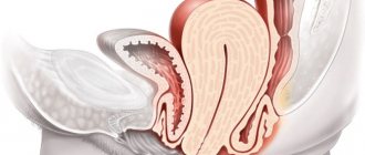
Quite often, signs of breast cancer are detected, such as the appearance of asymmetry due to changes in the size of the affected gland. Reduction, upward displacement, deformation and wrinkling of the mammary gland can be observed in the scirrhosis (fibrous) form of the tumor. On the contrary, the breast on the affected side becomes enlarged due to the rapid growth of the formation or due to edema, which forms due to impaired lymph outflow.
When the tumor spreads into the subcutaneous tissue, skin changes may be observed. The following symptoms of breast cancer are identified:
- “Platform” - the skin above the tumor becomes flattened; it is impossible to form a skin fold in this area.
- “Umbilization” - the skin over the lesion is wrinkled and retracted.
- “Lemon peel” is a characteristic appearance of the mammary gland due to lymphostasis.
Sometimes, when the tumor spreads to the surface of the skin, signs of breast cancer such as redness and ulceration may be observed. The presence of these symptoms indicates that the process is neglected.
Changes in the nipple can also be detected, but only in the later stages. In this case, the following symptoms of breast cancer occur:
- Forga's sign - on the affected side the nipple is higher than on the healthy side.
- Krause's sign - the nipple is thickened, the folds of the areola are noticeably pronounced.
Such a sign of breast cancer as pathological discharge is quite rare, but in some cases it may be the only symptom that is detected during examination. The discharge is often bloody in nature, serous and purulent are less common.
Depending on how the disease manifests itself, different clinical forms of breast cancer are distinguished. Most often - in 75–80% of cases - the nodular form occurs. In the early stages, the only symptom is usually a painless lump in the breast. If you divide the breast into four parts by horizontal and vertical lines, then in half of the cases the tumor will be located in the upper outer part.
Special forms of breast cancer have also been identified, which manifest themselves with typical symptoms. These include:
- An edematous-infiltrative form, which is characterized by enlargement and swelling of the gland, marbled skin color, and severe hyperemia.
- Mastitis-like. This type of breast cancer is manifested by hardening of the affected breast and increased body temperature.
- An erysipelas-like form, in which lesions are detected on the skin (sometimes ulcerations appear) that externally resemble erysipelas.
- The armored form is characterized by the presence of multiple nodes, due to which the gland shrinks and deforms.
- Paget's cancer affects the nipple and areola. Signs of breast cancer with this type of disease are: thickening of the nipple, changes in the skin in the form of redness and thickening, formation of crusts and scales.
Sometimes people, wondering what signs can be used to recognize the presence of a breast tumor, mistakenly look for symptoms of sternum cancer. This name is incorrect, since the sternum is the central flat bone of the chest and, even with metastasis of a malignant breast tumor, is almost never affected.
Comparison of similar symptoms with other breast diseases
If you find a lump in your breast, do not immediately think that these are cancer cells. There are many common breast diseases with similar symptoms.
Pain and lumps in the chest can occur with:
— Mastitis (inflammation of the mammary gland);
— Mastopathy of the mammary glands (small nodular or extensive diffuse compactions);
— Fibroadenoma (benign neoplasm).
The difference between mastitis and cancerous tumors
Mastitis occurs as a result of injury or infection entering the mammary gland through cracks in the nipple. Most often it occurs in primiparous women who are breastfeeding. Unlike oncology, mastitis develops very quickly literally in the first day after infection or injury.
Mastitis is characterized by:
- diffuse thickening in the chest;
- sharp bursting pain, aggravated by feeding;
— an increase in temperature, both local and general;
- purulent cavities and nodular seals may appear;
- pathological purulent or bloody discharge may be released from the nipple during lactation.
The difference between mastopapia and cancerous tumors
Mastopathy is a disease of non-inflammatory origin; it is a pathological proliferation of the alveoli and ducts of the mammary gland under the influence of hormonal imbalance (increased levels of estrogen, prolactin, decreased progesterone in the blood and gland tissues). There may be nodular and diffuse forms of mastopathy. Fibrocystic breast changes can develop into cancer. Read more about the diagnosis and treatment of breast cysts in a special article on our website. Upon examination and palpation, it is impossible to reliably distinguish mastopathy from cancer; additional diagnostics must be carried out.
Mastopathy is characterized by:
- upon palpation, compactions are determined in the form of nodules (like grains) or strands in case of diffuse lesions;
- often accompanied by menstrual irregularities and the onset of menopause;
- pain appears gradually as the compaction increases;
- Symptoms of mastitis may appear in the future.
The difference between fibroadenoma and cancerous tumors
Fibroadenoma is a benign neoplasm of glandular tissue, the causes of its occurrence are unknown.
There can be two forms of fibroadenoma: mature with clear contours and immature - loose. There is a possibility of degeneration into cancer cells. Differential diagnosis of cancer and fibroadenoma is difficult; additional diagnostic methods are required. Fibroadenoma is manifested by:
- single or multiple compactions in the mammary gland;
- pain is often absent;
- the skin is usually not changed.
Self-diagnosis of breast cancer
You should check your breasts yourself for the presence of nodules or any other changes once a month, after the end of menstruation. It is most convenient to carry out home diagnostics while taking a bath or while under the shower. You should tell your doctor about any changes that are discovered as soon as possible.
Procedure for performing breast self-examination:
- Undress from the waist up and stand in front of a mirror.
- Raise your hands up and place them behind your head. Examine your breasts carefully. Turn right, left side.
- Feel the mammary glands while standing with your index, middle and ring fingers folded together. Start at the upper outer part of the chest and move clockwise.
- Pinch the nipple with two fingers. Check to see if anything stands out.
- Feel the mammary glands again - now in a lying position.
70% of breast cancer cases are self-diagnosed by patients through breast self-examination.
Self-examination
Self-examination of your breasts should be done 1-2 times a week. This will allow you not to miss the development of the tumor. Most often, in the early stages, changes in the breast are almost impossible to detect palpably or visually. Women should be examined on days 6-7 of the menstrual cycle.
Visual inspection
Inspection of underwear . With neoplasms in the breast, there may be discharge from the mammary gland and have a purulent, sanguineous nature and, most importantly, a specific odor. When examining your body, this may not be detected, but when examining your bra, it is clearly visible.
Examining your body in the mirror . You need to choose a bright, warm room with a mirror. You need to pay attention to:
- Symmetry or asymmetry of the breast. Is everything the same as before? Both halves of the mammary glands should be at the same level.
- Raise your arms vertically (up), move them to the side, back. Lower your torso, turn left, right. The chest should move evenly and there should be no pain.
- Pay attention to the skin. There should be no peeling, hemorrhages or eczema on the skin.
Feeling
Palpation of the breast (feeling) should be done while standing. If the bust is large, more than size C (3), then you need to take a position lying on your back.
- Place your right hand behind your head. Using light circular movements, palpate the soft part, first the right, then the left with three middle fingers. You need to start from the top outer part, moving clockwise. This method will allow you to find any lumps in the breast. It is possible that with this method there may be discharge from the nipples and pain.
- Pinch each nipple at its base with your index fingers. See if there are any discharges.
- Take a position lying on your back. Feel each breast using light circular motions and gentle pressure. Start at the edge and work your way towards the nipple.
- Feeling the lymph nodes. Normally you shouldn't feel them. Raise your hand and feel the axillary and supraclavicular areas. If they are enlarged you will find small oval “balls” that may or may not be movable.
( 2 ratings, average: 5.00 out of 5)
Diagnostics
History taking
Diagnosing breast cancer begins with a conversation. At this stage, it is important for the doctor to evaluate the woman’s complaints and find out whether cases of breast cancer have occurred in her family, and if so, how often. This helps to suspect a hereditary form of cancer associated with mutations in the BRCA1, BRCA2, NBS1, CHECK, TP53 genes.
Inspection
Next, the doctor examines, palpates the mammary glands, checks to see if there are any nodes or lumps in them, or if the lymph nodes in the axillary, supraclavicular and subclavian areas are enlarged.
Diagnosis of a malignant tumor
After the examination, the doctor may refer the woman for a mammogram, an X-ray of the breast. Indications for this study are: lumps in the mammary gland, changes in the skin, bleeding from the nipple, as well as any other symptoms that may indicate a malignant tumor. Ultrasound examination is also prescribed to diagnose breast cancer. Mammography and ultrasound are complementary methods, each of them has its own advantages:
| Mammography | Ultrasound of the mammary glands |
| Allows you to detect pathological changes 1.5–2 years before the onset of symptoms. If there is bloody discharge from the nipple, ductography can be performed - X-ray with contrast of the milk ducts. This helps to obtain additional useful information. High sensitivity - accurate diagnosis of up to 90% of cancer cases. Ability to detect microcalcifications up to 0.5 mm. | Safety - there is no effect on the body from x-rays. Well suited for high density breast tissue in young women (up to 35–45 years). Allows you to distinguish cysts (cavities with fluid) from solid tumors. Allows you to assess the condition of regional lymph nodes. Well suited for monitoring needle position during biopsy. |
Magnetic resonance imaging is a highly informative method for diagnosing malignant breast tumors. It is used for lobular cancer, when mammography and ultrasound are uninformative, as well as for assessing the size and location of the tumor, which helps determine the tactics of surgical treatment. MRI can be used to screen women who carry abnormal genes associated with an increased risk of breast cancer and have a strong family history.
The final diagnosis is determined by the results of a biopsy. Tumor tissue can be obtained in different ways:
- It is imperative to examine the discharge from the nipple - tumor cells may be found in it.
- In a fine-needle biopsy, a needle is inserted into the tumor under ultrasound or mammography guidance.
- During a trephine biopsy (CORE biopsy), a special instrument resembling a thick hollow needle is used. It allows you to obtain more tissue and examine it in more detail.
- In a needle-gun biopsy, a needle is inserted into the exact location using a special gun.
- Stereotactic vacuum biopsy is almost as accurate as tumor biopsy during surgery, but it can be performed under local anesthesia without the need for general anesthesia. The procedure is carried out using a Bard Magnum pistol and a vacuum apparatus.
- An excisional biopsy is performed during surgery. The entire tumor is sent for examination.
- Sentinel biopsy is an examination of the sentinel lymph node during surgery. It helps determine whether the tumor has spread to regional lymph nodes and whether they should be removed.
Doctor of the European Clinic Portnoy S.M. talks about the role of biopsy in diagnosing breast cancer:
In the laboratory, cytological and histological examinations are carried out, that is, the structure of individual cells and tissues is assessed. Molecular genetic studies are currently available: they help to identify mutations that caused malignant degeneration and to select optimal antitumor therapy.
A biopsy can determine whether a tumor is cancerous, as well as determine its type and stage. In addition, examination of the biopsy material provides an answer to the question of whether the tumor is hormone-dependent, which also affects the treatment regimen.
Assessing the extent of cancer spread in the body
Once cancer is diagnosed, it is important to determine its stage and understand how far it has spread in the body. The following studies are used for this:
- Ultrasound and lymph node biopsy.
- Computed tomography and MRI - they help assess the size, location of the tumor, and lesions in other organs.
- Metastases in the liver are diagnosed using ultrasound.
- X-rays can help identify lesions in the lungs and bones.
- PET scanning is the modern “gold standard” for diagnosing metastases of malignant tumors.
Symptoms and first signs
The occurrence of a malignant focus is possible in any organ. In women, the most vulnerable place is the breast. Neoplasms can occur unevenly - in both glands or only in one. More often, initial seals appear in the upper outer part, less often in the lower part. Sometimes the malignant process begins in the nipple.
The first signs of breast cancer in the early stages include:
- the appearance of a compaction – usually small – up to 1 cm, painless;
- redness and enlargement of the mammary gland;
- the appearance of “lemon peel” on the skin of the chest.
A cancerous tumor in the mammary gland is prone to rapid growth and metastasis, so the initial symptoms intensify and pain may appear. Swelling under the armpit indicates the spread of the process to nearby tissues - in this case, the lymph nodes. At the same time, the ducts are blocked, and the hand begins to swell and increase in volume.
At the initial stage, among the symptoms of sternum cancer, women may experience discharge from the nipple - purulent or bloody. They are not like the milk that sometimes appears in young women who have already breastfed.
Breast cancer pain
Pain in the early stages, when a lump is detected in the chest, is usually absent with cancer. If the lump is painful, it is most likely a benign tumor that can be treated.
Although the tumor is very dense to the touch, almost like a stone, at first it does not cause concern. The pain comes later. According to the experience of oncologists, women extremely rarely complain about anything. If there is any pain, it is not related to cancer. This is how intercostal neuralgia can manifest itself.
In some cases, the tumor can disintegrate and the breast amputates itself, but in such cases the patients also do not feel pain. This is due to the fact that the nerve endings do not work and do not transmit the signal to the brain.
Pain occurs when the tumor grows and puts pressure on the spinal endings. Patients feel it in the back and collarbone. If metastases affect the bones, the spine and ribs may hurt.
Stages of breast cancer
Staging for breast cancer is based on the generally accepted TNM system. The T in this abbreviation indicates the size of the primary tumor:
- Tis is a “cancer in situ” that resides in the cells lining the milk ducts or lobules and does not invade adjacent tissue. This may be lobular, ductal, or Paget's carcinoma.
- T1—tumor diameter in greatest dimension is less than 2 cm.
- T2 - 2–5 cm.
- T3 - more than 5 cm.
- T4 is a tumor that has grown into the chest wall, skin, or inflammatory cancer.
The letter N indicates the presence of metastases in regional lymph nodes. N0 - there are no foci in the lymph nodes. N1, N2 and N3 - damage to different numbers of lymph nodes.
The letter M indicates the presence of distant metastases. One of two numbers can be indicated next to it: M0 - no distant metastases, M1 - distant metastases present.
Depending on the values of T, N and M, there are five main stages of breast cancer (within some of them there are substages):
- Stage 0: cancer in situ.
- Stage I: tumor in the mammary gland up to 2 cm in diameter.
- Stage II: a tumor in the mammary gland with a diameter of up to 5 cm or more, there may be metastases in the axillary lymph nodes on the affected side.
- Stage III: a tumor in the mammary gland up to 5 cm or more, can grow into the chest wall or into the skin, there are foci in the regional lymph nodes.
- Stage IV: The tumor can be of any size, it does not matter whether regional lymph nodes are affected. If distant metastases are detected, stage IV cancer is always diagnosed.
Where does breast cancer metastasize?
Metastases in breast cancer initially affect the closest lymph nodes. Their presence can be suspected by enlargement of the axillary, cervical, and thoracic lymph nodes. This type of cancer spread is not considered the worst, since it is quite slow in itself.
In addition to lymph nodes, it is possible to detect metastases:
- in the lungs, and then there are complaints of a constant cough, shortness of breath, and a decrease in the volume of working lung tissue;
- in the bones, and then there may be complaints of persistent aching pain in the lumbar region, in large joints, urinary and fecal incontinence, weakness in the lower extremities;
- when it enters the liver, complaints of pain appear in the area of the projection of this organ, body weight falls, appetite decreases, nausea and vomiting are often observed;
- in the brain, and then the patient will complain of severe diffuse attacks of headaches, decreased or complete impairment of visual functions, and dizziness.
Breast cancer treatment
The treatment strategy for breast cancer should be selected individually for each patient, taking into account factors such as tumor type, stage, and sensitivity of the tumor to hormonal therapy. The general condition of the patient is also taken into account. If the tumor is detected in the early stages and the correct patient management tactics are chosen, then the chance of completely curing breast cancer is very high.
Surgical method
The surgical method is the dominant one in the treatment of breast cancer. If the tumor is detected early, it is possible to perform an organ-preserving operation—sectoral resection. Performing such an intervention is accompanied by an increased risk of local recurrence, and therefore it is combined with other methods, for example, radiation therapy.
In later stages, breast cancer is treated with a mastectomy—removal of the entire breast along with nearby lymph nodes. The doctor may also decide to remove the second breast if there is a high risk of developing a malignant tumor in it.
A sentinel biopsy, or sentinel lymph node biopsy, may be performed to determine whether cancer cells have spread to the lymph nodes and to determine the extent of surgery . During surgery, a radiopharmaceutical or fluorescent dye is injected into the tumor to help visualize the lymph node that first receives lymph from the breast tissue. It is removed and histological examination is performed. If no tumor cells are found in the sentinel lymph node, you can limit yourself to removing the lesion in the mammary gland. Otherwise, excision of regional lymph nodes is indicated.
Radiation therapy
In order to improve the immediate and long-term results of surgery for breast cancer, radiation therapy is used as an auxiliary method. It can be used in the preoperative period to reduce the degree of malignancy of the tumor, damage and regression of micrometastases. However, more often radiation therapy is used after surgery, when it is necessary to destroy cancer cells that may have remained in the body.
Chemotherapy
To treat breast cancer, chemotherapy is used before or after surgery in order to completely cure the patient, to prolong life and improve its quality, or to reduce the volume of surgery. Each chemotherapy drug only affects cells in a specific phase of the cell cycle. Therefore, the most effective prescription is polychemotherapy - a combination of several drugs that have different effectiveness and mechanism of action.
The European Clinic uses the most modern, original European and American chemotherapy drugs for the treatment of breast cancer. We have the opportunity to create a “molecular fingerprint” of the tumor and, based on its analysis, select the most effective and safe combinations of drugs.
Drug therapy: hormonal and targeted drugs
A malignant breast tumor is recognized as hormone-sensitive if more than 10% of the cells in its composition have receptors for estrogen or progesterone. In this case, breast cancer should be treated using hormonal therapy. The more the tumor has hormonal receptors, the more effective such treatment will be.
This type of therapy includes several methods that stop the production of hormones and block their flow to the tumor. Today, drugs from the group of non-steroidal antihormones are increasingly used, which act only on the tumor and do not affect the mechanisms of hormone formation throughout the body. Therapy with hormonal drugs is prescribed both in the postoperative period to reduce the risk of recurrence, and as an independent treatment for inoperable breast tumors to control their growth.
Targeted therapy is also used to treat breast cancer - drugs directed against specific target molecules in tumor cells and minimally affecting healthy cells of the body. Targeted therapy is used either alone or in combination with other treatments, depending on which regimen works best for the patient.
Treatment methods
Several therapeutic methods are used to treat breast tumors. Their choice depends on the degree of development of the disease, the age of the patient and the nature of the education.
Partial removal
Surgery to partially remove the mammary gland is called a lumpectomy. The essence of the intervention is segmental removal of the tumor and part of the healthy tissue. Organ-preserving surgery is complemented by subsequent or one-stage plastic surgery. The advantage of the intervention is minimal trauma, but lumpectomy is not suitable in all cases of breast cancer. Surgery is excluded if there is a large scatter of oncological nodes or a large tumor size. The use of lumpectomy eliminates the occurrence of spontaneous bleeding and shortens the postoperative recovery period.
Mastectomy
Clinical recommendations from doctors often boil down to performing a mastectomy for breast cancer. The operation is performed using several methods, the most radical of which is the Halstead method. In this case, not only the affected breast is removed, but also muscles, fatty tissue and lymph nodes. In conjunction with surgery, reconstruction of the affected mammary gland is performed.
Irradiation
Radiation therapy involves the local application of high-energy X-rays. They kill cancer cells and stop metastasis. The use is relevant after surgery, helps to avoid relapses, but is sometimes used before surgery. Beams of rays are used for inoperable tumors for palliative purposes.
Chemotherapy
This type of treatment is necessarily used before and after a mastectomy; it helps to reduce the size of the tumor and prepare the patient for surgery. The essence of therapy is to destroy cancer cells and stop the development of metastases. Cytostatics are used for this purpose.
Hormone therapy
Hormone therapy for breast cancer is prescribed after a histological examination of the removed tumor. If receptors sensitive to certain types of hormones are found on the surface of pathogenic cells, the mammologist prescribes drugs that correct hormonal status and kill cancer.
Drug therapy
Excellent results are achieved by targeted (selective) therapy, which involves the use of drugs that affect tumor cells but do not affect healthy tissue. Many medications act on the principle of antibodies, so treatment is similar to immunotherapy.
To prevent relapses, lipophilic statins and beta-blockers are used. Usually their use is justified up to stage 3 of breast cancer.
Causes of breast cancer development
Despite the high level of modern medicine, doctors cannot reliably establish what exactly causes the development of a malignant breast tumor. However, a number of prerequisites have been precisely established that significantly increase the likelihood of breast cancer.
Bad habits
Smoking and drinking alcoholic beverages have the most negative impact on a person. This includes causing breast cancer. So try to get rid of these bad habits.
Chest injury
The mammary gland is a very delicate substance. And injury can lead to the development of malignancy. Therefore, breasts must be protected.
Abortion
Doctors trace a clear connection between a woman's abortions and the subsequent development of breast cancer. The more abortions there are, the higher this risk. Especially if the very first pregnancy was artificially terminated.
Refusal to breastfeed
Women who breastfeed their babies are less likely to develop breast cancer. And the longer breastfeeding continues, the lower this risk.
Deodorants – antiperspirants
Doctors have another hypothesis suggesting that the development of malignant tumors of the mammary glands can develop as a consequence of the systematic use of deodorants and antiperspirants.
Mastopathy
If a woman suffers from mastopathy, she should be especially attentive to her health. With this disease, small nodules begin to form in the mammary gland, reaching their maximum size before menstruation. However, they may disappear on their own immediately after menstruation. And it is these cells that form nodules that can transform into malignant ones. Therefore, treatment of mastopathy is necessary. You shouldn’t hope for “maybe” and wait for everything to go away on its own. Otherwise, there is a high risk that you will need another treatment - treatment for such an insidious disease as breast cancer.
After operation
The only method that increases the chance of survival is surgical removal of the tumor. Depending on the stage of the disease, a partial or complete mastectomy (removal of the breast) may be used.
With partial removal, most of the mammary gland is preserved. Removal is carried out by cutting the tissue in the areola area. In this case, the nipple and skin are not affected . This operation is considered less traumatic and leaves virtually no traces . The seam is located along the line of the areola and smoothes out over time, becoming invisible .
In case of extensive damage to the mammary gland, it is completely removed with the skin. During the operation, only a small part of the skin , intended to cover the wound area.
In this case, the surgeon makes an elliptical incision , from the inner lower edge of the chest to the center of the armpit. After breast removal, the skin is tightened and stitched with absorbable sutures.
What are the complications of non-alcoholic liver cirrhosis and those associated with drinking alcohol? The first symptoms of head cancer are described in this material.
Follow the link https://stoprak.info/vidy/mochevydelitelnaya-sistema/puzyr/otlichitelnie-osobennosti-opuholi.html prognosis for removal of a bladder tumor in women.
In the first few days, the seam has high edges . Its surface acquires a dark red glossy tint. As healing progresses, a scar forms , which is subsequently completely smoothed out . It can only be noticed by its shade, which is slightly darker than the intact surface of the chest.
Stages of the disease
Stage 0
At this stage of the disease, the presence of pathological cells is detected in the walls of the gland ducts. This is a non-invasive form of cancer because the cancer cells do not spread beyond the walls and do not penetrate into healthy tissue. Sometimes stage 0 is not considered an oncological process. However, there is a high probability that under the influence of unfavorable conditions or in the absence of proper treatment, the damaged cells will continue to change and cause the growth of a malignant tumor. The stage is asymptomatic.
Stage 1
It is considered the beginning of invasive oncology. Cancer cells form a small tumor, the size of which does not exceed two cm in diameter. It remains within the organ. When you palpate the gland, you can detect a small, painless lump. It should be taken into account that cancer is most successfully treated at this stage. Complete recovery occurs without excision of the mammary gland. Consequently, a woman retains not only her breasts, but also the possibility of natural feeding.
Stage 2
At this stage, oncology develops in four directions:
- The size of the tumor may remain the same, but cancer cells are detected in the lymph nodes in the armpit.
- A malignant neoplasm can grow up to 5 cm; single cancer cells are not detected in the axillary lymph nodes. The diameter of the formation is 2-5 cm, the appearance of cancer cells in the lymph nodes located in the axillary region is noted.
- There are no single cancer cells in the lymph nodes, the diameter of the tumor becomes more than 5 cm.
- Feeling the breast can help detect a small lump. There are usually no other symptoms.
Stages 0, 1 and 2 of breast cancer are considered early forms of the process.
In the early stages, it is possible to use gentle treatment methods to preserve the natural shape of the breast and the functionality of the mammary glands.
If the disease develops into a late stage, complete excision of the gland will be required. There is no need to give in to despair, since modern medicine has methods of surgical treatment with simultaneous breast surgery. If there are no contraindications, plastic surgery is performed immediately after tissue excision.
Radiation therapy
Radiation treatment after surgery plays an important role in the treatment of breast cancer and the prognosis of the disease. At the National Medical Research Center of Oncology named after. N.N. Petrov conducted a study of the role of radiation therapy after organ-conserving operations (sectoral resection with axillary lymphadenectomy) in patients with minimal breast cancer.
An analysis of ten-year relapse-free survival proved a higher effectiveness of treatment in the group of patients where postoperative radiation therapy was used.
Mammography
How to recognize breast cancer in women (a photo of the method is in this article) using mammography? This method is good at identifying tumors. The method is one of the main ones for determining the disease in the early stages. A routine inspection should be carried out once a year. The examination is carried out between the 5th and 9th day of menstruation. At this time, the hormonal effect on the breasts is least apparent.
Mammology is an x-ray scan of the breast. At the initial stage of cancer, a shadow of compaction appears in the tissues. The image reveals the tumor before its first signs appear. Mammography is not done only during pregnancy and lactation.
Diagnostic methods
Early cancer is difficult to detect visually. The mammologist examines and palpates the glands, but cannot confidently characterize the tumor. This requires instrumental diagnostics:
- mammography – allows you to find tumors of minimal size, the “gold standard” for diagnosing cancer due to its ease of use and high information content;
- Ultrasound is the best option for identifying non-nodal formations, examining lymph nodes, and complements mammography information;
- scintigraphy is a contrast study that involves the introduction of isotopes and with their help the detection of foci of malignant cells;
- biopsy – tissue is taken from the tumor for a comprehensive analysis, the type of cancer and the degree of its aggression are determined.
Hardware diagnostics are complemented by laboratory tests. The results of general and biochemical blood tests and tests for tumor markers are important.
Self-examination is simple and important
Considering the huge prevalence of the disease, every woman should not neglect self-examination. It is very simple, does not take much time and allows you to accurately monitor the slightest changes.
It is recommended to begin the examination at the age of 18–20 years, when the glands are already fully formed and the menstrual cycle has stabilized.
The examination session is carried out once a month. It consists of 7 steps.
Inspection with hands down. You need to stand in front of the mirror and carefully examine your breasts. Both glands should be approximately the same size (deviations are acceptable, but small), the skin color should be even, and there should be no discharge from the nipples.
If changes such as areas of reddened and wrinkled skin are noticed; the nipple protrudes strongly, or, on the contrary, is sunk; unusual spots, then you need to consult a doctor.
Inspection with hands raised. Place your hands behind your head and repeat the inspection.
Inspection with muscle tension. Hands on the waist (akimbo), tighten the pectoral muscles and examine the mammary glands.
Analysis of the condition of the nipples. Very carefully squeeze the nipple with your thumb and index finger. If any discharge appears, this is a reason for an unscheduled visit to the doctor.
Feeling while lying down. Lie down on a hard, flat surface. With the fingers of your straight right palm together, carefully feel the left gland, then with the fingers of your left palm – the right one. The entire gland is felt and then moved to the armpit. Suspicion should be caused by compactions, softened or painful areas.
Feeling while standing. Repeat the same steps while standing in front of the mirror.
Feeling the armpit. This can be done both lying down and standing. The reason for contacting a doctor should be enlarged or painful lymph nodes. Changes in the lymph nodes, even to the point of severe pain, do not always mean oncology. Much more often these are ordinary inflammations, the consequences of a cold or caries teeth. But in any case, it is necessary to establish the cause and treat the disease, no matter how “trifling” it may seem.
Self-control is one of the main components of the prevention of cancer, precancerous conditions and a host of other diseases that, although they do not pose a particular danger, still require treatment. It is very difficult to describe this procedure in detail. In addition, each woman has her own anatomical features, which she, out of ignorance, may mistake for a disease, and this will cause severe and completely unnecessary stress.
Therefore, in order to learn how to properly conduct a self-examination, you need to make an appointment with a doctor - and he will show you how and what to do, determine the position of the lymph nodes, and point out them. After all, when palpated, a lymph node is perceived as a lump, and you need to be able to distinguish a healthy node from pathological changes.
The location of the nodes is different for each person.
The doctor will also teach you how to properly palpate the glands. The fact is that those who are not familiar with this technique and do not know the details of the anatomical structure of the gland often make the following mistake - they take the gland into a fold (due to inexperience, this is very easy to do). In this case, the illusion of a thickening appears - and this is always stress, hassle, and sometimes thoughtless appeals to various “alternative healers” who will not miss the victim and will try to extract as much money from him as possible.
Too rough and sudden movements with inept self-examination can leave bruises on the delicate tissue of the mammary glands. Firstly, such an injury is completely unnecessary, and secondly, suddenly appearing pain or a spot can again greatly frighten a woman.
Therefore, before you begin regular self-examinations, you should definitely ask your doctor to show you how this is done.
If, during a self-examination of the mammary glands, a painful or painless lump is suddenly discovered, there is no need to panic and become depressed. In the vast majority of cases, a suddenly appearing tumor has nothing to do with cancer. Most likely this is a harmless cyst, associated either with age-related changes or with the body’s too rapid preparation for menstruation.
For example, the day before I had to be very nervous, or the woman was too carried away by products such as flax, fenugreek, and so on. But you still need to see a doctor. The specialist will adjust the diet and prescribe the correct treatment, which will quickly rid the breast of an unpleasant cyst.
Dear readers, I hope you would like to share the link to the article with your friends on social networks. To do this, click on our convenient buttons and the links will appear on their own.
Did you like the article? Share with friends on social networks:
This blog is read and used by 6,939 adherents of a healthy lifestyle and uses its advice and recommendations, so their health is in order, their mood is good, and their work is going well. Read it too.
I agree to the newsletter and accept the privacy policy.
You may also be interested
How to increase hemoglobin - diet and medications
Good day, dear readers of Alexey Shevchenko’s blog “Healthy image...
12 Read more
How to quickly reduce blood pressure at home - popular...
Good day, dear friends of Alexey Shevchenko’s blog “Healthy Image...
12 Read more
Why do people need sleep as a gift from the god Morpheus?
Hi all! Hello and stay healthy. Recently, a friend of ours, having...
64 Read more
How to quickly raise blood pressure at home - simple...
Good day, dear friends of Alexey Shevchenko’s blog “Healthy Image...
27 Read more
Prognosis and cure rate
The prognosis for the patient depends on the type of tumor. The overall cure rate for early cancer detection reaches 97%. The 10-year survival rate is more than 90%. Doctors note the close connection between the outcome of therapy and the patient’s mood and mental and moral state. Life expectancy depends on the patient herself. To prevent a relapse, you must strictly follow the doctor’s instructions, avoid self-medication, and undergo regular examinations. When diagnosing cancer pathology, patients can count on being assigned a disability (the group is determined based on the results of the examination).
Tumor markers
How to detect breast cancer using blood tests? To determine a malignant tumor, tumor markers are taken. The initial stage of the disease is detected by the CA 15-3 antigen. In 20 percent of the early stages of cancer, this mark rises.
This marker is used for the primary determination of a malignant neoplasm. The normal value should be up to 27 U/ml. Cancer antigen CA 27-29 has little sensitivity and can increase with pneumonia, cysts, etc.
Therefore, the presence of oncology is rarely determined by it. The CEA antigen has a normal value of 5 ng/ml. An increase in this level to 10 ng/ml indicates the presence of cancer. Tumor markers are prescribed as an addition to a comprehensive examination. If the tumor is not detected visually, the antigen indicators are not absolute.
Diagnosis of the disease
Even if you know all the symptoms of breast cancer, you should not diagnose the disease yourself - consult a doctor immediately. The doctor will carefully listen to the woman’s complaints, learn about a family predisposition to cancer, and conduct a manual examination of the mammary glands and lymph nodes. If necessary, other examination methods will be prescribed.
Mammography
This X-ray examination allows you to detect breast cancer one and a half to two years before the woman herself or even her doctor can do it. It is safe to say that mammography is the most reliable way for early diagnosis of breast cancer.
The principle of diagnosing a disease is simple and 100% error-free. Almost all malignant breast tumors have a large accumulation of calcium salts. They are formed as a result of the process of cell breakdown. In the image these clusters are visible as white lines.
However, mammography can be prescribed not only to diagnose breast cancer, but also to clarify the stage of the disease. Using X-ray examination, the size of the tumor is determined. By the way, the malignancy of a tumor is also easy to determine using mammography.
Ultrasound examination of the mammary glands
An ultrasound examination will allow the doctor to determine the structure of the tumor in the breast and the size of the tumor.
Biopsy
If necessary, after all the studies, the doctor prescribes a biopsy. A biopsy is the removal of a small area of the tumor for subsequent laboratory testing. The cells themselves are studied - whether they are malignant or benign, and where they come from - from the lobes of the mammary gland or the milk ducts. It is also determined whether there is invasion into neighboring tissues.
Using a laboratory test, it is possible to determine whether the neoplasm cells are sensitive to the influence of hormones such as progesterone and estrogen. If sensitivity is confirmed, the doctor may decide to prescribe hormonal therapy.