Tubular breast cancer
This type of breast cancer (BC)
is identified as a separate variant due to the characteristics of its clinical course.
Tubular breast cancer has a favorable prognosis; almost all patients with this cancer live 10 years or more.
The morphological hallmark of cancer is its tubular structure.
Cancer consists of rounded tubes lined with single-row monomorphic epithelium, evenly spaced against a background of fibrous stroma. Tubular breast cancer is always characterized by high histological differentiation.
True tubular cancer is detected in 2% of cases of invasive breast cancer. In cases of early cancer (T1)
the proportion of tubular cancer increases to 7%. Compared to typical ductal carcinoma, tubular carcinoma is diagnosed in older patients, is smaller in size, and rarely metastasizes to the lymph nodes. Often this type of cancer is associated with a radial scar, lobular neoplasia, and high-grade intraductal cancer.
In terms of histological structure, tubular breast cancer is similar to well-differentiated ductal cancer. The difference is that tumor tubes are built exclusively from one layer of cells, and cellular polymorphism in such types of cancer is weakly expressed.
There is no consensus among researchers regarding the required minimum proportion of areas with a typical tubular structure to establish a diagnosis of tubular cancer. For practical purposes, it is generally accepted that 90% of the entire tumor should be occupied by tubular structures. Cases of cancer having from 50 to 90% of the area a tubular structure should be interpreted as mixed cancer.
The structures of sclerosing adenosis, radial scar and microglandular adenosis are very similar to tubular carcinoma. For differential diagnosis in these cases, it is necessary to conduct an immunohistochemical study. In benign processes, a layer of myoepithelium around the lobular and ductal structures is necessarily detected.
In tubular breast cancer, estrogen and progesterone receptors are almost always detected; there is no expression of HER-2/neu (ERBB2) and EGFR.
Genetic abnormalities in this form of cancer are determined less frequently than in other types of breast cancer. When using the LOH and CGH methods, the most frequent changes were identified in loci 16q (loss), 1q (gain), 8p (loss), Zp FHIT locus, 1 lqATM locus. It is important to note that genomic changes in tubular carcinoma and other cancers appear to indicate genetic differences in tubular carcinoma (Figures 61-63).
Photo 61. Tubular breast cancer. Tumor cells form tubes, their lining is single-row. Hematoxylin-eosin, x 100
Photo 62. Tubular breast cancer. Hematoxylin-eosin, x 200
Photo 63. Tubular breast cancer. The tubes are of different sizes, the epithelium is predominantly single-row. Hematoxylin-eosin, x 200
The prognosis for tubular breast cancer is favorable; the life expectancy of patients approaches the life expectancy of the population. Metastatic lesions of the lymph nodes are diagnosed extremely rarely.
Tumor forms
In clinical practice, it is customary to distinguish several forms of breast carcinoma:
- Nodal. It is characterized by the formation of a dense and lumpy neoplasm in the breast, the size of which remains unchanged.
- Edema-infiltrative. The tumor is characterized by accelerated growth and a dough-like consistency. The neoplasm has no clear boundaries. The edematous-infiltrative form of cancer metastasizes already in the early stages of development. The pathological process affects the nipple and its areola.
- Erysipelatous. This form is characterized by the presence of hyperemic areas of the skin. The pathological process is acute and is accompanied by an increase in temperature.
- Mastitis-like. The tumor has no clear boundaries and is characterized by rapid growth. The skin with such a cancerous tumor turns red.
- Occult. The first signs indicating the presence of a tumor appear when it has metastasized to the lymph nodes.
The division of cancer tumors described above allows the doctor to select the most effective therapy.
Cribriform breast cancer
Cribriform breast cancer is a rare histological variant, accounting for 0.8 to 3.5% of all breast cancer cases. This type of cancer is more often detected in patients aged 53-58 years.
Cribrous breast cancer is characterized by a favorable clinical course and high 10-year survival rates (from 90 to 100%). A fairly typical sign of cribriform cancer is the presence of microcalcifications; in 20% of cases, a multifocal type of growth is determined.
The tumor is dominated by the characteristic pattern of ductal carcinoma with intraductal structures in the form of arches or sieve, and foci of invasion are noted. Some ducts have the appearance of a bizarre reptile, defining areas characteristic of intraductal papillary carcinoma and papillary carcinoma.
This histological variant of breast cancer is similar to salivary gland cylindroma, so this cancer is sometimes called lattice cancer or breast cylindroma.
Causes of stage 2 cancer development
As for the reliable reasons leading to the formation of breast cancer, there is no consensus among oncologists.
Experts include increased risk factors for the development of malignant tumor lesions of the mammary gland:
- age over 40–45 years;
- increased levels of sex hormones - estrogens;
- hereditary factor - the presence of breast cancer in the family in a mother, sister, grandmother;
- long-term use of hormonal contraceptives or hormone replacement therapy with estrogen;
- late first pregnancy (after 30 years) or absence of pregnancy and childbirth;
- long stay in a zone of increased radiation;
- the presence of mammary gland pathologies - fibroadenoma, fibrocystic mastopathy, etc.;
- endocrine disorders - thyroid pathologies, obesity, etc.;
- too early puberty and late onset of menopause;
- constant presence of fatty foods in the diet.
Types of tumor by degree of development
The first thing a doctor does when making a diagnosis is to determine the stage of development of the process. This pathology is the most common, but various types of breast cancer are often detected in women in late stages. This affects the results of treatment and its prognosis.
There are 4 main stages:
- 0 – 1 degree of tissue damage, in which changes are minimal. The surrounding lymph nodes are not affected, so in 98% of cases the disease responds well to treatment and does not cause relapses.
- Grade 2 is treated in 90% of cases. The tumor size is no more than 2 cm in diameter. The lymphatic system is not affected.
- Stage 3 damage is divided into several stages. Depending on this, the forecast is more or less positive. The surrounding lymph nodes are partially or completely affected. Based on the type of cancer and substage, treatment can be effective in 15 to 70% of cases.
- Grade 4 is characterized by the presence of multiple metastases. A positive result is achieved only in 10% of cases and depends on the qualifications of the doctor, as well as the financial capabilities of the patient.
The stage of the disease can be determined using various types of diagnostics: primarily biopsy, ultrasound and MRI.
Causes
Medullary breast cancer develops under the influence of similar factors as other malignant neoplasms. In most patients, the tumor occurs against the background of hormonal imbalance or pathologies of the reproductive organs.
A relationship has been established between the mutation of the BRCA 1 and BRCA 2 genes.
The transformation of breast cells into cancerous tumors occurs primarily under the influence of estrogen (ER type tumors). Transformation due to the activity of progesterone (PR type tumors) is also possible. Both factors explain why carcinomas often form due to hormonal imbalance.
On this topic
The effectiveness of targeted therapy in the treatment of breast cancer
- Olga Vladimirovna Khazova
- May 27, 2020
Tumors of the basal-like type are classified into a separate group. Such carcinomas grow from the basal layer of the mammary glands. Basal-like cancer is difficult to treat.
Possible reasons that provoke the development of a tumor process in the breast include:
- inflammatory diseases of the uterus and ovaries;
- early menstruation (up to 11 years);
- termination of pregnancy (abortions during the first pregnancy are especially dangerous);
- lack of lactation, early refusal of breastfeeding;
- absence of pregnancy;
- endocrine diseases;
- obesity.
Ionizing radiation and contact with carcinogenic substances can provoke a tumor process. Women who smoke are at risk for developing medullary cancer.
Macroscopic forms of breast cancer
The doctor determines the structure of the tumor during the initial diagnosis using ultrasound. This can be a nodular or diffuse form, as well as damage to the peripapillary region. The first 2 forms are much more common and require histological confirmation.
Nodular pathology
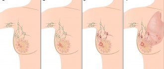
The most common form of breast tumor in women is nodular. When palpated, a dense area of tissue is determined - less often smooth, more often lumpy. The neoplasm is characterized by immobility, as it fuses with the surrounding tissues, in contrast to delimited benign tumors. Such cancer at the initial stage does not manifest itself as painful sensations, but has several signs by which it can be distinguished:
- the skin over the tumor changes color to dark red or brown;
- a depression in the skin or other irregularities appears;
- the presence of a “lemon peel” on the sore breast is characteristic;
- Discharge from the nipples is possible; if it is bloody, this indicates a malignant process.
Symptoms
Medullary cancer is characterized by asymptomatic development. The first signs indicating the course of the tumor process in the mammary glands occur when the neoplasm reaches 2-3 cm in diameter.
On this topic
- Oncomammology
How does breast cancer hurt?
- Olga Vladimirovna Khazova
- November 29, 2020
When palpating the problem area, dense, nodular areas are revealed. As the tumor process progresses, the clinical picture is supplemented by pain in the chest, enlarged local lymph nodes, and breathing problems. The neoplasm has a toxic effect on the body, which is why the patient experiences:
- pale skin;
- sudden weight loss;
- general weakness;
- increased fatigue and other symptoms of intoxication.
The tumor process causes abnormal discharge from the nipples. Due to the spread of cancer cells, the integrity of the skin on the chest is compromised. Over time, the size and number of wounds on the surface of the mammary glands increases.
If the tumor is localized near the areola, nipple retraction syndrome is diagnosed. This symptom becomes more pronounced when the patient places her hands behind her head.
Advanced medullary cancer grows into the tissue of the ligamentous apparatus. Because of this, cords form on the surface of the chest.
If the tumor metastasizes, the clinical picture is complemented by symptoms of dysfunction of the organs into which the cancer cells have penetrated.
Invasive and non-invasive
It is important to determine the histological type of the tumor in order to determine the treatment plan:
- Non-invasive breast cancer - intralobular or intraductal - is typical for the initial stages of the disease. Malignant cells are concentrated in separate areas and do not invade surrounding tissues. It may be benign carcinoma in situ, that is, local cancer. Under negative circumstances, the cells spread and a nodular or diffuse form occurs.
- Invasive cancer is characterized by the rapid spread of malignant cells beyond the duct or lobule.
Invasive and non-invasive breast cancer are distinguished by the degree of differentiation - well-differentiated and poorly differentiated. The most aggressive breast cancer is poorly differentiated, since during histological examination it is impossible to determine from which cells it developed. Therefore, choosing a treatment will be difficult.
Mucinous breast carcinoma (or colloid cancer) is one of the most dangerous types of tumors. Characterized by the presence of a large amount of mucus in the neoplasm.
Diagnostics
For lumps in the mammary glands, ultrasound and mammography are prescribed. Both methods help visualize the tumor and determine the location of the carcinoma. To determine the structural features of the tumor, MRI and CT are prescribed.
A biopsy is considered an important diagnostic measure. The latter is prescribed for suspected malignant tumors.
For medullary cancer, these diagnostic methods are not able to determine the type of tumor. To do this, it is necessary to conduct a histological examination of the problem tissues. The latter are sometimes collected through an open cavity formed in the mammary gland.
Medullary cancer is differentiated from other neoplasms based on five signs. Carcinoma consists of 75% malignant cells united in strands and separated by connective tissue.
On this topic
- Oncomammology
The role of hormone therapy in the treatment of breast cancer
- Olga Vladimirovna Khazova
- May 27, 2020
Histological examination of the problem tissue does not reveal glandular and tubular structures. The capsule of medullary cancer is diffusely infiltrated.
An important feature of carcinoma is the presence of one large and several small nuclei (nuclear polymorphism) in the cells, most of which are filled with cytoplasm. Medullary carcinoma usually does not grow beyond the original site. A pronounced layer of fibrous tissue separates the tumor from healthy cells.
Pathogenetic forms of pathology
The main cause of the disease is excess estrogen, but this only applies to some tumors. Other organs are involved in the pathogenesis of cancer.
Hypothyroid cancer
Hypothyroid breast cancer is one of the most rapidly developing. It is also called “young”, since the age range from 15 to 35 years is when a woman is not in the menopause stage. Estrogen does not play a role. If a woman has a thyroid disorder, is obese, or has ovarian disease, she may begin to develop breast cancer. This form of oncology quickly metastasizes, since the body is not able to resist the disease due to reduced metabolism and immunity. The prognosis after treatment is extremely unfavorable.
Ovarian cancer
Ovarian breast cancer, which occurs when the ovaries do not produce enough hormones, also has an unfavorable prognosis. A risk factor is the presence of cysts. It can also occur after childbirth, at the beginning of sexual activity. Breast cancer in this case develops quickly and metastasizes.
Hypertensive-adrenal
Occurs in women after menopause between the ages of 48 and 65 years. A risk factor is an increased level of cortisol, a hormone that is involved in the regulation of carbohydrate metabolism in the body. Glucose synthesis in liver cells increases, while in muscles it is weakened. Women experience increased body weight and appetite. Cortisol is produced by the adrenal glands, stimulates the process by adrenocorticotropic hormone of the pituitary gland. The pituitary gland, in turn, is affected by biologically active substances produced by the hypothalamus.
Most often, patients diagnosed with the pituitary-hypothalamic type of cancer have a tendency to hypertension. Immediately after menopause, they show signs of rapid aging. The prognosis is unfavorable.
Elevated levels of prolactin can lead to pituitary cancer. This hormone normally does not exceed 15 - 20 nanograms per 1 ml of blood, if the woman is not pregnant or breastfeeding. In pathological situations, the level can increase 10 times. If prolactin circulates in the body for a long time, it can cause breast cancer, obesity, cessation of menstruation, disorders of the thyroid gland, deterioration of vision and nervous system function. To determine the level of prolactin, it is necessary to take a sex hormone test at different phases of the menstrual cycle.
Treatment of poorly differentiated breast cancer
The peculiarity of this type of cancer is that the tumor is not sensitive to chemotherapy. If the disease is detected at a stage when surgery is not possible, the prognosis is poor.
The most effective is surgical removal of the tumor at stage 1 or 2. The earlier cancer is detected, the greater the chance of recovery.
Radiation therapy is prescribed before and after surgery. It allows you to reduce the size of the tumor and avoid relapse. The tumor is usually hormone-dependent, so hormonal therapy will help slow down its growth.
Methods of treating breast cancer
To combat breast cancer, a comprehensive approach is used, which involves the use of surgery, chemotherapy, hormone therapy and radiation therapy.
Recent Entries
Is it possible to give a mirror: how to protect yourself from bad omens It became known about the influence of cell phone towers on human health Is it possible to eat bananas bought in Russia?
The earlier the diagnosis is made, the easier and cheaper the treatment will be and the better the result.
In recent years, targeted therapy may also be included in this complex of treatment measures.
Chemotherapy and hormone therapy
Chemotherapy (treatment with cytostatic drugs) aims to destroy not only cancer cells within the tumor and lymph nodes, but also possible microscopic metastases throughout the body that have not been identified.
Chemotherapy may be:
- neoadjuvant, prescribed before surgery;
- adjuvant, performed after surgery.
Neoadjuvant chemotherapy is designed to first shrink the size of the tumor and/or lymph node to facilitate their subsequent removal. Adjuvant is prescribed for 21 days after surgery for the purpose of final “cleansing” of the body.
The following drugs are commonly used in chemotherapy:
- Cyclophosphamide;
- Doxorubicin;
- Vinorelbine;
- Cisplatin, etc.
If the removed tumor was hormone-dependent, the hormonal drug Tamoxifen must be prescribed to block the growth of tumor cells, and the menstrual cycle is forcibly suspended using an injection of a special drug.
A big disadvantage of chemotherapy is its high toxicity to healthy cells and tissues of the body.
Drugs used during chemotherapy - gallery
Cyclophosphamide
Doxorubicin
Vinorelbine
Cisplatin
Surgical removal of the tumor
Recently, oncologist surgeons in many cases have abandoned the practice of complete removal of the mammary gland (radical mastectomy), replacing it with sectoral resection (lumpectomy), when the tumor is directly excised with adjacent healthy breast tissue to a depth of 1 cm while preserving the organ.
Moreover, there are circumstances when it is impossible to avoid radical mastectomy:
- small breast size of the patient;
- germination of the formation into the pectoral muscle, skin;
- large tumor size;
- tumor involvement of the entire mammary gland.
In addition, the surgeon may decide to perform a “major operation” in cases where he considers it necessary. The patient should not perceive the loss of her breast tragically, since modern plastic surgery can help restore her to her former appearance.
In many cases, a plastic surgeon is immediately involved in performing a radical mastectomy, if there are no contraindications to this.
Women who are not planning breast reconstruction surgery are offered a wide selection of special underwear and modern silicone prostheses, which will fully compensate for the loss cosmetically and give the patient self-confidence, without giving others a reason to suspect anything.
Mastectomy followed by mammoplasty
During a mastectomy, as with a lumpectomy, lymph node dissection is performed - removal of the affected lymph nodes. If metastases have not been previously identified, only the sentinel axillary lymph node is excised for examination.
The sentinel axillary lymph node is so named because it is the first to receive lymph from the affected area.
Removal of axillary lymph nodes in most cases is the cause of a postoperative complication - lymphostasis, i.e. stagnation of lymph in the arm from the side of the operation. This phenomenon is present after both radical mastectomy and lumpectomy, since in both cases there is interference in the lymphatic system.
Lymphostasis of the arm is a common complication after removal of axillary lymph nodes.
To restore lymph circulation in the affected limb, the patient must begin to carefully develop her arm while still in the hospital.
You should know: the later you start the exercises, the more difficult it will be to restore the functionality of your hand.
A few months after lymph node dissection, new lymphatic vessels will begin to form, providing lymph drainage, but the condition of the arm will need to be monitored for the rest of your life.
A set of exercises to restore hand function after mastectomy - video
Radiation therapy
Irradiation of the surgical area is performed to prevent local relapse. In addition, with the help of radiation therapy, symptomatic (palliative) treatment of inoperable tumors and metastases is carried out.
How to recognize and treat allergic conjunctivitis:
Targeted therapy
Modern oncology has made a major breakthrough in the last two decades, introducing into clinical practice drugs called targeted drugs, which are capable of targeting a malignant tumor, blocking the growth of cancer cells and practically without damaging healthy tissues of the body. Unlike chemotherapy, this treatment does not cause death of blood elements, baldness, liver dysfunction, etc.
These medications can be used either independently or in combination with other types of treatment. The use of targeted drugs along with chemotherapy makes it possible to reduce the dosage of toxic drugs.
Targeted drugs also have side effects - fever and chills, but these factors can be eliminated by taking anti-inflammatory drugs.
For the treatment of hormone-dependent tumors, Aromasin, Femara are used, for hormone-independent tumors - Herceptin, Avastin.
Targeted drugs prescribed for breast cancer - gallery
Aromasin
Femara Herceptin
Avastin