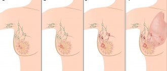
There are many questions regarding what invasive breast cancer is and what causes it. It is known to be the most common form of breast cancer. This is a malignant tumor that grows and develops in the tissues of the mammary gland. Invasiveness means that cancer cells spread through the lymphatic system through the bloodstream to other human organs. Invasive cancer almost always metastasizes.
The majority of reported cases of breast tumors are invasive breast cancer. No one is immune from this disease, but, according to statistics, it is most often diagnosed in middle-aged and older women. The prognosis for life with such a disease depends on many factors, but the main thing is at what stage it was diagnosed. Therefore, timely diagnosis and treatment will help to recover from the disease and avoid adverse consequences.
What is invasive cancer
Invasive cancer is a malignant neoplasm that grows outside the breast. The tumor quickly penetrates the axillary lymph nodes and spreads cells through the circulatory system to the liver, kidneys, and bone tissue.
According to medical statistics, breast carcinoma is diagnosed mainly in women aged 60-65 years. In the last 10 years, the number of patients with invasive cancer has increased by more than 30%.
Carcinoma grows from epithelial cells. This form of cancer occurs in 80% of women with tumors in the mammary glands. Sometimes carcinoma is detected in men.
It is difficult to prevent the development of carcinoma. Therefore, many countries have programs for the early diagnosis of tumor processes in the mammary glands. Carrying out such measures makes it possible to identify neoplasms of 1st and 2nd degrees of malignancy, which can be successfully treated in 95% of cases.
Types of Invasive Tumor
In medical practice, there are 3 key types of invasive forms of the disease.
- Ductal type - with such a tumor, the first pathogenic cell is formed in a single duct. This type of oncology is the most dangerous and occurs in most cases of mammary types of carcinoma. The affected tissue quickly penetrates the blood vessels or lymph nodes. Cells of this form of neoplasm provoke the creation of various atypical discharges from the lesion; with superficial cancer, the skin is deformed. Ductal cancer occurs regardless of the age category of patients.
Development of a cancerous tumor
Invasive ductal oncology is classified into 3 degrees of differentiation:
- High level - the nuclear structure in such tumor cells is identical. The stage is distinguished by a lower percentage of poor quality of the disease.
- Intermediate stage - the cellular structure of the tumor formation and the activity of the lesion is similar in nature to the non-invasive type of growth of small malignancy.
- Low degree - in this case, each pathological cell has a very different individual structure from other cells. These tissues are quickly distributed along the ducts, covering a deep layer of nearby materials and organs.
- Pre-invasive ductal type - there is no metastasis to nearby structures and organs. Active progression of the oncological process is observed in duct cells. However, there is a high probability of degeneration of the pre-invasive form into an invasive typology.
- Invasive lobular type - pathology is formed on the basis of cells of glandular lobules. This form of cancer can develop either as a single tumor neoplasm or as multiple nodules. With the lobular form of the disease, multiple tissue damage is possible. It is difficult to diagnose the lobular type of growth. Oncology is growing without signs indicating the presence of an atypical course in the human body.
An invasive tumor of a nonspecific type is characterized by the absence of a chance to determine this form of pathology. It is difficult to distinguish whether a ductal or lobular type of neoplasm has affected the patient’s body. Invasive unspecified cancerous growth is divided into the following types:
- Medullary - has the least degree of invasiveness of all existing varieties. Pathogenic cells spread slowly to nearby materials compared to other types. However, the tumor body quickly grows in size within its own membrane. Is detected in 10% of diagnosed diseases.
- Infiltrating ductal - infiltrating carcinoma rapidly penetrates into nearby tissues and begins to metastasize. In 70-75% of diagnoses with a malignant course, a breast tumor is diagnosed.
- Inflammatory - the symptoms of this type of oncology manifestation are similar in symptoms to the disease of mastitis. An atypical lump forms in the chest area. The skin over the affected area becomes suspiciously red. The described oncological form accounts for 10% of cases.
- Paget's cancer - as a result of the development of this form of the disease, the breast is affected, namely the nipple-areolar part. In terms of external manifestations, the disease is similar to eczema - an inflammatory process accompanied by the appearance of blisters, a weeping surface of the lesion and impenetrable itching.
The above types of cancer have one thing in common: in most cases (more than 65%), pathogenic cells depend on hormonal levels. The tumor contains estrogen-sensitive receptors. Therefore, treatment for invasive type of oncology includes hormone therapy. If the cancer develops before menopause, then such receptors are absent.
Basic diagnostic methods:
- The ultrasound method currently occupies a leading position. This is due to the fact that mammography is not used at a young age, and when diagnosing a pathological focus with the latter method, it is necessary for accurate visualization and differential diagnosis. This method allows you to evaluate not only the structure of the pathological focus and the gland itself, but also to clarify the density, size and shape. A thorough examination is also necessary to examine the tissue surrounding the formation; its shadow often indicates a malignant process. Using Doppler, blood flow is measured both in the tissues of the gland and the tumor.
- Mammography is a common, inexpensive screening method for regularly checking women's breasts. But its disadvantage is manifested by the fact that there is an age limit. Young girls are not recommended to use this method as a screening method; only after an ultrasound examination can it be used for a more accurate diagnosis. Mammography is prohibited during pregnancy, since it is in this situation that the likelihood of a malignant process increases. Since the method is an X-ray method, unlike ultrasound, one can see deposits of calcifications in the area of the ducts, which characterizes the shape of the infiltrate.
- Inspection is the initial diagnostic method. It is after this that the doctor can suspect carcinoma, as well as carry out a differential diagnosis with benign formations. Not only an external assessment is carried out, but also palpation of the organ. To do this, a strict sequence is observed; it is this that allows you to most accurately determine the ongoing process. Palpation should be carried out exclusively at the beginning of the menstrual cycle, no later than the 13th day from the start of menstruation. During menopause, this can be any day. Touching is carried out only with the pads of the four fingers of the hand strictly clockwise. A mandatory examination is required in two planes, first standing, and then the patient is asked to lie on the couch. The axillary lymph nodes are also examined.
- Puncture. This is a method for additional diagnosis of breast carcinoma. It is necessary when identifying an unquenched formation to study the composition of its contents. For more accurate sampling of material, a biopsy is performed under ultrasound control. The material is taken by puncturing the tissue with a needle and the tissue of interest is collected. After obtaining the cells, they are examined by microscopy.
- Tumor markers. This method is currently additional, but not always informative. To do this, venous blood is taken, in which, in the presence of carcinoma, a protein is produced that is foreign to the body. In response to its production, certain antibodies are formed. In modern laboratories there are two markers that reflect a malignant process in the mammary gland. These are CA 15-3 and 27-29, the first in turn may not always reflect the condition of only the mammary gland, with damage to the liver, ovary, uterus and its augers, as well as pulmonary and pancreatic tissue.
From the anamnestic data, much attention is paid to risk factors that affect the hormonal status of a woman. Clinical examination includes palpation of the lymph nodes and mammary gland in the supine, sitting, frontal and dorsal positions, bimanually. Reliable results of palpation of the breast are obtained in the middle of the monthly cycle. Instrumental research methods include:
- Mammography – allows you to detail the nature of the tumor and perform a targeted biopsy. The method is informative for preclinical cancer and diffuse forms of the disease.
- Ductography is an X-ray method of contrasting the milk ducts to detect intraductal pathology.
- Ultrasound of the mammary glands is informative and harmless, helps in diagnosing formations not detected by x-ray. A tumor biopsy is performed under ultrasound guidance.
- Computed tomography and the use of nuclear magnetic resonance to examine the breast, regional lymph nodes, liver, bones, and lungs.
- Cytological, histological and immunohistochemical studies are required.
To determine the degree of malignancy of the tumor and select treatment tactics, histological classification of breast cancer is used. The degree of differentiation of the primary tumor or G histopathological classification are taken into account. It provides the following gradations:
- Gx – degree of differentiation cannot be assessed.
- G1 – high degree of differentiation.
- G2 – medium degree of differentiation.
- G3 – low degree of differentiation.
- G4 – undifferentiated carcinoma.
To diagnose a breast cancer tumor and determine the exact size of the affected area, a woman is sent for an ultrasound examination of the breast. To confirm or deny developing carcinoma, the doctor prescribes a number of diagnostic measures. Diagnostics includes the procedure:
- Ductography is an X-ray examination of the chest area. The method involves the use of a contrasting element. The product fills the milk duct, which helps to examine in detail the clinical picture and characteristics of the compacted affected area.
- Puncture of the affected area of the breast and further biopsy - a sample of cellular material is sent for histological analysis to determine the form of the tumor.
- Immunohistochemical tests are intended to assess the sensitivity of pathological formations to female sex hormones. The final information obtained from the examination allows us to understand the likelihood of successful removal of a malignant lesion using hormone therapy.
To determine the stage of development of cancer, the patient is sent to undergo a CT scan of organs and structures susceptible to the development of metastases. If there is a suspicion of the existence of an oncological process in these systemic parts, it is recommended to conduct a histological examination. The method involves an early biopsy to collect a sample of tissue material. Oncology uses a system for determining cancer growth.
https://youtu.be/RjldJT4pils
Forms
Depending on the location of the tumor process, invasive cancer is divided into two types:
- Ductal . The most common form of carcinoma. Diagnosed in 80% of women. A tumor of this form initially grows from the tissues of the milk ducts.
- Lobular . Diagnosed in 15% of patients. This form of cancer affects the lobules of the mammary glands. At the initial stage of development, the neoplasm is detected as a lump in the breast.
On this topic
- Oncomammology
Breast puncture
- Natalya Gennadievna Butsyk
- November 29, 2020
There is a pre-invasive form of cancer. This carcinoma is localized in the ducts and does not grow into neighboring tissues. If left untreated, it transforms into invasive cancer.
Each of the above forms of tumor is divided into separate subtypes, characterized by their own characteristics.
Ductal form
The ductal form of invasive cancer consists of various cells that determine the histological composition of the tumor. This type of carcinoma is diagnosed primarily in older women.
The tumor process in the milk ducts develops asymptomatically. Due to its location, a malignant neoplasm cannot be detected by palpation. As carcinoma develops, the latter grows into neighboring tissues, causing the peripapillary zone to become deformed.
Depending on the histological composition, invasive ductal cancer is divided into:
- well differentiated (g1 or low grade);
- intermediate differentiation (g2 or average degree of malignancy);
- low-grade (g3 or high-grade malignancy).
Well-differentiated carcinoma consists of small monomorphic cells. A cancer tumor grows from various structures. The cell nuclei are the same size. Highly differentiated carcinoma is characterized by slow growth and in rare cases causes tissue necrosis.
The second type of tumor is similar in structure to low-grade. Neoplasms of the intermediate form develop from various structures and sometimes provoke necrosis of local tissues.
Poorly differentiated carcinoma is characterized by its relatively large size (more than 5 mm in diameter). The tumor consists of structures typical of intraductal cancer. This neoplasm provokes tissue necrosis. Poorly differentiated carcinomas cover the entire surface of the ducts.
Lobular form
Lobular invasive breast cancer develops mainly in older women. The neoplasm is well palpated. The tumor is most often localized in the upper part of the mammary gland. On palpation, the neoplasm is felt as a painless lump with uneven contours.
In 30-65% of patients, lobular carcinoma affects both breasts symmetrically.
As the tumor progresses, cancer cells invade the skin, causing the breasts to shrink. Histological examination of problem tissues reveals a pronounced fibrous stroma.
A characteristic feature of lobular cancer is the presence of chains consisting of 4-5 cells. With such a tumor, trabeculae form in the form of cords around healthy milk ducts.
Unspecified breast cancer
Unspecified cancer tumors include neoplasms of an unknown type. To determine the type of carcinoma, an immunohistochemical study is performed, which makes it possible to determine the nature of the lesion.
On this topic
- Oncomammology
Ovariectomy for breast cancer
- Natalya Gennadievna Butsyk
- November 29, 2020
Unspecified breast cancer is divided into several types:
- Medullary . Carcinoma practically does not grow into neighboring tissues. Medullary neoplasm reaches large sizes in the absence of treatment. This form is detected in 5-10% of patients with carcinoma.
- Inflammatory . The development of the tumor is accompanied by symptoms characteristic of mastitis. When palpating the mammary glands, compactions are revealed. As inflammatory carcinoma grows, the skin on the chest turns red and the body temperature rises. This tumor is also diagnosed in 5-10% of cases.
- Infiltrating . It is considered the most common type of unspecified carcinoma (found in 70% of patients). The cancerous tumor is characterized by aggressive growth. The tumor gives metastases in the initial stages of development.
- Paget's cancer . The tumor affects the nipples and areolas of the breast. The neoplasm causes symptoms characteristic of an allergic reaction.
The prognosis for such tumors is determined depending on the degree of malignancy. Invasive breast carcinoma of the nonspecific g2 type is less dangerous than well-differentiated cancer.
In the female breast, in addition to the above neoplasms, a hormone-dependent tumor often develops. These neoplasms often appear in patients during menopause.
In 60-70% of cases, hormone-dependent carcinomas involve estrogen receptors. Most cancers of this type respond well to treatment. A negative prognosis is observed in patients before menopause.
Classification
An invasive breast tumor begins its development in the epithelial cells of the mammary gland and then spreads beyond its boundaries. There are several forms of this oncopathology.
Invasive ductal form
One of them is invasive ductal breast cancer. It is characterized by the fact that the process of proliferation of cancer cells leaves the milk ducts and spreads to other areas of the mammary gland.
This type of cancer is otherwise called ductal carcinoma. It is believed that this form of pathology is the most common; it is diagnosed in 80% of breast cancer cases.
Pre-invasive ductal form
Another type is preinvasive ductal carcinoma. It differs in that the tumor, despite the fact that it does not spread to neighboring healthy tissues, can still change into another form and actively grow.
Invasive lobular form
Another type of pathology under consideration is invasive lobular cancer. This is a form of the disease in which cancer cells begin to develop in the lobules of the breast. The tumor does not form in the form of a lump, but as a kind of compaction. This form of cancer can also spread to healthy tissue and other organs.
According to the degree of differentiation, ductal carcinoma is divided into highly differentiated, intermediate and poorly differentiated. In the first case, cancer cells are small in size and have nuclei of equal size. This type of neoplasm mainly consists of cells that exist in an unchanged form, i.e., monomorphic. They develop in the ducts and take on the appearance of microcapillary or cribriform structures. Tissue necrosis occurs in ducts affected by cancer cells.
Poorly differentiated ductal carcinoma is distinguished by its ability to affect the entire duct in which it formed. Such a tumor can reach a diameter of more than 0.5 cm. Poorly differentiated cancer can quickly spread in the ducts and penetrate into neighboring structures. The intermediate type of ductal tumor is a condition between the two described above.
In addition, the classification of this pathology separately distinguishes invasive unspecified breast cancer. Its main difference is that based on the results of the biopsy it is impossible to accurately determine whether it is ductal or lobular.
In turn, it is divided into the following types:
- Paget's cancer;
- medullary cancer;
- inflammatory ductal carcinoma;
- infiltrating tumor.
Causes
Carcinomas in the mammary glands develop under the influence of many factors. Among the probable causes of the appearance of neoplasms, researchers identify genetic predisposition.
On this topic
- Oncomammology
How does breast cancer hurt?
- Olga Vladimirovna Khazova
- November 29, 2020
The risk of developing a tumor process in breast tissue increases in cases where cancer has previously been detected among a woman’s relatives.
Possible causes of carcinoma include hormonal imbalance caused by taking oral contraceptives and the onset of menopause. The high-risk group includes nulliparous women and patients with chronic pathologies of the reproductive organs. Also, the tumor process develops as a complication of frequent injuries to the mammary glands.
Mastopathy
Mastopathy is one of the most common causes of carcinoma. This pathological condition develops against the background of hormonal imbalance.
Mastopathy is characterized by constant pain in the mammary glands and abnormal discharge from the nipples. The development of the pathological condition causes the appearance of nodular formations in the chest.
Mastopathy is diagnosed mainly in women aged 30-40 years.
The growth of nodular formations provokes deformation of the mammary glands. Without treatment, the cells that make up the lumps transform into cancerous ones.
Fibroadenomas
Fibroadenomas develop mainly in young women. Neoplasms of this type are distinguished by a nodular structure and dense consistency. Fibroadenomas are benign. But against the background of hormonal imbalance, tumors can degenerate into carcinoma.
Dysfunctions of the reproductive organs
Frequent abortions can provoke the appearance of invasive breast cancer. Termination of the first pregnancy is especially dangerous. Abortion provokes hormonal imbalance, which causes inflammation of the reproductive system.
Due to an incomplete pregnancy, the growth of the mammary glands stops and the breasts begin to shrink. This process contributes to the formation of compactions, which over time transform into a cancerous tumor.
Refusal of breastfeeding, or early interruption of lactation, also provokes the appearance of lumps in the mammary glands.
The health of the female body is largely determined by the condition of the organs that produce hormones. This process is disrupted due to insufficient sexual activity, which over time causes the formation of a tumor in the mammary glands.
Mechanisms of tumor development
There are 4 mechanisms that cause the development of carcinoma:
- hypothyroid;
- ovarian;
- adrenal;
- senile.
The development of the hypothyroid mechanism is caused by excessive body weight, early menstruation (up to 11 years), the formation of follicular cysts in the ovaries or hyperplasia of the latter. These factors provoke hypothyroidism, which is common in girls under 30 years of age.
The ovarian mechanism is determined by the physiological processes occurring in a woman’s body after childbirth and during lactation. This factor causes invasive breast cancer in 50% of patients.
The adrenal mechanism triggers the tumor process after menopause. During this period, the concentration of low-density lipoproteins increases.
On this topic
- Oncomammology
The role of hormone therapy in the treatment of breast cancer
- Olga Vladimirovna Khazova
- May 27, 2020
The senile mechanism is caused by a weakening of the pituitary gland, which disrupts the hormonal balance. Tumors arising under the influence of this factor are distinguished by strict localization.
The least favorable prognosis is for carcinomas caused by ovarian and hypothyroid mechanisms.
Symptoms of breast cancer
The clinical symptoms and presentation of nonspecific breast cancer are varied. Symptoms of cancer are expressed depending on the age category of the woman, the size of the tumor and the intensity of the malignant course. The initial stages pass for a long time without signs of specificity.
In rare cases, pain or discomfort may occur during palpation of the mammary glands. The occurrence of pain symptoms or discomfort is not a 100% signal, which means the presence of oncological pathology. To accurately diagnose a specific invasive breast cancer, the doctor relies on the signs that accompany the atypical process in the gland:
- Orange peel factor.
- Wrinkled appearance in the affected area.
- A flat area is marked.
- The nipple becomes retracted or bulging and deforms from a normal, healthy state to an atypical one.
- The color of the skin changes, redness, itching, burning appears, and numbness of the epidermis occurs. The woman notes a change in the volume and shape of the mammary gland and fat layer.
- Local body temperature increases.
- The fold of the areola increases.
- Regional lymph nodes grow in the axillary, supraclavicular or anterior cervical part.
- A pea-shaped lump appears in the breast, or the neoplasm has ill-defined boundaries.
- Clear fluids or blood are discharged from the nipple.
- The structure of the epidermis changes, inflammation processes occur, the skin becomes dry, and ulcerative formations may develop.
- Pain in the area of the shoulder blade.
When pathological cells spread or metastasize to neighboring organs, symptoms appear that develop depending on the location of the secondary tumor focus. The division of cancer cells causes deterioration or lack of appetite, weight loss, exhaustion of the female body and poisoning.
Symptoms
The nature of the clinical picture of breast carcinoma is determined by several factors. In some patients, the course of the tumor process is not accompanied by severe symptoms.
In some cases, pain may appear on palpation of the affected gland (usually they first appear with cancer of the 3rd degree of malignancy).
The presence of neoplasms is indicated by lumps in the breast that do not disappear over a long period of time. The development of the tumor process over time causes deformation of the mammary gland (change in size, contours). In inflammatory carcinomas, the skin takes on a red tint. Young girls develop wrinkles on their chests due to tumors.
Some carcinomas cause abnormal nipple discharge that is not associated with lactation. In some cases, whitening of the skin in the problem area is recorded.
Reasons for the development of cancer pathology
What is the trigger for the development of invasive carcinoma is not known exactly. The pathogenesis of this type of cancer is associated with mutation of proto-oncogenes, which are converted into oncogenes and stimulate the division and growth of breast cells.
Groups of favorable factors for the development of invasive carcinoma have been identified:
- heredity,
- artificial abortions,
- the presence of benign tumors (fibroadenoma, cyst),
- mastopathy,
- lack of breastfeeding after childbirth,
- the onset of the first pregnancy after 35 years or no pregnancies,
- premature puberty,
- metabolic disorders, obesity,
- uncontrolled use of hormonal drugs,
- gland injuries.
Find out the instructions for using the drug Gordox for the treatment of pancreatitis of the pancreas.
Read about the symptoms and treatment methods for progesterone deficiency during pregnancy at this address.
Stages of tumor development
Cancer tumors go through 4 stages during their development. At the initial stage, the size of carcinomas does not exceed 2 cm. The tumor does not metastasize during this period.
At the second stage, the size of the cancer varies between 2-5 cm. Malignant cells at this stage often affect the lymph nodes located nearby.
The third stage is characterized by the appearance of pronounced symptoms. The size of the carcinoma exceeds 5 cm. The tumor at the third stage metastasizes to nearby tissues and organs.
The last stage is characterized by the spread of cancer cells throughout the body.
Occurrence of invasive ductal carcinoma
The mammary gland itself consists of lobules and milk ducts, which are surrounded by glandular, connective and adipose tissue.
Invasive ductal carcinoma occurs when normal cells in the ducts of the mammary gland, as a result of a gene failure, turn into malignant ones and begin to incorrectly divide and spread throughout the organ. Invasive ductal cancer means that the tumor has already spread to nearby breast tissue and has a risk of spreading to the lymphatic system and other parts of the human body.
Invasive ductal carcinoma is assessed by a pathologist using a biopsy in the laboratory, according to a special scale from 1 to 3, depending on the aggressiveness and growth of cancer cells:
- 1 – indolent form of cancer, which is characterized by slow growth;
- 2 – moderate growth of cancer cells;
- 3 – fast-growing form of cancer.
Diagnostics
In case of damage to the mammary glands, mammography is considered the most informative diagnostic method. The latter makes it possible to detect breast tumors of various sizes, including small nodules.
To determine the precise localization of carcinoma, ultrasound of the mammary glands is used. MRI helps determine the characteristics of the tumor.
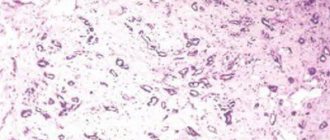
The type of tumor can be determined by a biopsy, which is performed using a thin needle. The procedure involves taking tissue from the problem area, which is sent for histological examination.
If the size of the tumor does not exceed 5 mm, ductography is prescribed, which makes it possible to determine the location of the tumor.
Women at risk should regularly palpate the mammary glands to detect lumps in the breasts.
Stages
The stages of tumor development differ depending on the size and shape, as well as the condition of nearby lymph nodes and the presence or absence of metastases:
- At the first stage, the size of the tumor does not exceed two centimeters, and there are no metastases;
- The second stage tumor does not exceed five centimeters in diameter, there are no metastases, but the lymph nodes above and below the collarbones, as well as in the armpits, are affected;
- In the third stage, the size of the tumor is more than five centimeters, there is no metastasis, but the tumor grows into the surrounding tissues and affects distant lymph nodes;
- In the fourth stage, many lymph nodes are affected, the tumor grows, and cancer metastases affect distant organs.
We recommend reading Multiple myeloma - what it is, causes, treatment, life expectancy
There are also four degrees of malignancy of the oncological process:
- G1 – well-differentiated carcinoma. In this case, breast cancer does not germinate, but develops very quickly.
- G2 is the second degree of malignancy of invasive cancer with little germination, but rapid tumor growth.
- G3 – new cells appear that differ from each other and differ from healthy tissues.
- G4 – the highest degree of neoplasm infiltration.
Knowledge of the stage of development and degree of malignancy of the oncological process helps to select optimal treatment methods.
Treatment
Treatment tactics for breast carcinomas are determined depending on the individual characteristics of the patient, location and type of tumor. Doctors often combine different types of therapy.
Hormone therapy
This type of therapy is used for hormone-dependent tumors or precedes surgery. This type of treatment is mainly prescribed to postmenopausal women.
On this topic
- Oncomammology
The effectiveness of targeted therapy in the treatment of breast cancer
- Olga Vladimirovna Khazova
- May 27, 2020
In this case, drugs that block the effect of estrogen on the tumor are more often used:
- " Tamoxifen ". The medicine prevents estrogen from binding to carcinoma receptors.
- inhibitors (Anastrozole, Exemestane). Prescribed during the postmenopausal period.
- Agnes gonadoropin-releasing factor. The substance suppresses ovarian function. Due to the effect of the drug, menstruation stops.
If the outcome is successful, treatment with hormonal drugs continues to prevent relapse of carcinoma.
Surgical intervention
Open surgery is considered the mainstay of treatment for breast carcinomas. During surgery, the tumor is completely removed. If necessary, tissue adjacent to the tumor is excised. In advanced cases, the affected mammary glands are removed.
During surgery, the doctor excises tissue from the axillary lymph nodes to check the extent of metastases.
After radical intervention, breast surgery is performed. This procedure allows you to restore the shape of the mammary glands.
Chemotherapy
Chemotherapy is used as a separate method of treating carcinomas, and in combination with surgery. This method can reduce the size of the tumor or prevent the recurrence of invasive cancer.
On this topic
- Oncomammology
5 Effective Breast Cancer Screening Methods
- Olga Vladimirovna Khazova
- May 27, 2020
Separate chemotherapy is used if the size of the carcinoma does not exceed 2 cm or contraindications to open surgery are identified. This approach uses antibiotics, antimetabolites, taxants (which prevent cancer cells from dividing), and alkylating agents.
Modern treatment methods make it possible to deliver drugs directly to the tumor (targeted therapy). This approach increases the effectiveness of the drugs and reduces the negative effect of chemotherapy.
Radiation therapy
Radiation therapy usually complements surgery. This approach is used to destroy any remaining cancer cells. Radiation therapy also helps prevent relapses of carcinomas.
The effectiveness of the method for tumors over 5 cm reaches 70%.
In cases where cancer is diagnosed at the last stage, palliative treatment is prescribed. This therapy is aimed at alleviating the patient's condition. As part of palliative treatment, medications are prescribed to relieve pain and other symptoms characteristic of carcinoma.
Treatment tactics
Invasive ductal carcinoma and other types of breast cancer are best treated with surgery. The extent of surgical intervention depends on the size of the tumor and the degree of damage to surrounding tissues. The operation can take place according to one of the following schemes:
- Removal of the tumor and adjacent tissues;
- Excision of the tumor and nearby lymph nodes;
- Partial removal of the mammary gland and lymph nodes;
- Complete amputation of the affected breast.
If the breast has been completely removed, then after the patient is completely cured, she can resort to plastic surgery to place a silicone implant. For invasive carcinoma that has reached the third or fourth stage, radiation therapy or a course of chemotherapy is given before surgery. This stops the development of the tumor and kills some of the malignant cells. After surgery, chemical and radiation therapy is also performed, which reduces the risk of relapse.
Forecast
The prognosis for carcinomas depends on the stage of development of the tumor process, the type and size of the tumor. Also, the effectiveness of treatment is determined by the age of the patient. The most favorable prognosis is for women under 50 years of age.
If the tumor does not metastasize to the lymph nodes, the 5-year survival rate among patients is 90%. For tumors at the second stage of development, treatment is successful in 70% of cases. Nonspecific type g3 cancer causes death in 53% of patients. A positive outcome of treatment for carcinomas at the last stage is observed in 16% of cases.
Ductal cancer is less treatable than lobular cancer. The prognosis for hormone-dependent tumors that respond to treatment is favorable.
The location of the carcinoma also determines the likelihood of death. If the tumor occurs on the outside of the breast, the risk is low due to the fact that such cancer is less likely to metastasize.
The prognosis for invasive cancer in men is usually favorable. This is explained by the fact that the majority of carcinomas in this group of patients are of the hormone-dependent type. Treatment for invasive cancer in men often involves removing the testicles followed by corticosteroids.
Treatment methods and survival prognosis
Several techniques are used to treat invasive cancer. They include systemic and local measures. The first include chemotherapy and hormonal medications. The second includes radiation treatment and surgery. Depending on the stage of the disease, the age of the patient, the shape and location of the tumor, one or another treatment regimen is selected. Most often, therapy is complex.
In the practice of treating such a disease, the following scheme has been developed:
- First of all, hormonal therapy is carried out. It is aimed at achieving a reduction in the size of the tumor. In addition, if tumor adhesions have formed, these drugs should promote their resorption.
- Surgical removal of a malignant neoplasm. Depending on the characteristics of the disease, the tumor and/or part of the mammary gland or the entire organ must be removed. Surgical treatment prevents the formation of metastases.
- After surgery, treatment is carried out with chemotherapy and radiation. It is aimed at preventing recurrence of the tumor and eliminating the possibility of developing metastases. Chemotherapy is carried out if the tumor is larger than 2 cm and it is not hormonally dependent.
When diagnosed with ductal carcinoma, the patient's life prognosis depends on various factors. If the disease was detected in the initial stages and provided that adequate treatment was started on time, the patient has about a 90% chance of full recovery.
Detection and initiation of treatment at stage 2 guarantees a 70% cure. If the tumor was detected at stage 3, then the chance of survival is no more than 40%. Late diagnosis of advanced disease has a rather unfavorable prognosis - only 10% survival rate.
The size of the tumor also matters. So, if it is no more than 2 cm in diameter, then the 5-year survival rate is guaranteed by more than 90%. Larger tumor sizes reduce this chance by 50%.
In addition, the prognosis is more favorable if the tumor is located closer to the outside of the breast. It is worse if the tumor is located in the center of the organ or in its internal structures. If carcinoma in the mammary gland has metastasized, the prognosis may not be in favor of the patient.
Causes and symptoms of breast adenocarcinoma
Breast adenocarcinoma is one of the most common malignant diseases found in women. It is generally accepted that the main reason for the steady increase in statistics is a decrease in the birth rate and a reduction in the duration of feeding. After all, one of the provoking factors of the disease is hormonal imbalance and infertility.
Breast adenocarcinoma is a malignant neoplasm that develops from glandular epithelial cells of the mammary gland.
The term “breast cancer” refers to two types of disease:
- malignant degeneration of squamous epithelium - squamous cell carcinoma;
- malignant degeneration of the glandular epithelium - adenocarcinoma.
The majority of cases of breast cancer are adenocarcinoma.
Causes
- hereditary predisposition (the existence of a gene has been proven that increases the likelihood of developing carcinoma);
- frequent/severe breast injuries;
- infertility;
- benign breast tumor;
- oncological pathologies of other organs;
- fibrocystic mastopathy;
- areas of atypical hyperplasia in the mammary gland;
- hormonal imbalance caused by various reasons - late childbirth (after 30 years), early puberty, late menopause (after 50 years), taking hormonal drugs in large doses to treat other pathologies.
Provoking factors include:
- bad habits - smoking and drinking alcohol.
- incomplete and incorrect nutritional diet, including foods high in preservatives, animal fats, dyes and other substances toxic to the body.
Symptoms
At the initial stages of the disease, a highly differentiated form of adenocarcinoma develops, in which only a weak cell mutation is observed - malignant formations are practically indistinguishable from healthy structures. At this stage there are no pronounced clinical manifestations of the disease.
Over time, the pathology begins to manifest itself with more pronounced symptoms, such as:
- sunken nipple;
- change in skin color in certain areas;
- changes in the shape and size of the mammary gland;
- swelling of the gland;
- discharge from the nipple (the contents can be mucous, purulent, bloody);
- enlargement of supraclavicular and subclavian, axillary lymph nodes;
- pain in the area of the tumor (occurs in the last stages of the disease).
Diagnostics
Women should be examined regularly by an oncologist. This will allow the disease to be detected in the early stages, which are often asymptomatic. Diagnosis begins with a general visual examination and palpation of the mammary gland.
If suspicion arises, the doctor prescribes additional diagnostic procedures:
- ultrasonography . A reliable and accessible method for detecting a tumor;
- mammography. As a result of the study, a high-definition image of all gland structures is obtained. Mammography can be performed using magnetic resonance imaging, x-ray and other methods, each of which has its own positive and negative aspects;
- biopsy It is the collection of biological material from a suspicious area of tissue. The material is sent for histological and cytological examination;
- Magnetic resonance and computed tomography can detect malignant neoplasms in other organs if metastasis is suspected.
Kinds
Adenocarcinoma is classified according to various parameters - degree of maturity, clinical signs, tumor location. Based on the degree of differentiation of malignant cells, three types of carcinoma are distinguished.
Well-differentiated adenocarcinoma of the breast
Malignant cells are practically no different from healthy structures; the structure of the tumor is similar to the structure of breast tissue. It responds well to therapy in the initial stages of the disease.
Moderately differentiated
This form of adenocarcinoma is similar in clinical features to the well-differentiated form.
The main difference is the clear difference between malignant cellular structures and healthy cells.
The course of the disease is usually of moderate severity. The risk of developing complications and associated pathologies is quite high. The tumor spreads to other organs through metastases.
Undifferentiated adenocarcinoma
Malignant neoplasm differs from healthy tissue at the cellular and tissue level. The degree of development of malignant cellular structures is quite primitive. It is difficult to attribute them to any type of tissue; therefore, it is impossible to establish the structure and origin of the neoplasm.
This type of malignant tumor grows very quickly, spreads to other organs in the early stages and is difficult to treat. Treatment prognosis is very poor and survival rate is low.
According to the localization of the pathological process, they are distinguished:
- ductal adenocarcinoma (tumor in the gland duct);
- lobular or lobular adenocarcinoma (tumor of lobular tissue).
Classification of adenocarcinoma according to clinical signs:
- inflammatory (mastitis-like). The tumor grows into the lymphatic vessels of the skin. Characteristic signs: redness of the skin, thickening of its structure, inflammatory lesions like erysipelas, high body temperature.
- medullary A large invasive malignant tumor, usually with a low degree of malignancy (poor ability to grow metastases);
- ductal infiltrative. Accompanied by the formation of nests and strands of malignant cells, surrounded by a dense collagen structure - stroma;
- papillary It is a non-invasive intraductal malignancy of low grade. It is extremely rare;
- Paget's cancer. Malignant lesion of the areola and nipple.
Treatment
Treatment tactics are determined by the form and course of the disease. There are several basic treatment methods, which doctors often combine with each other.
Surgery
A radical treatment method that can be carried out using one of two methods:
- mastectomy – removal of the entire mammary gland, surrounding lymph nodes and tissue. At the same time, aesthetic breast reconstruction surgery can be performed immediately;
- lumpectomy – removal of the tumor to the borders of healthy tissue, allowing to preserve the mammary gland.
Absolute contraindications to surgery are:
- inflammatory carcinoma of lymphatic vessels and lymph nodes;
- extensive swelling of the mammary gland;
- swelling of the hands;
- metastases in supraclavicular lymph nodes;
- distant metastases.
Hormonal treatment
Hormone therapy is used when malignant cells have receptors sensitive to sex hormones. Antagonists of these hormones have a negative effect on malignant structures, which leads to an improvement in the patient’s condition.
For premenopausal patients, the drugs of choice are:
- "Aminoglutethimide";
- "Tamoxifen";
- "Hydrocortisone";
- "Leuprolide acetate."
Postmenopausal patients are prescribed:
- "Megestrol acetate";
- "Aminoglutethimide";
- estrogens in high doses (“Diethylstilbystrol”);
- "Tamoxifen";
- "Leuprolide acetate."
Chemotherapy treatment
Cytostatic drugs act on malignant cells, as a result of which they decrease in size and die. Chemotherapy reduces the likelihood of metastases and relapse of the disease, and improves patient survival. Combination chemotherapy is more often used, including taking drugs for six months.
Most often, a combination of the following medications is prescribed:
- "Cyclophosphamide" (cyclophosphamide);
- "Fluorouracil";
- "Methotrexate."
If there is a high probability of relapse, the preferred course includes:
- "Cyclophosphamide";
- "Fluorouracil";
- "Doxorubicin hydrochloride."
For the treatment of a metastatic tumor, the following is prescribed:
In the absence of pronounced toxic effects, the drugs of choice should be prescribed in maximum doses.
Radiation treatment
Modern methods make it possible to precisely irradiate pathological tissue without affecting healthy cells. As a result, the tumor decreases in size, making surgical removal easier. Radiation irradiation is often combined with surgery to reduce the likelihood of relapse.
Ovarian adenocarcinoma can develop due to hormonal imbalance in the body and many other reasons. More details in the link provided.
You can find out what the prognosis is for highly differentiated adenocarcinoma of the uterus in this section.
The causes, symptoms, methods of diagnosis and treatment of low-grade gastric adenocarcinoma are described in this article.
Prognosis for breast adenocarcinoma
Five-year survival rate is the average that doctors usually report when breast cancer is detected. However, the prognosis of treatment depends on many factors. One of the main ones is the ability of a tumor to grow in size and metastasize (invasiveness). The presence/absence of concomitant pathologies has a significant impact on the effectiveness of treatment.
With small tumor sizes (up to 2 cm) and timely diagnosis, the prognosis is often favorable. Good treatment results are observed if the tumor does not grow into surrounding tissues, if there are no metastases, if the tumor is well differentiated.
5-year survival rate depending on the ability of cells to metastasize:
- for non-invasive tumors – 95%;
- with a poorly metastatic tumor, the 5-year survival rate is 80%;
- for moderately metastatic tumors and carcinoma with metastases to lymph nodes, the 5-year survival rate is only 60%.
5-year survival rate depending on the stage of the disease:
- Stage 1 (tumor less than 2 cm, no metastases) – 70–95%;
- stage 2 (tumor 2–5 cm, no metastases, or tumor less than 2 cm, and there are metastases in 4–5 lymph nodes) – 50–80%;
- Stage 3 (tumor larger than 5 cm, metastases in lymph nodes) – 10–50%;
- Stage 4 (tumor of arbitrary size, metastases in distant organs - bones, liver, lungs, etc.) - 0–10%.
10-year survival rate depending on the stage of the disease:
- Stage 1 – from 60% to 80%;
- Stage 2 – from 40% to 60%;
- Stage 3 – from 0 to 30%;
- Stage 4 from 0 to 5%.
therapycancer.ru
Types of carcinoma
This pathology represents a whole group of malignant neoplasms:
- lobular carcinoma (affects the lobules that produce milk)
- invasive ductal carcinoma of the breast (milk ducts are affected)
- nonspecific tumors of various types (this happens when a doctor in the laboratory cannot determine whether it is ductal or lobular carcinoma)
Each type is a dangerous disease that can lead to death. It is recommended to carry out preventive measures and regular examinations with a doctor in order to promptly detect the disease and begin treatment. According to statistics, this disease most often develops in women over 50 years of age. The older you are, the higher your risk of developing the disease.
Diagnostic tests
A gynecologist may suspect the disease during a routine examination of a woman. When palpating the chest, multiple or single compactions are noted. To identify the true nature of the tumor, location, size and degree of malignancy, doctors prescribe a set of laboratory and instrumental studies:
- The general condition of the patient’s body is determined using a clinical analysis of blood and urine. Changes in the formula indicate the presence of an inflammatory process.
- Ultrasound examination allows you to evaluate the structure of the mammary glands and the condition of the regional lymph nodes. The method is simple and quick to perform and does not require special preparation.
- Mammography uses X-rays to look at the breasts. All kinds of neoplasms and changes in the structure of the mammary glands are detected.
- Computed tomography and magnetic resonance imaging identify cancerous lesions throughout the body. Allows you to determine the method of blood supply to the tumor.
- The tumor marker test is informative for breast adenocarcinoma.
- A cytogram allows you to study the nature and structure of tumor cells. Determines the final diagnosis of the disease. Biopsy samples are taken in several ways:
- Scraping of affected tissue;
- Tumor puncture;
- Collecting fluid from the nipple;
- Tissue collection from ulcers on the surface of the skin.


