Health
- Anatomy of the uterus
- What are the stages of uterine cancer?
- What are the signs of uterine cancer?
- What is squamous cell carcinoma of the cervix?
- What is the prevention of cervical cancer?
- What test is there for cervical cancer?
- How is surgery performed for cervical cancer?
- When is radiation therapy indicated for uterine cancer?
Cervical cancer
– a malignant tumor of the female genital organs. This common disease ranks 4th among cancer problems in women. In most cases, the disease is asymptomatic and is accidentally discovered during an examination by a gynecologist. A woman may notice a slight discharge of blood after lifting weights, douching, or sexual intercourse.
11 out of 100 thousand women are diagnosed with this disease. This amounts to about 600 thousand cases per year. For some reason, the disease occurs 2 times more often in Latin women. There are two periods in a woman’s life when the risk of developing the disease is especially high: 35-40 and 60-65 years. And girls under 25 years of age practically do not get cervical cancer.
Since the 70s of the last century, mortality from cervical cancer has decreased by 70%. This is due to the fact that doctors massively examine patients for the presence of altered cells. Thanks to this approach, in most cases it is possible to recognize the disease in the early stages, when it can be completely cured.
Cervical cancer develops from the mucous membrane that lines the cervical canal. A tumor cannot grow from healthy cells. Therefore, the disease is always preceded by precancerous conditions. For example, this is cervical dysplasia. By starting treatment on time, you can protect yourself from cancer. The tumor can develop from condylomas and scars that formed after childbirth. It takes 2 to 15 years for these precancerous conditions to develop into tumors.
Cervical cancer is one of the few cancers that can be prevented. Regular visits to the gynecologist and a special vaccination will help protect you. PAP smear and other modern methods make it possible to detect the disease in the early stages.
Cervical cancer is caused by infection with the human papilloma virus. Increase the risk of a malignant tumor: early pregnancy, a large number of sexual partners, sexually transmitted diseases, inflammatory processes of the genital organs, smoking and long-term use of hormonal contraceptives.
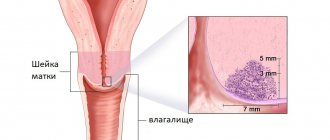
Anatomy of the uterus
The uterus is a hollow organ consisting of smooth muscle. It is located in the lower abdomen between the bladder and rectum. The uterus is suspended by ligaments that are attached to the walls of the abdomen and hold it in place.
The main function of the uterus is to ensure the attachment of a fertilized egg and create the best conditions for the growth of the fetus. During childbirth, the uterus contracts, pushing the baby out. The uterus is an organ that allows a woman to perform her main function - to give birth to a child.
The size of the uterus is small. Approximately 8cm high, 4cm wide and 2cm thick. It has the appearance of a flattened pear. Its wide part faces upward - this is the fundus of the uterus. In this area, it includes the fallopian tubes leading to the ovaries. The middle part is the body of the uterus. At the bottom it narrows, passing into the cervix, which descends into the vagina.
The uterus consists of three layers
:
1. Perimetry
- outer serous membrane. This is a layer of peritoneum that covers the organs in the abdominal cavity.
2. Myometrium
- middle muscle layer. It consists of three layers of smooth muscles, which are arranged longitudinally or in rings surrounding the uterus. Has a small amount of connective and elastic fibers.
3. Endometrium
- inner mucous layer. It, in turn, consists of two layers: basal and functional. The basal one is adjacent to the myometrium. It is responsible for the restoration of the mucous membrane after menstruation. Inside the cavity there is a functional layer. It consists of epithelial cells and glands.
Let's take a closer look at the structure of the cervix
, which interests us most today.
The cervix is mainly composed of smooth muscle, collagen and elastic tissue. Due to this structure, it is denser than the uterus. Its main task is to block the access of microbes to the uterus and not to release the child from it prematurely.
The length of the cervix is 3-4 cm. Inside it there is a cervical canal or canal of the cervix. It connects the interior of the uterus to the vagina. There are folds on the inner surface of the channel. They are needed to prevent vaginal contents from entering the uterus. The canal is usually clogged with thick mucus, which prevents germs and sperm from entering the uterus. But in the middle of the menstrual cycle, the mucus becomes more liquid. This is necessary so that sperm can enter the uterus and fertilize the egg.
The inside of the cervical canal is covered with columnar epithelium
, in which there are many
tubular glands
.
Inside the cervical canal there is a border where the squamous epithelium becomes cylindrical. The part of the cervix that enters the vagina (lips) is covered with squamous non-keratinizing epithelium
.
What are the stages of uterine cancer?
In order to choose the right treatment, it is necessary to know the characteristics of the tumor and the degree of its development. For this purpose, neoplasms are classified.
Cervical cancer develops from glandular and epithelial cells. Based on this, tumors are divided according to morphological characteristics
:
· Adenocarcinoma
- arises from gland cells that are located in the cervix.
· Squamous cell carcinoma
- arises from mutated squamous epithelial cells. This form is the most common.
Depending on the size of the tumor, its spread, the presence of metastases and the condition of the lymph nodes, the doctor determines the stage of cervical cancer
. Each stage has several substages, which are designated by Roman numerals and letters.
Stage 0
Cancer cells were detected only on the surface of the cervical canal. They do not penetrate into deep layers. This condition is also called cervical intraepithelial neoplasia.
A smear for oncocytology (PAP test) and a biopsy help to make a diagnosis and begin treatment. To perform a biopsy, a small piece of mucous membrane is taken from the changed area and examined in the laboratory.
Stage I
Cancer cells grow deep into the tissues of the cervix, but the tumor does not extend beyond the organ.
· I.A.
– small tumor from 0.5 to 7 mm. Does not apply to lymph nodes and other organs.
· I.B.
– the tumor can be seen with the naked eye. It has a diameter of 7 mm to 4 cm and penetrates deep into the cervix by more than 5 mm. Does not apply to lymph nodes and neighboring organs.
If a smear for oncocytology shows that a woman has altered (atypical) glandular epithelial cells on the mucous membrane of the cervix, then a diagnostic examination is prescribed. For diagnosis use: colcoscopy with biopsy. A colcoscope is a device that allows you to view the vagina and cervix with multiple magnification. At the same time, the doctor takes a sample of cells (a tiny section of mucous membrane) for analysis under a microscope. This is a very accurate method that accurately determines the presence of cancer.
Stage II
The tumor extends beyond the boundaries of the cervix and body of the uterus, but is not yet detected in the lower parts of the vagina and on the walls of the pelvis.
· IIA
– the tumor is about 4 cm in diameter, but does not extend into the periuterine space. It can affect the upper parts of the vagina. There are no cancer cells in nearby lymph nodes or distant organs.
· IIВ –
the tumor affects the tissues of the periuterine space (parametrium). Lymph nodes and neighboring organs are not infected.
For diagnosis, a colcoscope is used and a sample of mucosal cells is taken for a biopsy. Using biopsy forceps, the doctor removes 0.5 cm of tissue from the vaginal part of the cervix. To examine the mucous membrane deep in the cervical canal, diagnostic curettage is performed using a curette. The doctor also palpates all nearby lymph nodes and determines whether they are enlarged. This may indicate the presence of tumor metastases.
Stage III
The tumor affects the lower part of the vagina and is found on the walls of the pelvis. It can reach any size. The tumor compresses the ureters and interferes with the removal of urine from the kidneys to the bladder. Distant lymph nodes and organs are not affected by cancer cells.
· IIIA
– the tumor has affected the lower third of the vagina, but the walls of the small pelvis are clean.
· IIIB
– the tumor is found on the walls of the pelvis and impairs the patency of the ureters. Lymph nodes and distant organs are not affected by cancer. This substage also includes cases where there are metastases in the nearest lymph nodes.
Diagnosis at this stage consists of colcoscopy and wedge biopsy. This procedure takes a sample of cells from the deep layers of the cervix. Additionally, an examination using an endoscope of the bladder and rectum is prescribed. It helps determine whether there are metastases there. X-rays and computed tomography (CT) are used to examine distant organs.
Stage IV
The tumor spreads far beyond the cervix. Metastases are found in any organs and lymph nodes.
· IVA
– The tumor has spread to the rectum and bladder, which surround the uterus. It does not affect the nearest lymph nodes or organs located far from the cervix.
· IVB
– the tumor is found in distant organs: liver, lungs.
In order to diagnose what type of tumor the tumor is, a biopsy is performed. Magnetic resonance imaging (MRI) is used to detect metastases in distant organs.
Symptoms of pathology at a late stage
Advanced cervical cancer, the symptoms and signs of which a woman has ignored for a long time, is deadly. Experts divide all signs of the disease into general and specific. The first are:
- fast fatiguability;
- chronic fatigue;
- constant dizziness;
- noticeable pallor;
- lack of appetite;
- increased sweating,
- chills.
Specific symptoms of cervical cancer appear in the later stages of the disease. They indicate that the body is already extremely exhausted, the work of all its systems is disrupted, and the body cannot cope with aggressive malignant cells.
Must Read: Cervical Cancer
Signs of cervical cancer in women at a late stage of the disease are as follows:
- Severe pain that spreads to the entire pelvic and abdominal area. Such pain appears as soon as the pathology reaches the parametrial tissue. When malignant cells affect the nerve endings in the uterus, pain is localized in the lower back, hips and intestines.
- Lymphostasis is stagnation of lymphatic fluid in the lower extremities, resulting from compression of the lymphatic vessels. Lyphostasis leads to edema. In the later stages of cervical cancer, not only the legs, but also the woman’s genitals swell, as the lymph nodes become clogged and the functioning of the blood vessels is completely disrupted.
- Urinary retention - this symptom occurs due to the fact that swollen tissues compress the ureter. Poor functioning of the urinary tract does not allow waste products to leave the body. Soon the body becomes intoxicated, the kidneys cannot withstand the load and die.
- Intestinal dysfunction - constipation, blood in the stool, intestinal atony, fistula formation.
- Peritonitis is a symptom that develops in the last stage of cervical cancer. Inflammation of the peritoneum in a patient with oncology is extremely difficult to cure.
A woman may also suffer from other disorders of the body. It all depends on how strong the patient’s immunity was before the onset of the disease, what chronic pathologies she had, and whether she led a healthy lifestyle.
What are the signs of uterine cancer?
What does a woman experience with cervical cancer?
In the initial stages, the disease does not manifest itself in any way and the woman does not feel anything unusual. But then the tumor grows into the deeper layers of the uterus. This causes the following symptoms:
- Menstruation becomes longer, heavier, or more painful
- Heavy intermenstrual bleeding or spotting appears in the middle of the cycle
- Bloody vaginal discharge after douching, physical activity and visiting a gynecologist
- Copious mucous discharge, sometimes mixed with blood
- Pain during intercourse
- Aching pain in the lower abdomen
- Women during menopause may experience bleeding from the vagina.
Although these symptoms can appear with other gynecological diseases, they should alert a woman. This is a reason to see a doctor.
What a gynecologist can detect for cervical cancer
The first thing the doctor does is collect anamnesis. This means that the gynecologist asks about signs of illness that worry the woman. Finds out whether there were cases of genital cancer among her relatives.
After this, the doctor performs diagnostic procedures
:
1. vaginal examination using gynecological speculum
. At the same time, he assesses the condition of the cervix, fornix and vaginal walls. In approximately 95% of affected women, signs of cancer can be detected during a routine examination. A lumpy tumor covered with folds is visible on the neck, which bleeds. It often has ulcers and a coating of dead cells. Sometimes these changes extend to the vaginal vaults. If the tumor is located deep in the canal or does not grow outward, but into the thickness of the uterine wall, then the signs may be less noticeable.
2. Bimanual gynecological examination
. One hand of the doctor probes the uterus through the vagina, and the other through the anterior wall of the abdomen. With cancer, the uterus is enlarged, painful and denser. If metastases occur, it does not move well to the sides.
3. Oncocytology smear (Papanicolaou test, Pap test)
. This is a smear of the glandular epithelium (surface cells). If the mucosal cells have mutated, then when examined under a microscope this will be noticeable. In such cells, the structure of the cytoplasm changes and the nuclei become enlarged. A positive test result does not prove the presence of cancer, but serves as a reason for a more thorough examination. If atypical cells are detected, the doctor advises doing an analysis to detect the DNA of the human papillomavirus.
4. Colposcopy
is carried out if the test results are not very good (there are atypical cells or signs of human papillomavirus). A colcoscope is a device that resembles binoculars. It allows you to greatly enlarge the image of the vaginal mucosa and cervix. To make the changes more noticeable, the mucous membrane is treated with a solution of acetic acid. During the examination, the doctor can notice even the most minor changes and the slightest tumor. Ulcers, areas that rise above the rest of the mucous membrane, and warts can alert you.
5. Biopsy –
This is the removal of a tissue sample for careful examination under a microscope. The material is taken using special forceps, a curette, a scalpel or an electric loop from those places where there are signs of disease. To make the procedure painless, this area is treated with painkillers.
6. Palpation of lymph nodes
. The doctor checks the lymph nodes by touch, determines their size and density. This is done to identify metastases.
7. Additional Research
. The doctor may prescribe an ultrasound of the pelvic organs, x-rays, computed tomography and magnetic resonance imaging. If the tumor is confirmed, then to clarify its size and search for metastases, the following methods are used: cystoscopy, excretory urography, radioisotope renography, sigmoidoscopy.
Diagnosis and treatment
The doctor determines the diagnosis and determines the further course of therapy based on the examination and the results of additional studies.
At the beginning, a primary examination is carried out and a medical history is compiled. The gynecologist asks the patient about the symptoms that are manifesting, takes into account previous illnesses, injuries, abortions, etc. During a gynecological examination, the doctor notices visible pathological changes in the cervix. If an enlargement of the uterus, as well as the cervix, is detected, it is concluded that the cancer process is actively spreading. The greatest information about the extent of the tumor process is achieved through a rectovaginal examination. If the situation requires it, a consultation with an oncologist is scheduled.
Research carried out in the laboratory:
- Cytology of smear analysis. It is taken from the surface of the cervix. Under the influence of a special dye, atypical cells appear under a microscope; these are cancer cells;
- Virology. Study of the virus and designation of its oncostatus.
Diagnostics with special equipment:
- Ultrasound. Prescribed if there are objective reasons;
- Colposcopy. Diagnostics, used at the initial stage, uses it to determine the condition of the upper layer of the epithelium. The neck is treated with a special solution; observe the neck; depending on the color, healthy epithelium is determined or not;
- CT and MRI. In cases where it is necessary to check the pelvic organs, the peritoneum specialists resort to MRI services. Tomography helps to find the source of the tumor, the presence and size of metastases, their location and localization;
- X-ray based devices that examine vessels, lymph nodes, and venous threads. Additional methods to better detect problem areas and cancer areas;
- At the same time as colposcopy, a soft tissue biopsy is taken. A small piece is taken from the area of interest for subsequent histological analysis.
Four stages of development:
- stage zero – pre-invasive cancer. The epithelial layer is affected, which does not outgrow the basement membrane of the epithelium. Treatment: removal of the tumor layer;
- microinvasive cancer – resection of the uterus with appendages is performed. When the cancer is limited to the cervix, irradiation is performed followed by resection of the uterus;
- at this stage, serious pathological changes occur in the structure of the organ, the tumor spreads to the vagina and uterus. Existing options: vaginal, parametrial, uterine. This stage is fatal for half of patients;
- the tumor covers the pelvic area and kidneys. The chances of recovery are only 30%;
- metastases are spread to most internal organs, chances of survival are 10%
What is the prevention of cervical cancer?
Prevention of cervical cancer includes many points. There is a special vaccine that is designed to protect against the human papillomavirus (HPV), the main cause of the disease. It's called Gardasil. Used to vaccinate only women not infected with the virus.
Other methods are aimed at eliminating risk factors for developing cancer.
.
1. Treatment of precancerous conditions. First of all, this is cervical dysplasia, as well as erosion, papillomas and warts in this area. Such defects can become the basis for a tumor, since their cells can degenerate into cancerous ones.
2. Prevention of infection with human papillomavirus and genital herpes. These infections are transmitted sexually. Condoms help protect against them - they are also an effective remedy against other sexually transmitted diseases that increase the risk of developing cancer.
3. Refusal of promiscuous sex life. Studies have shown that if a woman has had more than 10 sexual partners in her life, the risk of developing cervical cancer increases 3 times.
4. If you have sex unprotected with a condom, the antiviral drug Epigen-intim will help reduce the risk of infection. This spray must be used to treat the internal and external genitalia.
5. It is necessary to tell girls about the dangers of early sexual activity and early first pregnancy (before 16 years of age). At this age, the mucous membrane of the genital organs has not yet fully formed, and its cells are actively growing and dividing. If they are injured, there is a high probability that they will begin to mutate.
6. If there is a need for gynecological manipulations: abortion, curettage, insertion of a spiral, then contact a qualified gynecologist. Poor performance of these procedures leads to the appearance of scars. And they can serve as the basis for a tumor.
7. When choosing hormonal birth control pills, you need to consult a gynecologist. Self-administration of these drugs can cause hormonal imbalances, which lead to the appearance of hormone-dependent tumors. Failure of hormone production can occur for another reason. Therefore, if you notice that your periods have become irregular, tell your doctor. He will prescribe the necessary tests and then prescribe treatment.
8. Quitting smoking helps reduce the number of carcinogens that affect the female body. These compounds cause cell mutation and the appearance of tumors.
9. Regular visits to the gynecologist help prevent the development of cancer or detect it in the early stages. Therefore, at least once a year, come for a preventive examination to the doctor, even if nothing worries you.
What test is there for cervical cancer?
To detect cervical cancer in the early stages, women undergo smears for oncocytology en masse.
It is also called
the Papanicolaou test or Pap test
after the scientist who invented it. To do this, a smear is taken from the cervical canal with a special brush. Then it is passed over a glass slide and an imprint is obtained. He is sent to the laboratory. There, the structure of cells is examined under a microscope. If deviations are detected in the structure of their nuclei or cytoplasm, then a suspicion arises that the woman may develop a tumor. In this case, additional research is required.
PCR swab
(polymerase chain reaction)
is
another test that shows whether you are at risk of developing cervical cancer.
It determines whether a woman is infected with the human papillomavirus
. Samples of mucus and cells are taken with a special probe and transferred to a test tube, which is sent to the laboratory. But it must be borne in mind that infection does not always cause illness. Papillomas, and especially cancer, may not develop if the immune system copes with the viruses.
Liquid cytology
– the third option, the most informative and accurate, but expensive. It allows you to determine whether the virus is present, how much there is and whether there are changes in the cells. The brush used to make a smear from the cervical canal is immersed in a special container with liquid. This solution of cells is then applied to a glass slide and examined under a microscope.
Early symptoms of cancer in women
In the early stages of tumor formation, the symptoms of cervical cancer are almost invisible. Women continue to lead their usual lives, only periodically noting a deterioration in their health.
The cancerous focus has a minimal size - in the thickness of the mucosa. Therefore, negative clinical signs of cancer occur during menstrual flow - they are more abundant, longer lasting, and are accompanied by severe discomfort in the perineum.
Sexual contacts with a tumor in the uterus at an early stage can result in spotting from the vagina - the woman notices bloody spots on her underwear. In the intervals between the main menstrual flows, periodic scanty flow may also be observed. They are what alert women to a malignant process.
How is surgery performed for cervical cancer?
There are many options for surgical treatment of cervical cancer. The choice of method depends on the stage of the disease. Let us introduce you to the basic techniques.
Laser surgery and cryosurgery
Prescribed for stage 0. In this way, cancer cells that lie on the surface can be removed. This is done only if the tumor has not grown deep into the tissue. Laser surgery burns or vaporizes cancer cells using a beam of laser beams. Cryosurgery uses liquid nitrogen, which freezes the diseased area and causes the death of cancer cells.
Conization
This small operation allows you to get rid of a tumor that has grown 1 mm in depth - stage I. During the procedure, a cone-shaped area is removed from the cervix. It is subsequently examined in the laboratory. If no changed cells are detected at the edges of the cone, then the treatment ends.
The procedure can be performed with a scalpel or a wire loop through which a weak electric current passes. After such a gentle operation, a woman can become pregnant and give birth to a child in the future.
Trachelectomy (amputation of the cervix)
Indicated for the treatment of stage I cervical cancer for women who want to have a child in the future. The operation is performed through an incision in the abdominal wall. The cervix and upper part of the vagina are removed. In addition, the doctor may remove nearby lymph nodes. After such an operation, the risk of recurrence of the disease is low. A woman can give birth to a child in 5-6 years. Childbirth is carried out through caesarean section.
Hysterectomy
This is an operation to remove the cervix and body of the uterus in stages I and II of cancer. In this case, the doctor tries to preserve the ovaries, lymph nodes and uterine ligaments. There are several options for uterine amputation.
· Open hysterectomy - the operation is performed through the anterior wall of the abdomen. After this, the woman spends a week in the hospital, and recovery will take about 6 weeks.
· Vaginal hysterectomy – removal of the uterus through the vagina. It is easier to tolerate than open surgery, there are fewer complications and there are no stitches left. The woman spends 2-3 days in the hospital. The recovery period lasts up to 3 weeks.
· Laparoscopic hysterectomy - removal of the diseased organ through small incisions (1-2 cm) using special equipment - a laparoscope. Such an anemic and high-precision operation allows you to avoid complications and fully recover in 2-3 weeks.
Radical hysterectomy
This is a treatment for stage II cancer. It involves removing the cervix and uterus, as well as the upper part of the vagina, fallopian tubes and ligaments, ovaries and lymph nodes.
· Removal of organs through an incision in the lower abdomen. The woman spends 5-7 days in the hospital; recovery takes about a month and a half.
· Removal of the uterus through the vagina. Lymph nodes and ligaments are removed using a laparoscope. A camera and surgical instrument are attached to the end of a special tube. The laproscope is inserted into the abdominal cavity through small holes in the abdomen. With this type of operation, there is less blood loss and healing is easier.
When choosing an operation, the surgeon sets himself 2 tasks: to preserve healthy organs as much as possible and to remove all cancer cells so that the tumor does not reoccur.
Exenteration of the pelvic organs
The most extensive operation performed for stages III and IV cervical cancer. In this case, all pelvic organs affected by the tumor are removed. This is the uterus, part of the colon, bladder, lymph nodes. After this, a bladder and a path for excreting feces are created from other parts of the intestine. Recovery after such an operation may take from 6 months to a year.
Contraindications
Serious illnesses may occur before surgery to remove the uterus. These include: severe diseases of the cardiovascular system and kidneys, lungs and liver, diabetes mellitus in the later stages, inflammation of the uterine appendages and pelvic tissue. With them, any surgical treatment is life-threatening. In this case, other treatment methods are prescribed: radiation and chemotherapy, interferon drugs.
Despite the statistics, stage and doctors’ forecasts, there is always a chance for recovery and a normal life. Remember this! The most important condition is your optimism and faith in a cure.
Treatment methods
The treatment program is developed individually, taking into account the size of the tumor, the stage of the disease, the age of the patient, the general condition of the woman, the degree of damage to the lymph nodes and other factors:
- In the initial stages of oncology development, there are options for organ-preserving therapy: excision of the affected area with a scalpel (or laser evaporation), removal of the cervix by ultrasound, cryodestruction (liquid nitrogen).
- If the tumor grows into the tissue (no more than 3 mm deep), surgical intervention is performed - hysterectomy without removal of the appendages in women (of childbearing age) and with removal of the appendages in women in the postmenopausal period.
- At stages 1-2 of pathology development, it is possible to use radiation therapy without surgery using two methods: through the vagina (intracavitary) or externally (remote).
- If metastases are detected in individual organs, chemotherapy is prescribed.
Treatment for cervical cancer is combined and includes surgery, chemotherapy and radiation therapy. In each specific case, treatment is prescribed individually, it depends on the stage of the disease, concomitant diseases, and the condition of the cervix
Important! Reviews from doctors show that for the most effective treatment, it is better to use complex therapy, combining several methods - chemotherapy, radiation therapy and surgery.
Radiation therapy for cervical cancer is used most often, especially if the patient has stage 3-4 cancer, when surgery is no longer relevant.
Chemotherapy against cancer is most often used as an addition to radiation, but in some cases it can be prescribed as an independent treatment method.
Life after cervical cancer in women with a favorable prognosis for cure undergoes changes: the recovery period is aimed at changing the diet, conducting breathing exercises (after surgery), avoiding stressful situations, physical fatigue, and infections.