The pancreas is an organ of the digestive system. In a normally developed organism, it weighs no more than 85 g and is anatomically located behind the stomach. The pancreas performs two functions in the body: endocrine and exocrine. When a disruption occurs in the functioning of an organ, the body’s immune system will malfunction and cease to cope with the development of pathogenic cells that progress in their development, and this is how cancer of the gland begins to arise.
Often, pancreatic cancer has invisible symptoms and manifestations of its development. In other cases, it manifests itself only in the last stages, when an enlarged focus of cancer begins to spread and create disruptions in the functioning of the body.
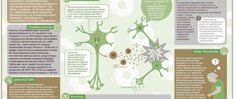
pancreas cancer
Subtypes of pancreatic cancer according to ICD
The pathology code for ICD-10 is C25 “malignant neoplasms of the pancreas.” Within the framework of the diagnosis according to ICD-10, subtypes are distinguished:
- C25.0 – Cancer of the head of the pancreas.
- C25.1 – Oncology of the body of the pancreas.
- C25.2 – Malignant formation in the tail of the pancreas.
- C25.3 – Ductal cancer of the pancreas.
- C25.4 – Malignant formations from islet cells.
- C25.7 – Oncopathologies of other parts of the pancreas.
- C25.8 – Complex pathological malignant process, including several types of gland lesions listed above.
- C25.9 – Malignant process in the pancreas of unspecified origin.
Malignant tumors of the head of the pancreas are up to 3.5 cm in size. They lead to obstructive jaundice. These tumors lead to duodenal stenosis and internal bleeding.
Cancer in the body of the pancreas gives consequences in the form of thrombophlebitis, phlebothrombosis, and diabetes mellitus. The pain syndrome at this location of the tumor is the strongest. During an attack, the patient leans forward, presses a pillow or knees to his stomach - this makes it easier to endure the pain.
A tail tumor of the pancreas is difficult to diagnose using ultrasound, since this part of the pancreas is located close to the lung, stomach and colon.
Detection of organ cancer is complicated by the fact that the pancreas is located deep in the body, so the external cancer process remains invisible.
Brief summary
Pancreatic cancer occurs less frequently in women than in men. Since they lead a healthier lifestyle, the first signs do not appear so quickly and acutely. Therefore, elderly ladies who detect signs of jaundice or ordinary pancreatitis should definitely visit an oncologist.
Representatives of the stronger sex aged 50 to 70 years are at risk. The reasons for the formation of a tumor in them are most often the following: poor diet in the form of fatty and spicy foods, cirrhosis, alcohol abuse, obesity.
Regardless of gender, the mortality rate for this type of cancer is the fourth highest among all cancers. The most common diagnosis is adenocarcinoma of the head of the pancreas; the body and tail of the organ are less commonly affected.
Causes of pancreatic cancer
Genetic failures are the root cause of the development of pancreatic cancer. But not every hormonal imbalance provokes the formation of cancer cells - if the body’s immune system is in order, the hormonal balance is equalized without oncological consequences. Common causes of pancreatic cancer:
- Chronic diseases of the pancreas (cholelithiasis, pancreatitis), cysts and benign tumors in the tissues of this organ.
- Oncology of other organs.
- Stomach operations.
- Dental diseases.
- Cirrhosis of the liver.
- Nonspecific ulcerative colitis and Crohn's disease.
- Diabetes.
- Sedentary lifestyle and lack of systematic physical activity.
- Excess body weight. Excess weight is often caused by an imbalance in hormonal levels, which leads to disruption of the formation of pancreatic enzymes, creating a favorable environment for the division of cancer cells.
- Tendency to food allergies. With frequent skin allergies, the body is predisposed to inflammatory processes. Digestive gland cells can become cancerous.
- Tobacco smoking and alcoholism.
- Working in hazardous industries, where the body is exposed to the toxic effects of asbestos, heavy metals, and inhalation of dye fumes.
- Human residence in a region with an unfavorable environmental situation.
- Frequent emotional shocks, stress, and a tendency to depression can provoke an oncological process in the body. These conditions are fraught with the fact that they disrupt a person’s usual diet, sleep and rest. Reactions to stress are individual: some people lose their appetite, while others “eat” it – both are equally harmful and lead to disorders of the digestive system. The body's compensatory mechanisms may fail.
- Genetic predisposition and mutations of the BRCA2 gene, dysplastic nevi, Lynch syndrome.
According to research, eating certain foods promotes the formation of cancer cells. These include bacon, ham, smoked chicken, coffee, carbonated drinks, grilled foods, and fatty foods.
Reasons for development
Smoking
People who smoke have at least 2 times the risk of pancreatic cancer than non-smokers. Smokers with more than 40 years of smoking experience may have a 5 times greater risk. Smokeless tobacco also increases the risk of this disease.
Obesity and dietary factors
Obesity, especially central obesity, is associated with a higher incidence of pancreatic cancer. Consumption of red meat, especially smoked and fried meat, also increases the risk of developing this disease.
Diabetes
Diabetes mellitus is a proven risk factor for prostate cancer. There has also been an association between the sudden onset of type 2 diabetes in an adult over 50 years of age and a new diagnosis of pancreatic cancer, although in these cases the diabetes is thought to be caused by cancer.
Chronic pancreatitis
Advanced chronic pancreatitis is a significant risk factor for the development of prostate cancer.
The risk of prostate cancer is even higher in patients with hereditary pancreatitis. It increases more than 50 times, and the cumulative risk level by age 70 is 40%.
This cumulative risk increases to 75% in individuals with a paternal family history of pancreatitis.
Genetic factors
Approximately 5-10% of patients with pancreatic carcinoma have a genetic predisposition to developing the disease.
Of these tumors
- 80-95% have mutations in the KRAS2 gene;
- 85-98% have mutations in the CDKN2 gene;
- 50% have mutations in p53;
- 55% have mutations in Smad4.
Families with BRCA-2 mutations, which are associated with a high risk of developing breast cancer, also have an excess risk for prostate cancer.
Types and signs of pancreatic cancer
Approaches to the classification of pancreatic cancer include different criteria. According to histology, pathology is divided into:
- Ductal adenocarcinoma, which occurs in the cells of the ducts. This type of pancreatic cancer is the most common.
- Glandular squamous cell carcinoma that arises from cells that produce enzymes.
- Giant cell adenocarcinoma is a neoplasm in the tissues of the pancreas, consisting of cystic cavities filled with blood.
- Cystadenocarcinoma, which looks like a transformed cyst.
- Mucinous adenocarcinoma is a non-aggressive form of pancreatic cancer. Rarely seen. This is the “female” type of cancer.
- Squamous cell carcinoma arises from the cells that form the duct. The disease is rare, but has an aggressive course.
- Undifferentiated cancer is oncology with the most aggressive course.
There is also a classification based on the characteristics of the structural tissues of the pancreas. So, the gland contains exocrine and endocrine tissue. Endocrine tissue is responsible for the production of hormones, and exocrine tissue produces digestive enzymes. In accordance with this, endocrine and exocrine pancreatic cancers are distinguished. Exocrine tumors are more common than endocrine tumors.
Endocrine tumors include neuroendocrine oncology, gastrinoma, insulinoma, glucagonoma, and somatostatinoma. As a rule, they are benign, but they can also be malignant.
Pancreatic lipomatosis
At an early stage, the disease is asymptomatic: there is no visibility of the pathological process. Painful sensations are often associated with disorders of the digestive system: general weakness, loss of appetite, abdominal discomfort, nausea, vomiting. The first signs can be recognized simultaneously with the onset of metastasis. The patient does not always pay due attention to them due to their similarity to disorders of the gastrointestinal tract. These include:
- Periodic pain impulses in the pancreas and discomfort under the ribs on the left side, not associated with food intake.
- Deep vein thrombosis in the legs. The presence of thrombosis is indicated by increased skin temperature of the legs, redness, swelling, and pain in the legs that are not associated with increased physical activity. There is a risk that part of the blood clot will break off and enter the vessels. There is a risk of developing arterial thromboembolism.
- Night discomfort in the pancreas.
- Paroxysmal pain in the navel, lower back, shoulder blades, stabbing and aching in nature.
- Mild yellowing of the skin and sclera of the eyes, which occurs due to blockage of the bile ducts by cancer cells. Blocked ducts can lead to liver failure and internal bleeding.
- Exacerbation of chronic diseases of the digestive system and loss of appetite.
- Increased fatigue, chronic fatigue.
If you ignore the symptoms of early stage cancer and delay a visit to the doctor, the tumor progresses. Each symptom becomes pronounced and developed. Specific oncological signs of oncology are:
- Aching or nagging pain in the abdomen, radiating to the back. The nature of the pain is similar to the pain associated with cholecystitis and pancreatitis. When bending forward, the pain intensifies. The maximum intensity of pain is reached at night. They are characteristic of cancer in the tail of the gland and in the head.
- Pain syndrome develops from periodic to permanent. The pain is localized in the left hypochondrium or in the navel area, indicating an oncological process in the head of the gland. Women experience pain in the ovarian area, men – in the prostate area
- Migrating thrombosis of peripheral veins.
- Skin changes in the form of itching and the skin becoming yellow-green. The symptom is characteristic of head cancer.
- Dark colored urine and light colored stool. Changes in the color of urine and feces are caused by the tumor compressing the bile duct. The gallbladder enlarges. A large amount of fluid accumulates in the abdominal cavity. The picture is observed with cancer of the head of the gland.
- Feeling of heaviness in the stomach, belching with a rotten smell.
- Internal bleeding due to tumor growth into the walls of the stomach.
- Decreased appetite and weight due to the production of small amounts of pancreatic juice. The symptom is characteristic of any tumor location.
- Diarrhea, nausea and vomiting as a result of compression of the duodenum and stomach by the tumor. Similar symptoms were reported by half of patients with pancreatic cancer.
- Changes in the functioning of the circulatory system, manifested by anemia, leukopenia, thrombocytopenia.
- Secondary diabetes mellitus and severe thirst due to changes in hormonal levels.
If, even with the listed symptoms, the patient does not seek medical help and does not begin treatment, or the symptoms are not classified as oncopathology, the cancer metastasizes to the liver, spleen and other organs. The entire body is already involved in the pathological process. Oncology grows into blood vessels and nerve endings. The patient has the symptoms listed above, in addition to them:
- Pain in the lumbar region, shoulder blades, and right hypochondrium is of a girdling nature. After eating fatty foods or alcohol, they intensify. Resistant to painkillers.
- Persistent hyperthermia is an unreasonable frequent change in body temperature without signs of acute respiratory disease.
- Intestinal bleeding.
- Marked weight loss.
- Insomnia caused by acute pain, which even painkillers cannot reduce.
- The stool becomes dark brown and greasy. The patient experiences difficulty in bowel movements.
- Yellowness of the sclera, skin, mucous membranes.
- Unbearable skin itching, due to which the patient cannot sleep. Unpleasant sensations in the skin are caused by blockage of the bile ducts by cancer cells. Epidermal cells are irritated by bile salts entering the plasma. The patient scratches the skin until it bleeds.
The danger of the described stage of oncology is the occurrence of intoxication of the body as a result of the release of decay products of cancer cells into the blood.
Operation: pros and cons
Despite the fact that the differential diagnosis of pancreatic cancer is diverse and allows one to identify a lot of pathologies by submitting material for laboratory testing, surgical intervention does not always confirm the development of a fatal disease.
The justification for the invasion of the body is data obtained through clinical, instrumental and other types of tests. However, all of them can only, to one degree or another, indicate cancer. It is often impossible to determine an accurate diagnosis and distinguish chronic pancreatitis from the early stage of oncology, since benign tumors can exhibit similar symptoms and look identical. Only based on the results of resection and examination of the removed parts can one speak with 100% probability of pancreatic cancer. Stage 4 is the only stage that is clearly determined by radiation methods, since it is manifested by metastasis to the following organs:
- kidneys;
- liver;
- lungs;
- intestines;
- spleen
- brain;
- bones.
Thus, making a decision to perform an operation is sometimes the only way to save a person’s life. Of course, the doctor pays special attention to the test results and only suggests resection if absolutely necessary. However, in the first stages of the examination, the role of tumor markers cannot be underestimated, the indicators of which determine the need for a thorough examination and subsequent radiodiagnosis.
Diagnosis of pancreatic cancer
To establish the cause of the development of a cancerous tumor, a comprehensive diagnosis is necessary. The oncologist collects and analyzes anamnestic information: heredity, past illnesses of the patient, and takes into account the impact of harmful factors at work.
To make a diagnosis, examination methods are used:
- Blood chemistry. This method is used to identify cancer markers in the blood: carbohydrate antigen CA 19-9, CA-242. In the presence of a malignant process, blood counts - bilirubin and bile acids - will be significantly higher than normal. If the cancer has already spread with metastases throughout the body, the level of protein in the blood is reduced. If the process of metastasis has begun, the results of a blood test will show a reduced level of hemoglobin.
- Testing for tumor markers is an immunohistochemical method to check a patient’s blood for the content of special cells formed during the progression of a malignant neoplasm in organs and tissues.
- Urine and stool analysis. When cancer occurs in the urine, pancreatic amylase is diagnosed. Elastase-1, alkaline phosphatase, and C-peptide will be present in the feces.
- Ultrasound, CT and MRI are procedures that help the attending physician decide on the need to perform surgery and determine its extent.
- Endosonography is an informative video method for examining the duodenum by moving a sensor through this organ. The method collects maximum information about the pancreas, since the duodenum is located in close proximity to the gland.
- X-ray, FGDS, sigmoidoscopy are methods that reveal the stage of metastasis and determine the localization of secondary tumors.
- Biopsy is a procedure for taking a sample of gland tissue for laboratory study and detection of a malignant process.
- Angiography is a diagnostic method based on staining blood vessels with a contrast solution to understand whether radical surgery is permissible.
Angiography of the pancreas
- Cholangiopancreatography is a method for diagnosing the pancreatic and bile ducts. The procedure has 3 types:
- Endoscopic retrograde cholangiopancreatography (ERCP). The probe is inserted into the duodenum, finds the opening through which the bile duct enters, and a radiopaque substance is injected through it. After these manipulations, the patient is sent for an x-ray. In the photographs, due to the radiopaque substance, the ducts are tinted, and it becomes possible to analyze their condition.
- Percutaneous transhepatic cholangiography is used in cases where endoscopic retrograde cholangiopancreatography is impossible for one reason or another. In this case, a radiopaque substance is injected into the ducts using a needle.
- Magnetic resonance cholangiopancreatography (MRCP). In terms of medical manipulations, this method is similar to MRI. It does not involve the introduction of any special instruments into the body. The peculiarity of the method is that it allows you to obtain accurate information about the localization of oncology, identify features, and determine the type. The disadvantages of the method include the fact that during MRCP it is not possible to carry out parallel diagnostics using auxiliary methods to clarify the clinical picture.
- Laparoscopy is a surgery that is used in cases of questionable data obtained by other diagnostic methods.
- The PET scanning method allows us to detect metastases in distant organs and tissues.
Based on a comprehensive analysis of laboratory data, the oncologist makes a verdict on the disease, makes prognoses and prescribes treatment.
Diagnostic measures
Pancreatic cancer is rarely diagnosed at stage 1. Only in 30% of cases is a tumor less than 2 months old. To diagnose this malignant neoplasm, the following studies will be needed:
- general and biochemical blood tests;
- general urine analysis;
- Ultrasound of the abdominal organs;
- tests for tumor markers;
- ultrasonography;
- radiography;
- CT or MRI;
- cholangiopancreatography;
- positron emission tomography;
- biopsy;
- cytological and histological analyses;
- laparoscopy.
Pancreatic cancer tumor markers CA-19-9, CF-50, CA-242 and CA-494 are necessarily detected in the blood of patients. Every second patient is diagnosed with cancer embryonic antigen. These tests do not allow for an accurate diagnosis. The greatest value is tissue research. The detection of atypical malignant cells in the gland confirms the diagnosis.
The tumor is visible on the screen during an ultrasound (ultrasound examination). This is a screening research method that allows you to determine the location of the tumor. Using ultrasound, you can assess the condition of other abdominal organs (gallbladder, spleen, liver) and exclude cholecystitis and hepatitis.
A detailed examination of the tumor is carried out using computer or magnetic resonance imaging. The latter is the most informative and safe. The advantage of MRI is the absence of radiation exposure. This research method allows you to detect a gland tumor measuring 2 cm or more, assess the condition of the lymph nodes and examine metastases.
To assess the condition of the duodenum and bile ducts, endoscopic retrograde cholangiopancreatography is performed. This study involves the administration of a contrast agent followed by radiography. Sometimes, if breast cancer is suspected, laparoscopy is performed. This is an invasive research method.
A blood test is required. During it, the following changes are revealed:
- acceleration of ESR;
- decreased levels of hemoglobin and red blood cells;
- increased alkaline phosphatase activity;
- increased levels of liver enzymes;
- bilirubinemia.
An additional diagnostic method is stool analysis. In iron cancer, undigested food fragments and large amounts of fat are often found. The patient assessment plan includes a questionnaire and physical examination. During the history taking process, the doctor identifies possible risk factors for developing cancer.
Stages of pancreatic cancer
Pancreatic cancer goes through a number of stages:
Stages of pancreatic cancer
- Stage 0. The process of oncology formation has been started: several cells have mutated. These cells are found in the lining of the pancreas. If they are examined using medical methods and removed, it will be possible to stop the process at the initial stage. Otherwise, the mutated cells will leave the mucous membrane of the pancreas and grow into the digestive organ. There are no symptoms of a malignant process at the initial stage.
- Stage 1. Oncology does not affect the appearance of the gland due to its small size. The stage is divided into two stages. 1A: Stage proceeds without metastases. The tumor is located within the pancreas. The size does not exceed 2 cm in diameter. Patients report digestive disorders as symptoms. If the tumor is located in the body or tail of the gland, signs of endocrine oncology appear. 1B: The tumor is larger than 2 cm, but it does not extend beyond the boundaries of the pancreas. If it is localized in the head of the gland, there is a slight yellowness of the skin. Associated symptoms are nausea and loose stools. As in stage 1A, signs of endocrine oncologies appear.
- Stage 2. It is divided into two subphases. 2A: Metastasis begins in the bile duct and duodenum, the size does not exceed 4 cm. Symptoms of endocrine tumors are present. 2B: The tumor increases in size and metastasizes to the lymph nodes. In this case, the lymph nodes enlarge and become painful when touched. Patients complain of severe abdominal pain, weight loss, loose stools, and vomiting. There are symptoms of endocrine tumors.
- Stage 3. Metastases spread to the stomach, spleen, large intestine, and possibly to nerves and blood vessels. At stage 3, damage to the lymph nodes is possible. With metastases to bone tissue, a person experiences pain similar to radiculitis. With metastases to the lungs, a cough with bloody sputum and shortness of breath appear. Their manifestation in the kidneys is detected by swelling of the legs, lower back pain, and increased blood pressure. A urine test reveals an increased level of red blood cells.
- Stage 4. The tumor spreads throughout systems and organs, resulting in signs of cancer intoxication. The symptoms of this stage are striking in their aggressive nature.
The pain syndrome at stage 4 becomes pronounced as a result of the effect of cancer cells on the nerve endings. Patients cannot sleep without taking a painkiller. The pain is increasing. Adopting the fetal position makes their course somewhat easier. If at first it is possible to get rid of pain with the help of paracetamol and other non-narcotic substances, now only opiates can drown out the sensations for a while.
As a result of a catastrophic disruption of the gastrointestinal tract, the body is exhausted. The spleen increases in size - the organ can no longer perform full immunofunction and efficiently filter blood. Blood tests show a buildup of toxins. Against the background of metastases, intestinal obstruction occurs. Up to 20 liters of fluid accumulate in the abdominal cavity, which is caused by metastases.
Secondary tumors form in the supraclavicular lymph nodes. Metastases in the lymph nodes lead to tissue cells dying and subcutaneous fat necrosis forming. Blood clots form in the veins, causing blockage of the veins and leading to a disease such as migratory thrombophlebitis.
Cancer at stage 4 cannot be cured. However, the critical clinical picture of stage 4 pancreatic cancer does not mean doctors refuse treatment. At this stage, therapy has specific features: the main goal of doctors is not to fight cancer, but to improve the patient’s well-being and contain metastases. Doctors are taking obvious measures:
- An operation is performed - complete or partial removal of the pancreas and parts of surrounding organs.
- Prescribing analgesics to relieve pain.
- Operations to prevent the occurrence of cancer complications. Through surgery, the intestines are restored and blockage of the bile ducts is eliminated, and internal bleeding is prevented.
- Chemotherapy is prescribed to prolong life by six months.
- Radiation therapy sessions are carried out, which destroys the protein of cancer cells and reduces the size of the tumor.
For stage 4 pancreatic cancer, the patient's life expectancy depends on the degree of spread of metastases and the organs they affect. General intoxication of the body has a direct impact on the patient’s well-being, undermining the body’s immunity and physical strength. How long a person will live once diagnosed depends on the body’s individual susceptibility to chemotherapy. An important role is played by the patient’s psychological mood, support from loved ones and care.
Based on the clinical picture of stage 4 pancreatic cancer, even for a person far from medicine, it becomes clear that survival at the last stage of the disease is unlikely.
Stages
There are 4 stages of development of this tumor. The division is based on the following characteristics:
- The size of the tumor.
- Spread to surrounding tissues.
- The presence of distant and regional metastatic foci.
Stage 0 is set if there is an area of accumulation of altered cells without germination into surrounding tissues (cancer in situ). In this case, regional lymph nodes near the gland are not affected and there are no metastases. At stage I, the neoplasm is small in size. In stage IA, the tumor diameter does not exceed 2 cm.
With grade IB, the size of the tumor is more than 2 cm. Metastases are not detected. Stage IIA is distinguished by the fact that the tumor has spread beyond the gland to the celiac trunk, bile duct, duodenum, or superior mesenteric artery. There are no metastases in other organs.
Stage IIB of the disease is characterized by the presence of a tumor of any size, but without invasion of the celiac trunk and superior mesenteric artery. In this case, single metastases to regional lymph nodes are possible. Stage III is characterized by tumor spread to the stomach, spleen and intestines, as well as damage to the celiac trunk. Regional metastases are present, but distant ones are absent.
At this stage, many patients consult a doctor. If the symptoms of the disease are ignored, a stage IV gland tumor develops. Atypical cells spread throughout the body. Secondary cancers can be found in the liver, lungs, bones, stomach, intestines and brain. This tumor is inoperable.
Description of the development of oncology according to the TNM system
The course of the disease can be described in accordance with the TNM system.
“T” denotes the main characteristics of the primary tumor.
- T1 – oncology is located inside the gland. Neighboring organs and tissues are not affected. There is no spread of metastases through blood vessels and nerves. T1a – tumor size is less than 2 cm. T1b – tumor size is more than 2 cm.
- T2 – initial metastasis to blood vessels and nerves.
- T3 – active spread of metastases to neighboring organs.
“N” is the degree of tumor metastasis to the lymph nodes.
- N0 – metastasis to the lymph nodes does not occur.
- N1 – initial spread of metastases to the nearest lymph nodes.
- N2 – secondary cancerous tumors in the lymph nodes.
- N3 – spread of metastases to distant lymph nodes.
“M” – the presence or absence of metastases in distant organs and systems of the body.
Taking into account the above described indicators of the TNM system, the degree of development of pancreatic cancer looks like this:
| N0 | N1 | N2 | N3 | |
| T1a | 1 | 2 | 3 | 4a |
| T1b | 1 | 2 | 3 | 4a |
| T2 | 3 | 3 | 4a | 4b |
| T3 | 4a | 4a | 4b | 4b |
| Any T, N and M1 | 4b | 4b | 4b | 4b |
Grade 4b, accompanied by extensive metastases, is common. Metastasis develops in different parts of the abdominal cavity, bone tissue, liver, and lungs. Complete recovery at the described stage is impossible, but through medical manipulations it is possible to reduce pain, improve the general well-being of the patient and prolong life.
Cancer cell metastases
Changes on ultrasound
Diagnosis of cancer of this organ requires a mandatory ultrasound examination. During this procedure, the specialist may notice the following changes:
- Increase in the lumen of the main duct of the organ.
- Presence of nodular structures.
- Enlargement of an organ or its affected part.
- Reducing the echogenicity of tissues.
- Uneven boundaries in the area of spread of the malignant process.
In addition to the pancreatic gland, it is worth paying attention to the condition of nearby organs and lymph nodes for blockage or the spread of metastases.
Pain relief and treatment of pancreatic cancer
There are cases where a discovered malignant neoplasm in the pancreas, initially mistaken for a focus of oncology, was in fact a powerful metastasis of oncology in another organ. As a rule, the focus of oncology in such a case is localized in the lungs, mammary glands, gastrointestinal tract, and prostate gland. Melanoma, leiomyosarcoma, osteosarcoma or Merkel carcinoma can also metastasize to the pancreas. In such cases, the doctor’s task is to prevent further spread of metastases simultaneously with the start of treatment of the main focus of cancer.
A feature of pancreatic cancer is a pronounced pain syndrome. Pain relief is carried out using 3 groups of drugs:
- Group 1: non-narcotic analgesics (analgin, naproxen and paracetamol). Used to relieve mild pain.
- Group 2: narcotic analgesics (tramadol, promedol and dihydrocodeine). Drugs are prescribed if non-narcotic analgesics do not work and the pain is moderate.
- Group 3: potent opiates (prosidol, fentanyl). Prescribed when narcotic analgesics have become ineffective due to an increase in pain.
Cancer treatment is a long process and expensive. When choosing methods, the characteristics of a particular case of disease and the individual capabilities of the patient are taken into account.
Targeted therapy and chemotherapy for pancreatic cancer
Targeted therapy is a modern variation of traditional chemotherapy that uses drugs that act only on cancer without negatively affecting healthy cells. The method is based on blocking the spread of cancer cells and preventing their division.
The method has virtually no contraindications and is easily tolerated by patients. Used when cancer is inoperable. However, the cost of treatment is significantly higher than chemotherapy courses.
Chemotherapy suppresses the formation of new tumor cells and destroys existing cells. Oncology of various origins and localizations is often treated using this method. However, in the treatment of adenocarcinoma, the technique shows positive dynamics only as an auxiliary one. Therefore, this method is used after testing other methods. For example, hormone therapy is more effective in treating pancreatic cancer. Hormonal drugs interact with estrogens on the walls of the pancreas, which has a healing effect and prolongs the life of the patient.
The targeted treatment method has side effects on the body - the toxic effect manifests itself in the form of nausea, vomiting, loose stools, baldness, and disorders of the circulatory and nervous systems. This type of therapy allows you to extend the patient’s life by 6-9 months, provided that it is used in cycles in accordance with the doctor’s instructions and in combination with other medical procedures.
The mechanism of the procedure is to influence the genetic structure of the cell and transform it. When a tumor's DNA is destroyed, the cancer cell cannot continue to divide and spread throughout the body. The cancer cell dies.
There are 2 types of chemotherapy:
- Monochemotherapy, treatment is carried out using only one drug.
- Polychemotherapy, for treatment, several medications are used, taken simultaneously or alternately.
The following drugs are used for chemotherapy:
- Gemcitabine (Gemzar) is a drug that reduces tumors and prevents the spread of metastases. Facilitates the course of the disease and relieves pain.
- Docetaxel (Taxotere) is a drug used in monotherapy. Slows down the process of cancer cell division and improves the general well-being of the patient.
- Fluorouracil and Cisplatin are drugs used in combination, but strictly taking into account the individual characteristics of patients, as they have multiple contraindications. If the drugs are suitable for the patient, the therapy prolongs the patient’s life by a year.
- Gemcitabine (Gemzar) and Fluorouracil are used in complex therapy. The action of the drugs slows down the process of cancer cell division and prolongs the patient’s life by a year or more.
The duration of the course is determined by the doctor, taking into account the individual characteristics of the patient.
Recommendations after chemotherapy
After chemotherapy, clinical recommendations must be followed:
- The intake of any medications and mineral-vitamin complexes after chemotherapy treatment must be agreed with the attending physician in order to avoid allergic reactions. Your doctor will help you choose antiemetic medications and medications to prevent baldness.
- It is necessary to drink plenty of fluids - this will speed up the removal of toxins from the body.
- Feelings of hunger should not be allowed to arise. You need to eat often, but in small portions. Nutrition should be balanced. It is not recommended to eat cold or too hot food. The diet should contain fruits, vegetables, eggs, seafood, lean meat, and dairy products. Drinking alcohol after chemotherapy is prohibited.
- Drinking ginseng tincture will help normalize your emotional background.
- Consulting a psychologist will help you cope with stress, learn to enjoy the new day, and rethink your own life. Classes with a psychologist will allow you to master relaxation techniques and create conditions for creative self-realization.
Radiation therapy and innovative treatment methods
Radiation therapy is used to kill cancer cells and reduce the size of the tumor. Indications for it are individual. The doctor prescribes it as an independent method in preparation for surgery or as an additional remedy in the postoperative period.
Innovative methods of combating oncology are actively used abroad - the introduction of a weakened Listeria monocytogenes vaccine to a patient along with radioactive particles. Testing of the method showed good results: the bacterium exclusively affects metastases without affecting healthy structures. Becoming a carrier of radioactive particles, the microbe transfers them to cancer cells, killing the latter.
Foreign scientists are also working towards the development of stimulants for the human immune system, which would teach the body to effectively resist cancer. A drug with similar action has now been developed - Ipilimumab. It belongs to the group of monoclonal antibodies.
Traditional methods of treating pancreatic cancer
Folk remedies can slow down the pathological process and relieve pain. The condition for positive dynamics is compliance with the doctor’s instructions and complete courses of treatment. Common methods of alternative medicine are:
- Vodka and vegetable oil. Mix 30 ml of vegetable oil and a similar amount of vodka. Carefully move the mixture by vigorously shaking the tightly closed container. The resulting mixture is drunk once within 15 minutes. before meals 3 times a day. The course of treatment is 3 times 10 days with a five-day break. After the third course there is a break of 14 days. In between courses, they control weight, monitor hormone levels by taking a blood test and consult with an oncologist. The scheme is repeated many times over several years. Mandatory condition: the number of meals should not exceed 3 times a day.
- Treatment with tincture of Djungarian aconite 2.5%. Treatment begins with a single drop of tincture, adding 1 drop every day, the dose is adjusted to 30 drops per day, then the dose is gradually reduced to 1 drop per day. After 30 min. After the tincture, you need to drink a herbal mixture of orris root, chamomile, hops, calendula flowers, dill seeds, marsh calamus root, cinquefoil root with the addition of 1.5 ml of 10% tincture of the common flower.
Remember, alternative medicine is good as an adjuvant treatment. Folk remedies are not an alternative to surgery.
Surgery for pancreatic cancer
Surgery is considered the most effective method of treating oncology. But there are many contraindications to the procedure for pancreatic cancer. Often surgery is contraindicated in the presence of metastases, and pathogenic cells form early and spread quickly. In addition, pancreatic cancer is usually detected in elderly people, when the patient runs the risk of not undergoing surgery due to age-related changes in the body.
Surgery for pancreatic cancer is complicated by the fact that the organ is surrounded by other vital organs in the abdominal cavity. To perform a high-quality operation, the surgeon must be highly qualified.
Surgery for pancreatic cancer is prescribed when the process of spreading metastases has not yet begun. As a rule, the pancreas in the latter case is completely removed. If the operation was performed before the formation of metastases, the patient is said to have recovered.
When the tumor metastasizes to other organs, removal of the pancreas removes the focus of the cancer, but does not eliminate the disease completely. Partial removal of the pancreas is possible in order to prolong the patient’s life and relieve pain. These medical procedures increase the effectiveness of further chemotherapy treatment.
Sometimes an anastomosis is installed between the jejunum and the gallbladder to drain bile. The procedure requires surgery, but of a different nature. Anastomosis is used in cases of deep tumor invasion to alleviate the patient’s condition.
A safe method that can be used to cure cancer is the cryogenic treatment method, in which tumor cells are exposed to low temperatures, due to which they freeze out and die. The method shows high effectiveness in relieving pain and does not cause complications. But in Russia the number of doctors who know the described technology for treating oncology is extremely small.
It should be noted that adenoma increases the risk of postoperative complications. In case of a successful outcome of the operation, to prevent relapse and as maintenance therapy, the patient needs lifelong intake of insulin and enzymes that compensate for the absence of the organ that produced them before the operation.
Ultrasonography
When the manifestation of pancreatic cancer, symptoms that clearly indicate problems in this organ, begin to bother the patient, he goes to the doctor. In the first stages of examining a patient, the specialist includes not only a survey and general tests, but also an ultrasound of the abdominal cavity. Sometimes painful sensations indicate one organ, but in fact another one located nearby is suffering. This method allows you to localize the possible focus of the disease and help the doctor select further methods of diagnosis or therapy.
An ultrasound may show enlargement of any part of the pancreas or a change in its contour. During ultrasound, special attention is paid to the head of the gland, since in 80% of cases it is there that a new formation is observed. In the tail part, cancer manifests itself much less frequently. However, it happens that an examination reveals a tumor of the entire tissue, which in fact may not be an oncological disease, but an acute form of pancreatitis.
Ultrasound also helps to visualize the nature of the changes and the structure of the gland. Typically, with this form of cancer, the tumor is hypoechoic and has no internal echo structures.
Prevention of pancreatic cancer
Quitting smoking helps prevent the development of any pathological process in the body, including pancreatic adenocarcinoma.
It is necessary to establish a diet: accustom the body to frequent but small portions of food. This will help prevent the development of cancer and get rid of excess weight. A prerequisite for a healthy diet is a complete abstinence from alcohol.
If, due to the nature of your professional activity, it is not possible to exclude exposure to heavy metals and asbestos, it is recommended to follow safety rules and be sure to use personal protective equipment.
Effective prevention of pancreatic cancer is a healthy lifestyle and proper balanced diet. Paying attention to health, undergoing periodic medical examinations and treating identified diseases is a sure way to protect the body from cancer.
Oncologists recommend once a year an ultrasound examination of the abdominal cavity and organs behind the peritoneum, even for healthy people who are not at risk for pancreatic cancer. If a person is at risk for pancreatic oncology due to 2 or more factors, then in addition to an annual ultrasound, doctors recommend taking a blood test to detect the CA 19-9 marker.
CT scan
This study is carried out using x-rays that pass through organs and tissues. Since they all have different densities, just like cancer formations, the device manages to transmit the image layer by layer. The final display allows you to visualize those organs that were subjected to tomography and their structure. A specialist can evaluate not only the size of the pancreas, but also various deposits, inflammation and swelling. It should be noted that the radiation intensity of a CT scan is much less than that of a conventional X-ray. When preparing for this type of examination, you need to keep in mind that contrast is often used. Therefore, the presence of contraindications to the use of iodine-containing drugs must be communicated to the attending physician. You should also notify your doctor if you have any allergic reactions to medications.
Prognosis for pancreatic cancer
The prognosis for the outcome of the disease depends on the time of detection. The danger of the disease is that even with early diagnosis and the patient’s compliance with the doctor’s recommendations, complete recovery occurs only in 15% of cases. 20% of patients are doomed to die within 5 years after surgery, even if all clinical recommendations are strictly followed.
The key point in prognosis is the possibility of surgical removal of the tumor. It should be remembered that with extensive metastasis, surgical intervention will not bring a noticeable improvement in the situation. After a verdict on the stage of the disease is made, patients live for 3 years. Maintenance therapy is prescribed to relieve pain. If medical recommendations are ignored, death will occur within 2-3 months.
Compliance with clinical recommendations, chemotherapy, radiation therapy, ensuring the removal of bile from the body, taking painkillers, and psychological assistance helps to prolong the patient’s life and ease his well-being.
If the patient turns up at the first signs of the disease, the pathological process can be stopped and the disease will end in healing. Statistics indicate a favorable outcome in 3/4 of patients.
Regardless of the stage of cancer, the prognosis of the outcome of the disease is largely determined by the patient’s mood. Faith in the best, complete trust in the doctor and the methods used, and an optimistic attitude are the components thanks to which many patients have coped with cancer, even when doctors have doubts about the success of treatment.
Biopsy punctures
A biopsy involves taking a small piece of diseased tissue from the patient from an area where a tumor may develop. It is usually removed surgically by making a small incision. New technologies now make it possible to perform a biopsy by removing a small number of cells using a syringe. When taking a puncture, it is imperative that the tumor area is monitored using translucent devices: ultrasound, x-ray, tomography. This allows precise positioning of the tissue harvesting device.
Important information: Symptoms of pancreatic insulinoma
Endoscopy is also used for punctures, but due to its pain and complexity, it is preferred to perform surgical intervention or by syringe in initially diagnosed patients.
Only a biopsy and clinical tests to determine the nature of the tissue can help establish an accurate diagnosis. This procedure is mandatory.
Diagnostics
Various diagnostic methods are used to determine the stage of cancer. The most informative is CT. To clarify the diagnosis, a puncture biopsy is performed using ultrasound or CT. If the tumor is located only in the pancreas, it is not so easy to detect it at an early stage.
Diagnostic methods:
- the abdominal cavity is examined using ultrasound;
- A CT scan examines the entire organ in detail and reveals exactly where the tumor has formed;
- a biopsy is performed using an endoscope or needle;
- cholangiography or angiography is performed, that is, a special contrast agent is injected into the ducts or vessels and pictures are taken;
- blood tests to detect which tumor markers have increased (CA19-9 and CEA).
Important! Pancreatic cancer accounts for 3% of all cancers. In 60% of cases, the tumor affects the head of the organ, 10% – the body, and 8% – the tail. In 23% of cases - the entire organ.
Symptoms of the disease
At an early stage, the patient feels symptoms that are quite easily associated with a number of other diseases. These may be common constipation, malaise and weakness. Diabetes is very adept at masking a more serious illness. The first signs of pancreatic cancer in women and men in the early stages can be detected by untested studies by a number of specialists.
Thus, many sick people complained that almost six months before the main stamp of the disease began to appear, they suddenly felt an aversion to their previously favorite foods and drinks. A small number of people experienced an attack of pancreatitis six months before diagnosis. The first symptoms of the disease often manifest themselves only in the later stages, since in this case there is already compression of neighboring organs.
Pain in pancreatic cancer is the main symptom that manifests itself among the first. This symptom indicates that the disease is metastasizing and spreading to the nerve endings. Pain sensations appear depending on the location of damage to one of the organs. The pain intensifies after eating junk food, as well as when lying on your back.
Thrombosis is a significant symptom that manifests itself as swelling and redness of the legs. The patient's temperature also rises. If the tumor reaches the bile duct, obstructive jaundice may occur. The skin and mucous membranes become yellow, the urine darkens, and bloating occurs. Over time, the skin turns from yellow to a sharp greenish tint.
Some people experience intoxication, which occurs when harmful substances enter the blood. The symptom appears in the early stages of the disease, but is often mistaken for another disease. The person begins to lose weight sharply, feels tired and has a complete loss of appetite. At the same time, weight often goes off even with normal nutrition. Metastases from pancreatic cancer can manifest themselves in the form of a severe cough, shortness of breath, chest pain and even hemoptysis. Another striking external symptom is the patient’s lethargic appearance.
Important information: What are reactive changes in the pancreas in a child
Cancer undergoes the following stages of development:
- The tumor is located only in the pancreas area, has a maximum size of 2 cm and is quite easily removed surgically.
- The tumor spreads to other organs, but is still subject to surgical intervention.
- Cancer grows into the circulatory system and affects the lymph nodes. Surgery is still possible, but not in all cases.
- Metastases pass into the abdominal cavity and are no longer removed from the body.
Treatment of the disease
How to treat cancer? The first thing to do is to completely get rid of all infected cells in the body. If this is not possible, it is necessary to stop further growth of the tumor, preventing it from metastasizing, and reduce the symptoms of pancreatic cancer in women and men. A variety of methods are used for treatment.
Surgical intervention is often practically not used, since it implies a number of serious conditions and further complications. First, the cancer must not spread to neighboring organs. Secondly, there must be a complete absence of metastases.
This operation involves complete or partial removal of oncology, and sometimes other affected organs are also removed. Since this procedure is unusually difficult and complex, tumor formation is most often removed surgically in young people, since it is their body that recovers most quickly after serious external interventions. After the operation, the recovered person takes enzymes and insulin for life. Surgical procedures are advisable in only 10% of cases of diagnosing the disease, since cancer develops at an extraordinary speed and there is a high risk of major deterioration after the procedure.
Chemotherapy for cancer is inappropriate and is prescribed in addition to radiation therapy to reduce the patient's symptoms. Hormonal treatment quite often stops developing tumors, since such drugs contain chemical elements that prevent the further spread of metastases. With regional therapy, drugs are injected into the gland, and with systemic therapy, drugs are injected into a muscle or vein.
Important information: Deformation and bending of the pancreas
Radiation treatment is prescribed after the operation in order to finally eliminate all remaining carcinomas in the cells that the surgeon’s scalpel could not reach. In internal therapy, irradiation occurs using special needles. External radiation involves manipulation, which is carried out using special technologies.
The cancer clinic with biological or immunotherapy only undergoes a series of tests to confirm its feasibility. The synthesized substances are injected directly into the immune system, causing them to begin fighting harmful cells. At the initial stage, they start with non-narcotic substances, later they move on to narcotic ones, including potent ones. Pancreatic cancer with metastases is practically untreatable.
Doctors eliminate only the symptoms of the disease using the above-mentioned treatments.
There are no traditional methods to combat pancreatic cancer. Yes, some medicines may contain certain plants, but they also contain many other ingredients. Under no circumstances should you try to get rid of such a serious disease on your own using dubious methods. This can pose a serious risk to life. The selection of the right medical center to combat the disease will also play an important role.
TAB
Fine needle aspiration biopsy is the removal of a liquid substance from the affected area using a needle for subsequent cytological examination. It is used to determine the nature of the lesion - the nature of the tumor or inflammation.
The material is collected under the control of ultrasound, computed tomography or endosonography. Such control ensures that the needle accurately hits the suspicious area and also prevents puncture of the vessel.
Indications for fine needle biopsy:
- asymptomatic cystic tumors of the pancreas, the nature of which cannot be assessed by other methods;
- suspicion of autoimmune pancreatitis;
- suspicion of pancreatic lipomatosis;
- suspected pancreatic lymphoma.
The listed diseases most often do not require surgical treatment, but some of them require specific therapy.
TAB is also advisable if surgical treatment of prostate cancer is impossible for any reason. In such cases, chemotherapy and sometimes radiation therapy are performed to slow down the tumor process, so to assess the sensitivity to such treatment, it is necessary to find out the type of malignant tumor.
The use of FNA in the diagnosis of prostate cancer is limited for the following reasons:
- impossibility of high-quality sampling in the case of a sclerotic, hard tumor;
- the accuracy of the method is not very high, especially for small tumor sizes;
- the likelihood of contamination of the peritoneum or puncture canal with cancer cells.
If no tumor cells are found in the aspirate obtained during FNA, this does not mean the absence of a tumor. If, according to the results of MSCT and EUS, the tumor is considered resectable, a fine-needle biopsy is most often not required.
Forecast
Due to the rapid growth and metastasis of the tumor, the prognosis for pancreatic cancer is more than disappointing. Life expectancy with such a diagnosis depends on the type of cancer, stage, initial state of health, and treatment measures. With active treatment, the maximum lifespan of the patient is about five years.
- without surgical intervention when the last stage is diagnosed, a person lives no more than six months;
- a course of chemotherapy prolongs life expectancy from six months to nine months;
- radiation therapy without surgery gives the patient another year to live;
- after radical surgery, life expectancy can vary from one and a half to two years (in rare cases, up to five);
- surgery along with radiation therapy gives one and a half years;
- when diagnosed with stage 4, only four percent of people live more than a year and two percent live another five years.
Naturally, a painful existence with pancreatic cancer can hardly be called life.
Preventive measures
Compliance with preventive measures does not provide a 100% guarantee against cancer. Prevention allows you to reduce the risk of the disease, since most external factors influencing its occurrence are reversible.
First of all, doctors recommend giving up bad habits such as smoking or frequent drinking of alcoholic beverages. A proper, balanced diet is necessary, containing fish products, dairy foods, fruits, vegetables containing vitamins E, C. If you frequently eat nuts, it is important to take into account that stale nuts are hazardous to health due to aflatoxin contamination. It is better to exclude foods containing nitrates from the diet.
Exercise will be beneficial, as lack of physical activity leads to the development of cancer. It is enough to devote at least half an hour to sports every day, and the result will be obvious.
Drinking enough water helps remove pathogenic microbes from the body, which influence the development of pathologies.
Preventive measures include timely examinations every 12 months, if there are no symptoms. If there are signs that give cause for concern, it is better not to delay the examination. Only by combining all prevention methods will it be possible to achieve a positive result and strengthen the body. Pancreatic cancer can be cured only if you follow all the doctor’s instructions, undergo the necessary operations, and use all methods of traditional and alternative medicine. You cannot give up, give up, or wait for cancer to completely consume your body. Modern medicine has the opportunity to help cancer patients. It is forbidden to self-medicate so as not to aggravate the condition of the body. Any procedures must be agreed with the attending physician; therapy should not be carried out without his recommendations.
Therapeutic measures
Curing pancreatic cancer is more difficult than any other pathological process. Cancer as a disease progresses rapidly and is difficult to cure, even in the early stages, so every effort should be made to combat this disease.
A less dangerous stage 1 tumor is removed surgically. In addition to the tumor itself, lymph nodes that are located near the site of cancer development are removed. After removal, irradiation is performed in order to avoid the tumor from reappearing or moving to another location.
Therapeutic measures will directly depend on the degree of development of the cancerous tumor in the gland, since the stage of the disease determines the measures that will need to be taken and the duration of therapy. Several methods of therapy are used simultaneously. It is impossible to get rid of cancer only through surgery, chemotherapy or radiation. There are several types of cancer surgeries that are effective methods in the fight against pathology.
Surgical therapy
Whipple surgery is performed only during the development of the initial stage of cancer. When performing this type of operation, the duodenum, lymph nodes, and part of the stomach will be partially removed. The gallbladder and the pancreas itself are removed. It is extremely difficult to decide on this method of therapy, but its implementation shows high effectiveness in the fight against a terrible disease. There is little time to think, since during this time the number of cancer cells continues to increase.
For a disease that has not spread beyond the body of the gland, complete resection is performed. Developed cancer in the tail or body is removed using distal resection. The operation involves complete removal of the body and tail. The head is not touched. An operation is performed to remove the central portion of the gland. The tail and body are sewn together using an intestinal loop. This type of operation is called segmental resection.
If the formation is unresectable, a person’s life is made easier using palliative surgical methods. When they are carried out, areas of the tumor are removed so that the pressure on nearby organs is reduced. Nerve endings and metastases, if any, are removed. They get rid of a section of the intestine, bile ducts, and perforation of the section.
Inoperable types of tumor that block the bile duct will not be removed. An endoscopic stent is produced, which involves installing a tube into the bile duct. The bile will be discharged through the tube into the small intestine; if this is not possible, then the bile will be transported through the tube into a sterile plastic container.
In the case where the tumor does not allow food from the stomach to enter the intestines, bypass surgery is performed. The two digestive organs are sutured together, the neoplasm is not affected. After surgery, radiation or chemotherapy is a mandatory measure.
Use of chemoradiotherapy, radiation therapy
Such measures are mandatory, since only with the help of radiation and chemical drugs can the risk of a recurrent tumor be reduced. Due to the fact that new cancer cells that have not acquired their immunity to the drugs are blocked, there is a possibility of a long stage of remission. In the late period of disease development, chemotherapy can only alleviate the condition.
Polychemotherapy and monochemotherapy methods are used as this type of therapy. Monochemotherapy involves the use of one drug; this type of treatment provides less than a 30% chance of recovery. When choosing polychemotherapy, the chance increases to 40%.
During therapy with these methods, patients experience side effects. These include hair loss, weakness, nausea, and an increased risk of contracting infectious diseases.
Radiation treatment is performed if the tumor is inoperable, before surgery, and after it to reduce the risk of tumor regrowth and the development of metastases. The treatment itself occurs using bremsstrahlung radiation, fast electrons, and gamma radiation.
Targeted treatment
Not long ago, a new type of therapy began to be used. Targeted therapy is one of the branches of chemotherapy, but with positive dynamics in treatment, the degree and number of side effects decreases. The composition of the drugs affects the affected cells. Living, healthy cells are not affected, so this type of treatment is easier to tolerate. The cost of such drugs is higher than others, but their effectiveness has been proven.
Treatment
It is quite difficult to cure a patient, since most begin to seek help already at stages 3-4. The possibility of eliminating the tumor through surgery is diagnosed in 5% of patients, while the mortality rate of radical types of operations is about 50%.
There are such methods for treatment:
- Surgical method. It consists of two types of surgical intervention:
- Radical type (for example, removal of the gland, various types of modifications of pancreaticoduodenectomy, total pancreaticoduodenectomy, Whipple operation, minimally invasive surgery, cryogenic method);
- Palliative type, in which the tumor tissue itself is not removed, but only the consequences of the disease are eliminated (bloodless bile drainage, elimination of the infected part, suturing of the affected vessels, etc.).
- X-ray surgical method - this method is similar to a palliative type of treatment. It is used to decompress the bile duct in jaundice.
The following radiation therapy options are used for treatment:
- Bremsstrahlung;
- Remote gamma therapy;
- Fast electron emission.
Irradiation courses are divided into:
- Preoperative;
- Intraoperative;
- Postoperative.
- Chemotherapy is a method used in severe cases when the tumor tissue has begun to metastasize to nearby organs.
The most popular treatment systems are based on mitomycin, doxorubicin and fluorouracil.
Among the new laboratory developments that are popular are:
- Docetaxel.
- Irinotecan.
- Gemcitabine.
- Paclitaxel.
- Raltitrexed and others.
Chemotherapy is of 2 types:
- Monochemotherapy, when only one strong drug is used as treatment;
- Polychemotherapy – more than one drug is used at the same time, they are combined or used alternately.
Laboratory diagnostics and detection of tumor markers
In the early stages of development, only one laboratory test can confirm cancer: a blood test for the presence of tumor markers. For the pancreas, these are carcinoembionic (CEA) and carbonic anhydrate (CA-19-9 antigens).
At later stages, in addition to this analysis, the following is carried out:
- General clinical blood test, which reveals a decrease in hemoglobin and red blood cells, an increase in the erythrocyte sedimentation rate.
- Biochemical analysis - disturbance of the protein composition of the blood, increase in alkaline phosphatase and gammaglutamyl transpeptidase.
- General urine analysis - increased glucose, appearance of diastase (pancreatic amylase).
Classification of the disease
Taking into account the individual features and functions of the organ, there are subdivisions of pancreatic oncology:
- according to the anatomical and histological properties of the affected structures;
- according to the location of the source of the disease.
Varieties
Depending on what tissue is damaged, it can be represented by the following varieties:
- mucinous cystadenocarcinoma, originating from a degenerated glandular cyst;
- giant cell adenocarcinoma with cystic degeneration of the structure;
- acinar cancer;
- glandular-squamous, combining origin both from the cells of the gland ducts and from the sections that produce enzymes;
- squamous, emanating from the duct structures;
- ductal adenocarcinoma, which affects the cells lining the ducts;
- undifferentiated cancer, the most prognostically dangerous.
Involvement of endocrine-active zones leads to the occurrence of:
- glucagonoma (producing excessive amounts of glucagon);
- insulinoma (producing excess insulin);
- gastrinomas (with hyperproduction of gastrin, a hormone that stimulates the functions of the stomach).
By localization
Due to the division of the organ into anatomical and functional zones, the lesion is distinguished:
- heads;
- tail;
- bodies.