A disease in which cells from different organs of the gastrointestinal tract, such as the small or large intestine, gradually form in the gastric epithelium is called gastric metaplasia. The problem occurs against the background of another disease in which healthy epithelial cells die. As a result, the newly formed tissues perform the physiological function of the intestines with the loss of basic gastric properties.
Failure in the formation of stomach cells is a transitional disease.
Description
Gastric metaplasia characterizes a transitional state and not an independent disease. Gradual modification of stomach tissue occurs with constant exposure to unfavorable factors, such as nutritional disruptions, drug or alcohol abuse.
The gastric mucosa reacts to the irritant with an inflammatory process, which without treatment becomes chronic. An atrophic change occurs in cells that gradually lose their functional ability . At this stage, gastric metaplasia begins in most cases. If appropriate measures are not taken, transformation into dysplasia occurs, characterized by changes in cell nuclei and cytoplasm, and this leads to cancer. Metaplasia is also called the benign course of tissue atrophy. This process is considered reversible if proper therapy is prescribed and all recommendations are followed.
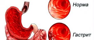
How is it diagnosed?
The earlier the diagnosis is made and gastric metaplasia is detected, the greater the chances of a favorable outcome. Advanced pathology is dangerous due to irreversible consequences that will worsen digestive function.
A biopsy of the mucous membrane of the digestive organ is necessary to make an accurate diagnosis.
It is possible to cure gastric metaplasia after making an accurate diagnosis and determining its type. Pathological proliferation is determined through a comprehensive examination. If you suspect a deviation, you need to contact a gastroenterologist, who will take an anamnesis and prescribe the following diagnostic procedures:
- biopsy of the gastric mucosa;
- blood analysis;
- ultrasound examination of the abdominal organs;
- fibrogastroduodenoscopy.
Features and risks
Intestinal metaplasia occurs:
- complete small intestine;
- incomplete colon.
https://youtu.be/i4xGNiA6UzI
The forms differ from each other in their tendency to malignancy. The first focal type is not precancer, since the altered cells retain their functions. The second type tends to become malignant due to impaired cellular differentiation, which is similar to dysplasia.
The danger of metaplasia lies in the difficulty of differentiating the disease due to the lack of specific signs. The clinical picture is fully consistent with the symptoms of focal pathology that caused cell atrophy. For example:
- In the chronic stage of gastritis with high acidity, “hunger” pains, increased heartburn, and “night hunger” appear.
- With an ulcer - clearly localized pain on an empty stomach. Symptoms will worsen in spring and autumn.
- With reflux syndrome with the reflux of intestinal contents into the stomach - a bitter taste in the mouth, widespread pain, vomiting.
- When a bolus of food is expelled from the stomach back into the esophagus, there is a strong burning sensation in the digestive tract, sour belching.
The only reliable method for diagnosing focal disease is fibroesogastroduodenoscopy, performed simultaneously with a biopsy. With its help, the stomach and intestines are carefully examined and the type of irritant, the localization of the tumor, the prevalence of the focal form are determined, and the malignancy of the process is confirmed or refuted.
Kinds
General typical classification of metaplasia:
- I - complete, small intestine, mature;
- II - incomplete, colonic, immature.
Based on the degree of distribution and area of areas with modified epithelium, they are distinguished:
- mild - damage to 5% of the organ surface;
- moderate -20%;
- pronounced - over 20%.
Based on the severity of the atrophic process in the gland, the following types are distinguished:
- A - insignificant;
- B - intermediate;
- C - complete.
By the nature of the lesion:
- Pyloric or antral form of development, which can be: focal with the replacement of part of the glands of the fundus against the background of inflammation and disruption of the process of cell renewal; diffuse with the distribution of atrophy from the pyloric to the fundus of the stomach, the antrum can be captured; the process takes place without destruction or death of cells.
- Ciliated shape. The appearance of cells of this type in the stomach indicates the development of adenocarcinoma.
- Pancreatic form with the appearance of small-grained cells with acidophilic and basophilic endings.
https://youtu.be/3IqlVAfuqlU
Development of pathology
Pathological disruption of the cellular activity of the digestive organ occurs mainly in older people. Constant renewal of cellular material is a natural process inherent in the genetic apparatus of gastric cells. The intense impact of negative factors on the mucous membrane causes an increase in the rate of their formation. When the proliferation rate of gastric cells exceeds the permissible rate, failures of the genetic program occur, resulting in the formation of intestinal cellular structures instead of gastric ones. The functionality of glandular tissue also changes, performing the role of intestinal absorption.
There are 2 types of intestinal pathology of the stomach.
- Small intestinal, also called mature. Found in most cases in patients suffering from gastritis. The stomach contains a full set of enterocytes: columnar enterocytes, goblet epithelial cells, exocrinocytes with acidophilic type granules, enterochromaffinocytes of various types: EC, ECL, I, S, D. Goblet epithelial cells fill the space of the stomach, being framed by bordered epithelial cells. The specificity of the histology of metaplasia is the presence of acidophilic exocrinocytes, especially in large numbers, accumulating in the recesses of the folded surface along with borderless epithelial cells.
- Intestinal metaplasia of the stomach of the immature type is characterized by the formation of goblet epithelial cells combined with prismatic epithelium, reminiscent of columnar cells of the large intestine. Endoenterocytes characteristic of the small intestine are not detected. A cytogenetic study reveals polyploidy of the nuclei and an increase in the intensity of the regulatory function of the nucleus on metabolic processes in the cytoplasm. When prismatic and endocrine epithelium are identified, mixed mucosal metaplasia is established. Immature (colic) metaplasia is diagnosed much less frequently. When cells characteristic of the small and large intestine are detected, a conclusion is drawn about a transitional form and the beginning of the development of a precancerous condition. A malignant neoplasm of the stomach is detected by histological examination in nineteen out of twenty patients with colonic metaplasia. Benign cell metamorphosis in the colonic form of metaplasia occurs in only one out of ten patients. In the absence of timely treatment, colonic metaplasia ends in death.
The degree of the disease is marked in Latin letters (A, B, C) and is described as minor, incomplete and complete.
Focal intestinal metaplasia, depending on the scale of the lesion, is divided into:
- weak - with the scale of damage to the surface of the mucous membrane up to 5%;
- medium – covering less than 1/5 of the entire surface of the stomach;
- strong – when spreading over more than 1/5 of the mucosal surface.
Based on the nature of the pathology, there are 3 types of metaplastic processes:
- With the pyloric type of disorder, focal and diffuse lesions are distinguished. The focal direction of the pathology reveals lesions of some glands of the fundic type. The renewal of glandular glandulocytes occurs with failures, the results of which are the formation of enterocytes. With a diffuse nature, the glandular cells of the stomach become smaller, since new glandular glandulocytes are involved in pathological processes, the process proceeds unidirectionally from the pyloric to the fundic. The antrum of the stomach is also subject to pathological renewal.
- Ciliary pathology often has an unfavorable prognosis, as it is detected during the development of malignant carcinoma. The appearance of ciliated epithelium in the gastric region is not considered normal, but a malignant neoplasm cannot be guaranteed. About a third of ciliary pathologies accompany benign gastric metaplasia.
- In pancreatic pathology, the glandulocyte is stained unevenly over its surface: one pole of the cell is stained with eosin dyes, the other pole is stained with basophilic dyes.
Complete metaplasia
Foci of mature metaplasia are formed from all cellular structures of the intestine, which contain sialomucins, sulfamucins, enterocytes (goblet, borderless, bordered). When filling the epithelium of the stomach lining, goblet nuclei alternate with bordered ones. A special feature of this type is the presence of Paneth nuclei with apical granulation in the deep pits of the wall. In most cases, small intestinal metaplasia occurs with underlying chronic gastritis. Another name for the problem is enterolization.
Small intestinal is sometimes combined with incomplete metaplasia. They can be located in the same gland or in different parts of the gastric wall. Enterolization is considered to progress to colonic metaplasia. In chronic inflammation, the focus of complete metaplasia turns into incomplete metaplasia in 11% of cases.
Incomplete metaplasia
The immature form of the pathology is characterized by the replacement of gastric nuclei with alternating goblet-shaped and prismatic ones. No other names are found. The epithelium becomes polymorphic with a tendency to increase nuclear-cytoplasmic influences. There is a disruption in the process of maturation and differentiation of the glands, which is characterized by the uniformity of the upper and lower layers.
The incidence of detection of this type of metaplasia is increased in benign diseases of the stomach. Almost all cases of cancer developed precisely because of metaplasia of tissue damage. Therefore, the immature form is a precancerous condition, characterized by a high mortality rate when detected late.
In gastric cancer, colonic metaplasia is found in 94% of cases. Due to the high frequency of coincidences of malignant transformations, pathology of this type requires a differentiated assessment when diagnosing intestinal metaplasia.
Causes
The causal factors influencing the development of gastric pathological processes have not been fully studied. To date, a group of the most significant provoking criteria has been identified, leading to the onset of intestinal metaplasia in 100% of cases. The group includes:
- constant irritation of the stomach walls;
- chronic inflammation of the epithelial membrane;
- prolonged depression or constant psycho-emotional overload;
- inflammatory processes in the esophagus;
- ulceration of the epithelial membrane;
- intestinal dysfunctions, manifested by frequent reflux of gastric and duodenal contents into the esophagus.
Foci of metaplasia are often diagnosed with low acidity of digestive juice in the stomach. Lack of acid leads to selective changes in healthy microflora in the digestive organ. The process is accompanied by the destruction of gastric bacteria with abundant contamination of intestinal microorganisms.
https://youtu.be/H0xVa03Qaak
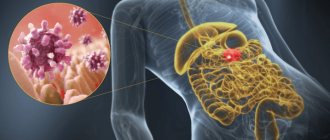
Infection of the mucous membrane with Helicobacter is considered a dangerous phenomenon that provokes many pathologies in the gastrointestinal tract, including metaplasia. The microbe secretes proenzymes that add salts of nitric and nitrous acids to form carcinogenic products - nitrous substances. These substances combine with other carcinogens from ingested foods. Serious damage to gastric tissue occurs, which rapidly progresses, provoking the development of a malignant tumor. The pathological process intensifies when the rules of a healthy diet are not followed, for example, due to the abuse of salted food and strong alcohol.
Basics of nutrition in pathology
As in the case of other diseases of the digestive tract, improvement and recovery is possible only by following the recommended diet:
- Food that irritates the gastric mucosa is excluded from the diet - spicy and salty dishes, smoked foods, fresh milk, yeast bread, pastries and flour sweets. Various types of sour, canned and pickled foods are also contraindicated;
- alcohol and salt are extremely dangerous!!!;
- Under no circumstances should you starve - this provokes the development of ulcers; eat small portions more often, forget about overeating;
- You will also have to give up the habit of overeating at night: the damaged mucous membrane of the stomach, just like you, needs rest;
- It is best to cook dishes using steam or water, and do not eat the food immediately after it is ready while it is hot, but let it cool slightly until it is warm.
Symptoms
Intestinal metaplasia, which affects the gastric mucosa, does not have specific symptoms. The clinical picture of the problem corresponds to the causative factors that provoked the atrophy of the glands. In pathologies with an imbalance in the acidity of the digestive juice, the patient will complain of the following sensations:
- constant burning in the gastrointestinal tract;
- "hunger" pains;
- severe hunger during sleep.
With reflux pathology with constant reflux of bile into the stomach, the patient will complain of symptoms such as:
- diffuse pain;
- vomiting;
- bitter taste in the mouth.
During diagnosis, a disorder in the muscular activity of the sphincter in the antral zone of the stomach will be detected, and reverse peristalsis will appear. Histological analysis will reveal focal metaplasia located in the antrum of the organ. If the provoking factor is an ulcer, the following are noted:
- local pain with high intensity;
- “hunger” pains that subside after eating;
- periodic exacerbation of pain, mainly in spring and autumn.
During an endoscopic examination, characteristic complications such as scars, perforations, and bleeding will be revealed.
If the provocateur is a hormonal imbalance, the course of the pathology will be asymptomatic. The problem is discovered accidentally during a blood test for another reason.
With helicobacteriosis, the symptoms will be typical of chronic gastritis. Along with general symptoms, when examining a biopsy taken from the mucous membrane, waste products of Helicobacter will be detected. The data will be confirmed by a respiratory test, which involves analyzing the patient’s exhaled air, as well as by examining stool. General symptoms of metaplasia, characteristic of all patients:
- the appearance of constant or periodic pain in the epigastric region;
- belching sour or bitter;
- periodic attacks of nausea and vomiting;
- loss of appetite.
Prevention
The main thing is to exclude factors that may affect the process of metaplasia development. And this:
- Smoking and drinking;
- Binge eating;
- Use of GMO products in food;
- Unsanitary conditions in caring for the genitals.
A non-standard method of prevention is vaccination against cervical cancer. The question of whether to take it should be decided by a woman individually with her doctor.
So, cervical metaplasia has a positive prognosis for treatment if it is noticed in time and treatment is started in a timely manner. The main thing is to prevent the development of cancer.
Metaplasia is the process of replacing differentiated cells while maintaining the appearance of the tissue.
Its peculiarity is the development of changes under the influence of unfavorable internal or external factors.
After their influence ceases, the tissues can again restore their previous structure.
Diagnostics
Detection of the problem is possible with fibrogastroduodenoscopy, an endoscopic method for examining the internal cavity of the stomach. The procedure is carried out using an endoscope with a camera and a probe for collecting biological material for histological analysis.
With metaplasia, the biopsy specimen reveals characteristic tissue changes at the cellular level. The presence of sulfamucin, a substance capable of absorbing carcinogens, is detected. The formed compound is the main provocateur of the development of oncology.
https://youtu.be/4Br2J4CUL4o
At the same time, changes in the composition of self-antigens in cells are detected. A carcinoembryonic antigen appears due to decreased differentiation of cell nuclei. This can be confirmed by blood test results. An additional method for detecting pathology is the method of chromatic endoscopy. During the procedure, the condition of the affected tissues stained with methylene blue is assessed. Changed tissues as a result of exposure to the dye acquire a specific color. The method determines the degree of metaplasia. When conducting a comprehensive diagnosis, the size and location of metaplasia is clarified.
What is cervical metaplasia?
The very concept of metaplasia implies changes in the cells of the cervix, which lead to pathology. Simply put, cervical metaplasia is a precancerous condition. If you do not urgently treat this pathology, then in almost 100% of cases it will develop into cancer.
This disease most often occurs in patients who have reached the age of fifty. Metaplasia is very rare in young girls. At a late stage, this disease is quite difficult to cure; it is easier to eliminate it in its infancy. This is why all women undergoing menopause are advised to regularly visit a gynecologist.
Let's discuss in detail all the nuances of cervical metaplasia, so that when faced with it, we have a complete understanding and plan of action. See below for a photo of this pathology.
Medicines
Directions of drug therapy:
- Preventing the return of a bolus of food from the stomach into the esophagus. This will prevent focal or diffuse inflammation in the lower esophagus, causing gastroesophageal reflux disease.
- Destruction of infection that has developed due to the contamination of the gastric and intestinal mucosa with Helicobacter.
- Prevention of cancer development.
The treatment regimen is developed by the attending physician based on the diagnostic results obtained. In order to restore beneficial gastric microflora, drugs like Linex are prescribed. It is recommended to take synthetic and natural immunomodulators and herbal medicine recipes. Appointed:
- proton inhibitors to stabilize the acidity of the digestive tract in conditions of its increase: “Rabeprozole”, “Omeprozole”, “Pantoprazole”;
- antacids to neutralize hydrochloric acid: Maalox, Phosphalugel;
- H2-blockers of histamines to suppress secretory activity: Cimetidine, Ranitidine;
- gastroprotectors to regulate gastric acidity and prevent mucosal destruction.
Drug therapy is carried out with dynamic monitoring of the patient’s condition and recovery processes in the stomach. If the conservative method is ineffective and metaplasia progresses, surgery is prescribed.
What is epithelium?
The gastric epithelium is a collection of columnar cells. They are called single-layer columnar epithelium. Against the background of prolonged irritation of the mucous membrane of the esophagus by gastric juice, for example, with gastroesophageal reflux, when the contents of the stomach are thrown into the esophagus, the risk of developing the gastric variant of esophageal metaplasia increases.
The essence of the disease is the replacement of the stratified squamous epithelium of the esophagus with a single-layer cylindrical epithelium, characteristic of the stomach. The part of the esophagus in which the mucous membrane of the organ passes into the stomach is most susceptible to tissue replacement. It is in the area of connection of the two organs that the most intense irritation with hydrochloric acid occurs against the background of gastroesophageal reflux.
Surgery
Surgery is a radical method of eliminating the problem. The goal of surgical intervention is to prevent the development of a malignant tumor. There are:
- Abdominal intervention, in which the affected area is completely removed. The advantage of this method is that it minimizes the risk of carcinogenic formations.
- Laparoscopy is a minimally invasive intervention that involves a small depth of the lesion. It is carried out using an endoscope.
Advantages:
- short rehabilitation period;
- minimum complications;
- low degree of epithelial injury;
- visual aesthetics of seams.
Treatment with folk remedies
Alternative medicine offers many effective methods to combat and prevent the onset of formation of foci of metaplasia. Treatment with folk remedies is carried out by consuming decoctions and tinctures of medicinal plants and herbal preparations, which reduce the manifestations of inflammation and relieve pain . Recipes:
- Collection with chamomile, yarrow, calendula, marshmallow root. You need to take 5 g of each herb, mix it, pour 500 ml of boiling water. The product is infused in a thermos for 1 hour. The drink is taken after filtering 4 times a day, half an hour before meals. Quantity of 1 dose - 20 ml.
- A decoction of flaxseeds. You need to take 25 g of seeds, pour boiling water, cook for 5 minutes. After settling for 2 hours, the drink is drunk before each snack. Dosage - 30 ml.
- Tincture with St. John's wort. 15 g of crushed herb is poured into 250 ml of boiling water and infused in a thermos for 12 hours. The drink should be consumed after filtering, bringing the volume to 250 ml half an hour before meals. Dosage - 50 ml. The course is 2 weeks, repeated after a 7-day break.
Diet for metaplasia of the gastric mucosa
Effective treatment of foci of metaplasia on the mucosa is made by following the correct regimen and diet. Important Recommendations:
- Exclusion from the diet: dairy products, foods that irritate the gastric mucosa, such as fried, spicy, over-salted foods.
- Refusal to drink alcohol in any form, coffee, carbonated drinks.
- Fractional meals - up to 6 times a day. In this case, the last meal should be 2.5 hours before bedtime.
- Including salads from fresh vegetables and fruits in the daily menu.
- Consumption of all available types of cereals and cereals.
- Food should be warm, since hot food irritates and burns the mucous membrane, and cold food can provoke vasospasm and increase the production of hydrochloric acid due to long digestion.
Forms of the disease
The disease has three forms. They do not in any way affect the process of the disease, but they have a different prognosis for recovery. Another point that led to the division of metaplasia into types is that the epithelium in the cervix has different types.
- Immature metaplasia of the cervix. This form is the least favorable for the patient’s health. It is very difficult to attribute these pathological cells to a specific type of epithelium. In the smear, the doctor observes pathological cells that are small in size and randomly located in the epithelium. The cytoplasm of the cervical cells is disrupted.
- Squamous metaplasia of the cervix with dyskaryosis. Dyskaryosis is a phenomenon of abnormal division in the nucleus in the form of mitoses. This type is more differentiated compared to immature metaplasia.
- Squamous metaplasia of the cervix. The epithelium has the characteristics of a normal cell. Only the arrangement of the cells is abnormal. Problem cells are located in the cervical canal area.
Diet
Proper nutrition allows you to avoid most gastrointestinal pathologies and, accordingly, prevent the development of foci of intestinal metaplasia. Nutrition and diet are considered correct if the following conditions are met:
- reducing the amount of carbonated drinks, spicy and smoked foods, salty and refined foods that negatively affect the condition of the mucous membranes;
- increasing the amount of cereals, bread, vegetables, fruits consumed;
- consumption of boiled lean meat.
Limiting the consumption of harmful substances
Reducing the amount of alcohol consumed will reduce the negative impact on the stomach and avoid damage to the epithelium in the membrane. If abused, gastritis will be a mild problem, and the most dangerous will be a malignant tumor.
If you do not follow basic rules, a gentle nutritious diet, and refuse to lead a healthy lifestyle, you can provoke the development of serious pathologies. If any suspicious signal occurs indicating a malfunction of the stomach, intestines or the entire gastrointestinal tract, you should consult a specialist for advice.