Muscle atrophy is a process that develops in muscles and leads to a progressive decrease in volume, resulting in their degeneration. In other words, gradually the muscle fibers, due to prolonged immobility or inactivity, become thinner, their number decreases, and in severe cases they completely disappear.
Prevalence of the disease, history of the study
SMA: what kind of disease is it? Spinal muscular atrophy is a rare disease called orphan. It occurs sporadically in the population and is genetic in nature.
Around the world, one baby out of 6,000-10,000 receives a diagnosis of spinal amyotrophy (SMA). Such pathologies have been poorly studied and are difficult to diagnose and treat. Including spinal amyotrophy. About 30% of children with orphan diseases do not survive beyond 5 years of age.
Spinal atrophy was first described in the works of Werdnig at the end of the 19th century. He discovered pathological changes in the muscles, peripheral nerves and spinal cord in children with motor function disorders. Werdnig's descriptions note that atrophy in the spinal cord is symmetrical and affects the anterior horns of the structure + anterior (motor) roots. Later, Goffman identifies the disease as a separate nosology.
Why does spinal atrophy appear? Scientists say this is the result of a mutation in the SMN1 gene. Every 40th person on the planet has a defect in this gene. Spinal muscular atrophy can run in families in clinically healthy people. The probability of having a sick child between two carriers is 25%.
Spinal amyotrophy appears in childhood. It is accompanied by atrophy of the muscles of the lower extremities, the inability to walk, sit, or hold up the head. Sucking, swallowing, and respiratory function are impaired. Such defects cause severe developmental delays in children.
Etiology
Clinicians distinguish two types of etiological factors of muscle atrophy - primary and secondary. The primary form of the disease is hereditary, and any neurological pathologies can only aggravate the pathology, but will not become a provoking factor.
Secondary etiological factors include the following:
- constant physical stress, which is a consequence of excessive physical exertion in sports or due to the nature of work;
- infectious pathologies;
- nerve ending injuries;
- myopathy;
- pathologies of motor cells of the brain;
- infectious diseases with typical etiology.
In addition to pathological processes that can lead to muscle atrophy, general predisposing factors for the development of this pathological process should be identified:
- disturbances in the functioning of the peripheral nervous system;
- paralysis;
- mechanical damage to the spine;
- lack of proper nutrition and rest;
- damage to the body by toxic substances;
- disruption of metabolic processes in the body;
- long bed rest.
It should be noted that quite often this symptom can be observed after severe trauma to the musculoskeletal system or being immobile. In any case, rehabilitation after such pathologies should be carried out only by a qualified medical specialist. Self-medication (in this case we are talking not only about taking medications, but also about massage, exercise therapy) can lead to complete disability.
Causes and factors of the disease
Why does spinal amyotrophy appear? What risk factors can be identified? What is the main cause of the disease? The disease is genetic in nature and is transmitted in an autosomal recessive manner. In this case, both parents must have the defective gene. In this case, spinal atrophy occurs in the child in 25% of cases.
How do genes influence the development of the disease? Spinal amyotrophy occurs when there is a deficiency or complete absence of the SMN protein. It ensures the survival of motor neurons. Its deficiency is the main cause of muscle atrophy in SMA. Brain cells die and there is no signal from them to the muscles.
When the long arm of chromosome 5, on which the SMN 1 gene is located, is deleted, the protein is not produced. Spinal amyotrophy develops.
There is a copy of the SMN2 gene. It's not functional. But in the absence of the main gene, SMN2 makes it possible to produce the necessary protein, albeit in small quantities. The more copies of the SMN2 gene a patient has, the milder spinal amyotrophy occurs.
Risk factors:
- Family history is significant for stillbirths;
- The disease was detected in close relatives;
- Familial infant mortality cases;
- Spinal amyotrophy in an older child.
The likelihood of muscle atrophy occurring in younger children is 1:4.
Amyotrophy
Have you ever seen a documentary about astronauts returning from space, they barely move on the ground, there is no gravity in the spaceship. The cosmonauts don’t walk there, but they fly. I don’t know if I’m calling their movement in the spacecraft correctly, there is very little space in the apparatus, they hardly move. They have simulators on which they train so that the muscles do not completely atrophy.
Second example. Some of you broke your arm or leg, remember you were put in a cast, you didn’t move your limb for 1 month, when the plaster was removed a month later, your muscles atrophied so much that you could not bend your arm or leg. Here is an example of muscle atrophy, but after 2 - 3 months your arm or leg recovered and you could already walk, run, or do anything with your arm normally.
What does this muscle memory rely on, you can, in a short time, return to the shape that you had before, a short time is not a week or a month, a little more. But if you compare you and a person who has never done bodybuilding before, then you will pump up much faster to the size you had than him.
So don’t worry too much if you quit training for some reason, you can return to this sport of bodybuilding and start training again and quickly restore all your performance thanks to muscle memory.
Similar articles :
Link for your site:
Show article to a friend:
As we age, exercise becomes an increasingly important aspect of optimal health and longevity. As you probably know, protein is essential for proper muscle growth and maintenance, but this study found that as you age, your body becomes less able to use protein from food to build muscle.
How to recognize SMA
Muscle atrophy in children diagnosed with SMA (spinal muscular atrophy) can begin at different times. The most severe form of muscle atrophy in newborns appears in the first six months of life. The baby is lethargic and sucks poorly. At 3-4 months the child does not roll over on his own and does not attempt to crawl. With spinal muscular atrophy, the baby's posture resembles a “frog”.
There is a type of disease that appears after 7-18 months. Muscle atrophy leads to regression of acquired skills in the child. The baby, who was crawling and starting to get up, suddenly becomes inactive. Over time, he stops sitting up straight. With spinal atrophy, reflexes from the upper and lower extremities disappear.
Symptoms of muscle atrophy with spinal muscular atrophy may appear closer to two years of age. Patients have already mastered the skills of standing and walking. At the same time, muscle atrophy confines them to a wheelchair. Intelligence, urination and defecation functions are preserved. SMA after 2 years is the mildest type of the disease.
How is muscle atrophy determined independently? With spinal atrophy, the muscles decrease in volume, become soft and flabby.
Muscle atrophy (spinal muscular atrophy) has the following symptoms:
- Appears in infancy or childhood;
- Accompanied by disturbances in walking, running, and standing;
- Tremors and fasciculations (twitching) are detected;
- The “rollback” of motor skills is determined;
- With spinal atrophy, no impairment of intelligence or autonomic functions is detected.
If a child has these signs, he should be consulted with specialists. The diagnosis of SMA is made based on DNA testing.
Lower extremity lesions
The main clinical manifestations of leg muscle atrophy:
- Weakness in the legs increases; when walking, patients quickly get tired, cannot lift and hold their legs in weight, often fall, it is difficult for them to sit down and get up from a chair, or climb stairs;
- There is marked muscle thinning, and the limbs appear thin compared to the body. With an asymmetrical lesion, one leg is thinner than the other;
- The extensors of the foot are often affected, causing the foot to sag. When walking, patients raise their knees high, while their soles fall flat to the ground, making a characteristic slap sound - this gait is called a “cock’s gait”;
- Tendon reflexes are reduced.
Pelvic girdle and hips
With atrophy of the thigh and gluteal muscles, the following is noted:
- weakness in the legs during physical activity. Difficulties arise when climbing stairs, running, jumping, or getting up from a squatting position. When trying to take a vertical position, the patient seems to climb on his own, resting his hands on his own hips or surrounding objects (standing up with a “ladder”);
- weight loss in the thighs is not always pronounced due to deposits of fatty tissue;
- change in “duck” gait, when the patient rolls from side to side when walking;
- with hypotrophy of the quadriceps femoris, hyperextension of the legs in the knee joints is observed.
Description of spinal muscular atrophy
SMN protein deficiency leads to the death of motor neurons in the anterior horn of the spinal cord. Following the disruption of innervation, muscle atrophy begins. Spinal atrophy begins in the muscles of the lower extremities. Muscle atrophy affects the muscles of the lower leg, thigh and foot. The proximal muscle groups are most quickly affected.
Muscle atrophy is most severe in young children. The muscles responsible for sucking, breathing, and swallowing quickly become involved in the disease process (spinal atrophy). Muscle atrophy leads to loss of basic life skills. With type 1 SMA, life expectancy does not exceed 1 year.
How long do children with SMA (Werdnig-Hoffmann muscular atrophy) live? Using mechanical ventilation and enteral nutrition methods, you can save a child’s life for 1-2 years. It should be remembered that independent breathing and feeding of the baby is impossible. Hoffmann-Werdnig syndrome is the most severe form of SMA.
Late forms of spinal atrophy are easier. Muscle atrophy begins in the lower extremities. The patient complains of cramps and severe weakness. Tremors and fasciculations may appear. Symptoms are symmetrical on both sides. The muscles in the torso area gradually atrophy. Respiratory and respiratory muscles are rarely affected.
Atrophy of the muscles of the upper extremities (with spinal atrophy) begins last. Muscle atrophy first affects the shoulders and girdles. Later, the disease spreads to the forearms and hand muscles. Tremors and painful spasms also appear on the upper extremities.
Muscle atrophy in patients leads to disability. Each person develops the disease differently. Muscle atrophy may not manifest itself for a long time during active gymnastics and exercise therapy. Progressive atrophy of muscle tissue appears only in types 1 and 2 of the disease.
Spinal atrophy (SMA) and the diagnosis of PSMT have similar symptoms. Spinal syndrome develops when there is injury only in certain muscle groups.
Symptoms
At the initial stage of development, atrophy of the muscles of the back or other parts of the body manifests itself only in the form of increased fatigue from physical activity. As a consequence of this, the patient may experience pain.
As symptoms develop, the clinical picture may be supplemented by the following symptoms:
- with atrophy of the muscles of the limbs, tremor is observed;
- restriction of movements of arms, legs, torso;
- change in habitual gait;
- loss of sensation in the limbs;
- low blood pressure.
If the cause of atrophy of the thigh muscles or other parts of the body is an infectious process, then the clinical picture may be supplemented by the following symptoms:
If the cause of spinal muscle atrophy is damage to the nervous system, then the overall clinical picture may be supplemented by the following signs:
The severity of symptoms in spinal atrophy will depend entirely on the severity of the injury or the degree of deterioration in muscle tone. Therefore, you should consult a doctor at the first signs of damage to muscle tissue. In this case, serious complications can be avoided.
Symptoms of SMA in a child and an adult patient
The most severe clinical picture develops with type 1 sma (spinal muscular atrophy in children). SMA develops in a child from 6 months. The following signs are characteristic:
- Lack of active motor skills in infants with spinal muscular atrophy. The newborn is lethargic, apathetic, eats poorly;
- The baby cannot roll over, sit, or crawl;
- The sucking reflex decreases, frequent choking on food appears, and there are difficulties in releasing secretions from the lungs.
The prognosis for spinal muscular atrophy in children with this type of disease is unfavorable. The disease can be diagnosed by characteristic clinical signs, results of genetic analysis and electromyography.
Dubowitz syndrome develops between 6 and 18 months. A healthy child who could crawl, sit, and walk gradually loses his skills. Over time, weakness of the respiratory muscles appears, limbs, spine and chest are deformed.
Kugelberg-Welander syndrome appears after 18 months. The child can already stand, walk, and perform complex motor actions. Patients cannot cope with running or walking up stairs. Later, chewing and swallowing disorders may occur.
In adults, muscle atrophy appears after 35 years. Its symptoms often resemble other neurological diseases. The disease significantly impedes activity and leads to disability. An adult cannot move without a stroller, and his socialization decreases. The disease has virtually no effect on life expectancy.
Differential diagnosis
Genetic analysis allows you to confirm or refute the presence of the disease. But in 5% of patients with SMA it turns out to be negative due to the atypical location of the mutated gene. In this case, the diagnosis is difficult to confirm based only on the clinical picture.
Diseases with which SMA should be differentiated:
- Childhood botulism;
- Duchenne muscular dystrophy;
- Neuropathy;
- Myopathy (metabolic, congenital);
- X-linked SMA with respiratory disorder.
For differential diagnosis, electromyography, computer and magnetic resonance imaging, and blood tests for hormones are used.
Symptoms of different forms of the disease
There are 4 types of typical SMA and several atypical forms of the disease. The genetic disease is characterized by mutations on chromosome 5 in the form of deletion of the SMN1 gene. With atypical amytrophies, the genotype can be very diverse.
Typical SMA begins in childhood. Atypical amyotrophy Kennedy appears at the age of 30-50 years. Both boys and girls suffer from SMA. And Kennedy's disease is typical only for males.
Signs of Kugelberg-Welander disease
The onset of symptoms appears after the age of 2 years. The child complains of fatigue when walking and running. Unsteadiness and instability of movements appear. The patient's activity gradually decreases, complex motor skills (running, climbing stairs, sports games) are inaccessible to him.
Over time, the patient is forced to move in a wheelchair. He develops contractures of large joints, atrophy of the thigh muscles, and deformation of the chest and spine. This may be accompanied by the appearance of pain. Corrected by timely administration of gymnastics and exercise therapy.
Typical signs:
- Pterygoid blades;
- “Duck” gait;
- Tremor of the tongue and upper limbs;
- Absence of tendon reflexes;
- Atrophy of large muscle groups.
Amyotrophy Kennedy
The disease is typical for adults over 30 years of age. Mostly men are affected. No cases of pathology have been described in women. The first manifestations are characterized by fatigue of the calf muscles and thigh muscles. The patient notices that he cannot walk or stand for a long time.
Atrophy progresses slowly. For about 10 years after the first signs of pathology appear, patients can continue their usual lifestyle. Later, the disease spreads to the upper extremities: tremor appears, the muscles of the head and neck atrophy.
Kennedy amyotrophy is characterized by endocrinological changes. In adult men, a lack of sex hormones is determined, testicular atrophy and a decrease in libido occur. The pancreatic tissue is affected and diabetes mellitus develops.
A characteristic symptom of the disease is fasciculations of the perioral muscles (twitching of the corners of the lips and pulling them out with a tube).
Distal MCA and SMA Vulpian
Distal SMA develops between the ages of 20 and 50 years. The disease causes atrophy of the muscles of the distal parts of the upper extremities (hands, forearms). Later, problems develop on the legs. They involve the feet and legs. Over time, atrophy of all the muscles of the limbs occurs.
SMA Vulpian is characterized by atrophy of the muscle groups of the shoulder blades and lower leg muscles. The syndrome develops after 20 years, but the likelihood of atrophy persists up to 40 years. The disease causes limited mobility of the shoulder joints. The shoulder blades protrude and resemble wings. Hence, a characteristic sign of Vulpian SMA is the symptom of “wing-shaped” shoulder blades.
SMA of this type allows a sick person to remain active and maintain mobility for 30-40 years. Only when the function of the extensors of the feet is impaired and the muscles of the legs are atrophied is the ability to move lost. In general, this is a favorable type of amyotrophy that does not lead to early disability and does not cause death.
Symptoms
Each type of SMA has its own special symptoms, but there are some symptoms that make it possible to combine disparate diseases into one group. This:
- Increasing muscle weakness and atrophy.
- With a disease that appears after 1-2 years, degradation of already achieved abilities, for example, running and walking, is noticeable.
- Tremor of fingers. Trembling is also observed in the tongue.
- Skeletal deformation.
- Preservation of intellectual and mental health in the majority of patients.
Possible complications of SMA
The most common form of complications is respiratory disorders. Patients experience atrophy of the chest muscles. If breathing stops, begin immediate resuscitation and call an ambulance. With the first type of muscular atrophy, respiratory arrest occurs after 1 year of life. Artificial ventilation prolongs the life of a child.
Aspiration pneumonia is the second cause of fatal complications of the disease. In children with muscle atrophy, the cough reflex is impaired and sputum production is difficult. To effectively evacuate it, it is better to have a suction device at home. Breathing exercises are effective; influenza immunization is mandatory.
Pneumonia is infectious and is treated with antibiotics.
In children with SMA, the swallowing reflex is impaired. Frequent choking occurs on solid and then liquid food. If solid food enters the respiratory tract, acute respiratory failure may develop. In this case, people caring for the child should know effective assistance techniques. The Heimlich maneuver adapted for children is used. After a case of aspiration of a hard object, be sure to show the child to the doctor.
It's easy to lose the muscle you've built
These conclusions fall into the category of common sense, according to the principle “do not let good things go to waste.” As we age, exercise becomes an increasingly important aspect of optimal health and longevity.
How to Prevent and Reverse Muscle Wasting
This study explains the biological processes that cause muscle wasting as you get older.
As you probably know, protein is essential for the proper growth and maintenance of muscle mass, but this study found that as you age, your body becomes less able to use protein from food to build muscle.
They also found that in older adults, insulin no longer prevented muscle tearing between meals and at night, as it typically does in younger patients .
This double whammy adds significant muscle wasting in sedentary older adults. And poor blood flow, which prevents nutrients and hormones from being properly delivered to your muscles, can be a big factor.
Exercise is a natural remedy for poor circulation , and the team confirmed that three weight training sessions per week for 20 weeks rejuvenated blood flow in the extremities to the point that it was identical to that of the younger group !
Types of SMA
| SMA, type | Genotype | Clinic |
| 0 (zero) | The SMN gene is missing 1 1 copy of the SMN2 gene | Severe form of the disease Death in utero or in the first month of life |
| 1 (first) spinal atrophy type 1 Werdnig-Hoffmann disease | Deletion or mutation of SMN1 2 copies of SMN 2 in Werdnig disease | Floppy baby syndrome Severe course Death in the first 2-3 years of life (spinal muscular atrophy type 1 is currently being treated, life expectancy results after treatment will be published in the next 5 years) |
| 2 (second) spinal atrophy type 2 Dubowitz disease | SMN1 after mutation turns into SMN2 SMN 2 is present in more than 3 copies | Children aged 1 to 2.5 years are affected. Motor skills are reduced. Survival with 009sma up to 10-14 years. |
| 3 (third) spinal atrophy type 3 Kugelberg-Welander disease | More than 3 copies of SMN2 There may be mutations in the main gene | Appears after 30 years Mobility is maintained for a long time Does not affect life expectancy |
| 4 (fourth) spinal atrophy type 4 Kennedy's disease | Long repeat of three CAG nucleotides in the androgen receptor gene X - linked inheritance | Only men get sick Symptoms start from 30 to 50 years |
As you can see, the symptoms of Werdnig-Hoffmann spinal atrophy are the most severe hereditary form of the pathology. With Werdnig's disease, the "flaccid" child syndrome develops. With type 1 SMA, the baby is motionless, lies in the “frog” position, muscle tone is reduced in all groups (typical of spinal atrophy), joint hypermobility (typical of Werdnig’s disease).
A child with Werdnig-Hoffmann spinal amyotrophy is not able to acquire motor skills. Characterized by frequent shallow breathing and the inability to take a deep breath. With Werdnig-Hoffman disease, the baby does not perform functional muscle tests.
Spinal myotrophy of Werdnig Hoffmann, as well as other types of muscular spinal atrophy, not only the first type, have pathogenetic treatment. The drug "Spinase" can improve the motor skills of children even with a severe form of SMA. The course of application is lifelong. The drug is used in the treatment of Werdnig's disease, Dubowitz's disease and other types of amyotrophies.
Features of manifestations in children
Since atrophied muscles in children are in most cases caused by genetic factors, the first symptoms can be detected in utero - late and weak movements of the fetus are noted; after birth, such children usually die in the first few weeks of life due to paralysis of the respiratory muscles.
With the development of atrophy in infancy, the so-called flaccid child syndrome is formed; in such children, a characteristic “frog pose” is noted - with widely separated hips and a flat stomach, a pronounced decrease in general tone and motor activity, with some diseases there is a disorder of sucking, swallowing and breathing.
Muscle dystrophy in an older child is manifested by disturbances in motor activity and specific deformities of the limbs.
Main types of treatment
Treatment of spinal amyotrophy includes:
- Therapeutic gymnastics;
- exercise therapy;
- Physiotherapy;
- Compliance with diet and nutrition;
- Support with vitamins and nutrients.
Spinal muscular atrophy is now treated with the drug Spinaza. It was registered in 2019 and approved in 2019 as an orphan drug by the Russian Ministry of Health. When starting therapy, the degree of atrophy matters. Its effectiveness is higher when the drug is started early.
Atrophy of muscle mass in SMA occurs as a result of the destruction of the nerve cells that innervate the muscle. The defect is caused by the absence of a protein that ensures the survival of motor neurons. When cells in the spinal cord die, they do not send signals to the muscles. As a result, muscle involution occurs.
The drug Spinase treats spinal muscular atrophy by activating the dormant SMN 2 gene. This oligonucleotide stimulates the production of the essential SMN protein to normal levels. This helps slow down the death of motor neurons.
The drug should provide a breakthrough in the treatment of muscle atrophy. How long do children with muscular atrophy type 2 live? No more than 5-7 years. And now the child will live much longer. Doctors promise a complete reduction of symptoms with early treatment. Such encouraging results were obtained from testing the drug and its use in practice. The presence of two (Risdiplam is not yet registered in Russia) medicines will help provide assistance to children diagnosed with type 2 SMA.
Possible therapeutic strategies
The future is already here and now there are 2 working drugs for SMA. True, the price of an annual course of treatment costs about $750,000. Some countries provide support to families with such a diagnosis. For example, in Italy and Greece, the treatment of a child is paid for by the state.
In our country, the budget of the Ministry of Health does not allow such expenses. But there are charitable foundations that organize treatment for patients with SMA. If the disease occurs in children, treatment should begin as early as possible. This is the key to the success of high-quality disease therapy.
Drug therapy, diet and child nutrition
Modern domestic medicine suggests using drugs that improve neuromuscular conduction (alpha-lipoic acid, l-carnitine, alpha-glycerophosphocholine).
To improve the conductivity of nerve endings, vitamins and dietary supplements (thiamine, pyridoxine, amino acid complexes) are used. In addition, nootropics and vascular drugs are prescribed.
For muscle atrophy, a diet rich in plant and animal protein is recommended. They use porridge, lean meat, cereals, and fermented milk products. Spinach, kohlrabi, and broccoli are rich in B vitamins. Your child's diet should include fresh fruits and vegetables.
It is better to feed the child homemade food. You need to cook stewed, boiled, baked foods. You can steam dishes. Be sure to stick to your meal plan.
Small portions should be used, but multiple meals (5-6 times a day). In the diet, vegetables and fruits should account for 50%, meat and white fish 35%, cereals and other carbohydrates 15%.
Physiotherapeutic methods including massage
Electromuscular stimulation courses help stop muscle dystrophy. EMS machines are available for home use and allow you to train different muscle groups. This does not require any effort from the patient. The device is attached to the skin and sends impulses into the tissue.
Other techniques (magnet, laser, electrophoresis, bioptron) can be used to treat pain due to contractures, curvature of the spine or chest. Light stimulating massage is also indicated for SMA. It allows you to improve blood supply to individual muscle groups. Used in courses of 7-10 sessions.
Folk remedies
Treatment with folk remedies:
- Using a compress of reed panicles;
- Treatment with calcium tincture;
- Use of Echinops.
A compress of reed panicles is used for atrophied muscles. Pour boiling water over two handfuls of panicles and leave for 30-45 minutes. Gauze is soaked in the decoction and placed on the atrophied limb. After the lotion has cooled, massage the limb from the distal to the proximal.
Calcium tincture is prepared from fresh eggs. They need to be washed under water and filled with acid. It is better to use lemon juice from freshly squeezed fruit. In this case, calcium is washed out of the shell and enters the solution. It takes 5-6 days to dissolve. Then the solution must be taken after meals, a teaspoon 3 times a day.
Russian mordovnik is used internally. A 1-2% tincture from the plant is used. It should be taken in courses after meals, 20-30 drops.
New in the treatment of spinal muscular atrophy
There is now an effective drug to treat the disease. The problem for families with SMA from Russia is that the course of treatment with Spinaz is very expensive and is not available to domestic medicine. Janssen is a pharmaceutical company that is going to introduce the drug to the Russian market and ensure the availability of Biogen products.
Risdiplam or RG7916 is an investigational drug for the treatment of SMA. Clinical trial data were presented at the International Neurological Congress. Doctors noted encouraging results from its use. An application to register the drug with the FDA has already been submitted.
Patient care
Relatives are caring for the child. It's often hard work. Therefore, the help of loved ones, volunteers and other caring people is needed. Both the patient and his relatives need psychotherapy sessions and consultations with a professional psychologist. This helps cope with burnout when caring for a sick child.
- To rehabilitate the patient, it is necessary to use anti-gravity positioning. It is used while walking, in bed and in the arms of parents. Its rules are taught by rehabilitation specialists. Special devices are used (high headboard, pillows, anti-decubitus systems);
- Gymnastics, physical therapy, stretching, exercises are performed by patients every day. When exercising, the patient needs an assistant who knows the exercise complex. Daily training of muscle groups is the key to maintaining long-term activity;
- Technical means of rehabilitation facilitate the movement of a patient with SMA and help socialization. For this purpose, strollers, beds, and verticalizers are used;
- Splints, orthoses, and corsets are used to correct posture and maintain joints. They must be made individually for each patient;
- Hydrotherapy is beneficial for patients with SMA. It is advisable to do it daily. The water should be warm (37-38 degrees). Exercises can be combined with swimming.
The patient’s relatives must be proficient in positioning and massage techniques, and know the physical therapy complex. Also, loved ones need to be able to provide first aid in an emergency. Parents must skillfully cope with additional means of rehabilitation and care (aspirator, ventilator, device for sanitation of the oral cavity). Schools and courses on caring for the sick are organized for them.
Treatment prognosis and possible complications
The treatment prognosis before the advent of Spinaz was disappointing. Children with SMA type 0 died in the first days of life. The life expectancy of the babies was no more than a month. With the first type, the child needed constant respiratory support. The average life expectancy was about 1 year, less often up to 2 years.
The effectiveness of Spinaz gives hope to families with SMA. Testing of the drug showed not only improved function, but also the possibility of full recovery. The results of long-term observations will be available after 2022.
Typical complications of SMA: bronchopneumonia, aspiration pneumonia, acute respiratory failure. A sick child needs constant care and round-the-clock supervision. Only in this case can emergency assistance be provided to the baby.
Treatment
The treatment process depends on the form of atrophy, the cause of its occurrence, as well as the age and general condition of the patient. First of all, it is necessary to identify the cause of the disease and get rid of it so that the muscles do not continue to deteriorate. Treatment is also prescribed to maintain muscle function and eliminate symptoms of the disease.
Therapy is prescribed with medication. For atrophy, the following medications are indicated:
- Disodium salt of adenosine triphosphoric acid;
- Vitamins B and E;
- Prozerin.
The patient is also prescribed a course of physiotherapy, massage and physical therapy. Such procedures help normalize muscle tone, strengthen them, and restore normal motor activity. A visit to a psychologist may also be indicated, especially in children with mental retardation.
Treatment of atrophy is permanent; it should not stop, otherwise the disease will begin to actively progress. Therefore, the patient must adhere to the doctor’s recommendations throughout his life, attend physical therapy, do exercises and monitor his diet.
It is recommended to eat the following foods:
- Fresh village milk and fermented milk products. It is recommended to give preference to non-pasteurized products, without preservatives, dyes and harmful additives.
- Chicken eggs.
- Meat, fish and seafood.
- Vegetables, fruits and berries. It is worth giving preference to seasonal products.
- Nuts, seeds.
- Mushrooms and berries.
- Cereals.
- Natural spices and herbs.
In general, food should be healthy, balanced and tasty. The diet should contain a large amount of vegetables and fruits, protein, so that the body receives a sufficient amount of necessary substances. It is worth giving up alcohol, caffeine, sweets, salty and junk food.
What's expected in the future
Despite breakthroughs in treating the disease, drug development for muscle atrophy continues. Scientists are looking for a way to completely get rid of the defective gene. To achieve this, the following areas of genetic engineering are being developed:
- correction and replacement of the defective SMN1 gene;
- increased activity of the SMN2 gene;
- motor neuron protection;
- muscle protection.
Genetic engineering uses the technology of introducing “vectors”. A special viral “vector” is developed in the laboratory. It enters the body and integrates into the damaged DNA fragment.
After this, the SMN1 gene is activated and produces the necessary protein. Vector engineering technology must be tested to detect long-term side effects.
A diagnosis of SMA is not a death sentence. It is important not to give up, but to seek help for a sick child. Daily care from loved ones, physical therapy and gymnastics, and the participation of medical personnel and doctors will help cope with the disease.
Prevention
There are no targeted preventive measures, since this is a symptom and not a separate ailment. In general, you should adhere to the rules of a healthy lifestyle and prevent those ailments that can cause such a disorder.
Muscle atrophy is a process that develops in muscles and leads to a progressive decrease in volume, resulting in their degeneration. In other words, gradually the muscle fibers, due to prolonged immobility or inactivity, become thinner, their number decreases, and in severe cases they completely disappear.
Planning for future offspring
Many parents, after a child with SMA appears in the family, are afraid of a repetition of the situation and do not dare to plan to increase their offspring. To avoid SMA disease in your next child, you can carry out prenatal diagnosis (it allows you to detect the disease in early pregnancy) or use the IVF method with the selection of a healthy embryo (IVF + PGD - preimplantation genetic diagnosis).
There are two types of prenatal diagnostics:
Chorionic villus biopsy - examination is carried out at 10-12 weeks of pregnancy. Samples of the tissue projections that form the placenta are removed. The cells are examined for genetic disorders. The risk is 1 in 100 or 1%.
Amniocentesis - examination is performed after the 14th week of pregnancy. Amniotic fluid containing fetal cells is collected. The cells are examined for genetic disorders. The risk is 1 in 200 or 0.5%.
Important: If there are two carriers in a family, then the probability of having a child with SMA is 25% for each pregnancy
Risks for parents who are carriers of SMA:
- 25% - birth of a child affected by SMA;
- 50% - birth of a child carrier of SMA;
- 25% - the birth of a healthy child.
When planning a future child, consultation with a geneticist is necessary.
Diagnosis and treatment methods
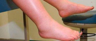
Massage of the lower extremities to maintain muscle tone
First of all, a thorough examination of the patient is carried out to determine his hereditary predisposition to atrophy. A full examination is prescribed to identify chronic diseases, as well as a biopsy of nerves and muscles.
Treatment is always prescribed individually. It all depends on the cause and course of the disease, the age of the patient. A complete cure is impossible, but with the help of drugs and physiotherapy it is possible to return the patient to normal life, relieve pain symptoms, and restore metabolic processes.
The patient must always be treated comprehensively.
Be sure to take not only medications, but also vitamins and minerals that help normalize metabolism.
Such patients must be prescribed a massage. It maintains muscle tone and improves blood flow to a weakened lower leg. You can also use special elastic ankle bandages.
Important! Therapeutic massage should only be performed by a specialist. What it will be and how long the course will last can only be decided by the attending physician.
Why is this happening?
Leg muscle atrophy occurs for a number of reasons:
- The development of a general destructive process as a result of the aging of the body.
- Disturbances in the functioning of endocrine organs and hormonal changes.
- Chronic diseases of the digestive system.
- Connective tissue diseases.
- Damage to peripheral innervation (polyneuritis) due to complications of infectious diseases or helminthic infestation.
- Circulatory disorders of any origin (vascular pathology, atherosclerosis).
- Lack of stress on the leg muscles (due to disability or serious illness).
- Congenital pathologies - genetic abnormalities or fermentopathy.
- Insufficient or improper nutrition.
- Excessive physical activity in the absence of adequate rest.
- Complication after injury.
Therapeutic exercise and physiotherapy
A set of exercises for restoring atrophied muscles is designed so that the load is applied in doses, with a gradual increase. At the same time, muscle mass increases and functionality returns.
At the first stage, exercises are carried out to prevent fatigue or pain. All movements are performed in slow mode. It is important to understand that at the moment the body is weakened and does not have the strength it had before the defeat.
It is recommended to start with exercises in water. This allows you to make all muscle groups work with minimal load. In addition, water will reduce pain. Regular exercise in the pool helps restore tone in the affected areas of the body, and the memory of functioning returns to the muscles. Damaged muscle tissue relaxes. It is advisable to carry out all exercises with an instructor, who will tell you the correct execution and can advise additional movements for better results.
While in the pool, perform the following actions:
- Walk in the pool in a place where the depth reaches your waist;
- Over time, increase the depth and duration of walking;
- In order to maintain balance, a stick or a special water bar is used.
Walking in the pool does not pose a threat to health; on the contrary, the muscles develop and strengthen. The lower body also becomes significantly healthier. At this time, objects taken to maintain balance are used to train the upper half.
Raising your legs to hip height gives good results. Push-ups from the pool floor allow you to develop the muscular system of the upper limbs. To do this, you need to go to the wall of the pool, put your hands on the floor and lift your body out of the water to the position of outstretched arms, then smoothly return to the water. If this exercise cannot be performed, then a simpler version is used: place bent elbows on the floor and lean towards the wall.
After sufficient strengthening of the muscles, they begin to perform exercises without using water space. In this case, the first classes are carried out under the supervision of a doctor or an experienced instructor, then the second stage is allowed to be performed independently.
The movements are performed from a lying position. This includes raising your legs, training your arms, and stressing the whole body.
Why does atrophy develop?
The circumstances due to which muscle atrophy occurs are of two types. The first type refers to burdened heredity. The condition is aggravated by neurological disorders, but they do not provoke atrophy. The secondary type of pathology is associated with external root causes: pathologies and trauma. In an adult, the muscles will begin to atrophy first in the arms.
In children, myofibers atrophy due to:
- Neurological disorders, for example, autoimmune pathology causing muscle paresis (Guillain-Barré syndrome).
- Benign pseudohypertrophic myopathy (Becker myopathy). Appears due to complicated heredity in adolescence and in young people 25–30 years old. This is a mild degree of atrophic changes with damage to the calf myofibers.
- Injuries during childbirth, difficult pregnancy.
- Spinal paralysis in a child caused by infection (poliomyelitis).
- Stroke in a child. Microcirculatory processes in the vessels of the brain are disrupted due to blood clots or hemorrhage.
- Abnormal development of the pancreas.
- Chronic inflammation of muscle tissue.
The main causes of muscle atrophy in adults:
- A job in which a person experiences constant overexertion.
- Incorrectly selected physical education classes, when the load is incorrectly calculated according to the person’s weight.
- Endocrine dysfunctions. If a person is sick, for example, with diabetes, then metabolic processes are disrupted and polyneuropathy occurs.
- Poliomyelitis infection or other infectious pathologies that cause movement disorders.
- Oncological processes of the spinal column, causing compression of the spinal nerve fibers. Their nutrition and conductivity are disrupted.
- Paralysis after injury, infarction changes in the brain.
- Vascular disorders and disorders of the central nervous system, PNS. Oxygen deficiency occurs, muscles starve.
- Chronic intoxication syndrome, which occurs during prolonged contact with chemical toxins, alcohol, drug intoxication.
- Physiological aging, due to which muscles atrophy.
Manifestations of atrophy will occur due to an incorrectly selected diet; if you starve for a long time, the body experiences a deficiency of nutritional components, the muscles lack protein, and they disintegrate. In a child, the processes of dystrophy and degeneration of myofibers develop after surgery. The rehabilitation process is delayed, the child is forced to be immobilized for a long time, and atrophic changes in muscle tissue occur.
Diagnostics
If you suspect the development of a muscle atrophy process, you should immediately seek medical help. The doctor's specialization during the initial examination will depend on the current clinical picture and general condition of the patient.
Electromyography
The diagnostic program consists of the following:
- physical examination with general history;
- clinical and biochemical blood test;
- electromyography;
- hormonal studies;
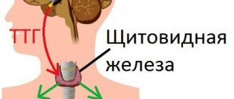
- Ultrasound of the thyroid gland;
- muscle tissue biopsy;
- nerve conduction testing;
- CT and MRI.
Additional diagnostic methods will depend on the current clinical picture and the patient’s condition at the time of seeking medical help. It is important that if the patient took any medications to eliminate symptoms, the doctor should be notified about this before the diagnosis begins.
Result
This work is not easy, but the reward is very great - a real rollback of time, the reconstruction of a face, its youthful structure. The face becomes relaxed and youthful. The tissues in the middle part of the face are smoothed out, nasolabial folds are significantly reduced. An additional bonus is that the condition of the gums improves, neck pain and headaches decrease. Background stress that you are used to not noticing will be reset: you will feel it being erased from muscle memory.
And at the same time, this is a great opportunity to balance your teeth, chewing muscles and temporomandibular joints, for which dentists will only praise you.
By the way, visiting the dentist is the first step to start working with the chewing apparatus. It's time to treat and insert teeth, raise or correct the bite, since the condition of the oral cavity and teeth has a direct impact on the entire posture and gravitational vertical of the body. Don't skimp on dentists, especially those with osteopathic specialization!
Functional states and recommendations
Considering the progressive nature of the disease, the scope of medical care for a particular person is best chosen based not on the type of disease, but on the functional state of the patient - it is determined by what physical abilities he has preserved. For different types of SMA, the age at which these conditions are achieved is different, but the recommendations for patients are the same. It is important to take into account the individual nature of the disease and the characteristics of a particular patient.
Recumbent
Average age of onset of the condition: SMA I – from birth; SMA II – over 12 years; SMA III – over 30 years old.
MOVEMENT
Main problems
- Limitation of mobility: independent movements are minimal or completely absent;
- Pressure ulcers: the risk increases with the patient's weight and age;
- High risk of developing contractures and other deformities, as well as scoliosis;
- Chest deformity;
- High risk of developing joint arthrosis;
- High risk of developing osteoporosis;
Secondary complications
- Pain syndrome;
- Impaired functioning of the respiratory system;
- Disruption of the digestive system;
- Disruption of the circulatory system;
Diagnosis of musculoskeletal disorders
- Regular examination by a physical therapist or podiatrist is required;
- Decreased joint mobility is the first sign of the development of contractures;
- Redness of the skin is the first sign of bedsores;
- Pain syndrome can be determined by the patient's complaints. If his speech is impaired, it is necessary to pay attention to non-verbal signs: facial expression, body movements, crying and the inability to console the patient, groans. The most common behavioral reactions to chronic pain: forced position, reluctance to change it, a frozen, suffering or indifferent facial expression, decreased interest in what is happening around, excessively quiet behavior, increased irritability, decreased mood, sleep disturbance, anger, loss of appetite;
- Symptoms of arthrosis are pain when the joint is loaded (subsides when the joint is at rest), decreased mobility, crunching, and muscle tension in the joint area. Periodically, a joint affected by arthrosis may swell and become deformed over time.
Solutions
- Correct positioning allows you to provide the patient with the necessary comfort, reduce the risk of secondary complications, the development of deformities and contractures, and ensures the safety of the patient;
- Using special U-shaped mattresses: - mattresses filled with millet take the shape of the patient's body, distribute weight evenly, and help maintain the correct body position - mattresses filled with holofiber help maintain the desired posture;
- Regular changes in body position and massage can reduce or avoid the occurrence of bedsores;
- Vigorous physical activity allows a person to retain existing skills longer. It is possible to imitate natural movements using special simulators. Swimming helps the patient reduce the influence of gravity and allows him to perform those movements that he does not have enough physical strength to perform on land;
- Preference should be given to active exercises, encouraging the patient himself to perform the range of movements available to him and use techniques to increase their volume;
- To facilitate patient care, it is recommended to use special devices and means: orthopedic bed, multiroller, electric lift, bathing chair, etc.;
- You should carry a small child with his head on your shoulder so that it does not fall, and also provide an anti-gravity position to reduce the risk of accidental aspiration. To move a teenager and an adult patient, it is necessary to use special devices;
- The use of orthopedic devices will avoid or slow down the development of deformities and contractures;
- It is recommended to use specially selected and adapted technical means of rehabilitation (TSR), for example, an adapted wheelchair (in some cases with an electric drive), a verticalizer;
- Changing the position of the body in space is an important mechanism of habilitation. To make this possible, special supports for sitting or standing are used. The main principle is verticalization by age. Contraindications – severe scoliosis or contractures in the legs, large amounts of sputum;
- To reduce pain, drug therapy can be used as prescribed by a doctor.
Necessary equipment
Positioning
- U-shaped millet mattresses for children up to 110 cm;
- U-shaped mattresses with holofiber for patients above 110 cm;
- Square pillow with ear cutout;
- Functional bed.
TSR
- Active-passive type simulators;
- Verticalizer;
- Stroller (with or without electric drive);
- Orthopedic chair.
Orthopedics
- Rigid corset;
- Orthoses;
- Splints.
BREATH
Main problems
- Weakness of the respiratory muscles and deformation of the chest lead to inadequate independent breathing and insufficient ventilation of the lungs;
- Impaired cough function and removal of mucus from the respiratory tract;
- Swallowing impairment, danger of aspiration of food and saliva, and, as a result, high risk of airway occlusion, hypoxia or suffocation;
- During respiratory diseases, a strong regression of respiratory function occurs.
Diagnosis of breathing disorders
- Chest deformity;
- Shortness of breath, rapid breathing;
- Blue discoloration of the skin in the area of the nasolabial triangle and nails;
- Decreased oxygen level in the blood (drop in saturation);
- Restless behavior;
- To monitor the dynamics of respiratory functions, diagnostics are required every 3-6 months.
Basic diagnostic procedures
- Overnight pulse oximetry;
- Change in vital capacity (VC) (for patients older than 5-7 years);
- Transcutaneous capnometry;
- Polysomnography;
- Cardiorespiratory monitoring.
To monitor the dynamics of respiratory functions, diagnostics are required every 3-6 months.
Solutions
- Correct posture is the key to safety and control of breathing. It is important to ensure that the patient is in an anti-gravity position so that saliva flows out of the mouth under the influence of gravity;
- Daily breathing exercises using an Ambu bag;
- It is important to ensure the patency of the airways, if necessary, clearing them of sputum;
- Drainage of sputum. It is recommended to use an electric suction and cougher, or manual coughing techniques in combination with an Ambu bag;
- If swallowing is impaired, installation of a nasogastric tube or gastrostomy tube is recommended to reduce the risks of aspiration and airway obstruction;
- If indicated, ventilation may be used (invasive or non-invasive);
Important Attention! The use of oxygen without respiratory support is contraindicated in neuromuscular diseases. Oxygen should only be used in emergency situations as a short-term measure or as palliative therapy in terminal conditions.
Required equipment (safety kit)
- Ambu bag - for daily breathing exercises and assistance in critical situations;
- NIV. Non-invasive ventilation (NIV) or invasive ventilation (IVL) devices - to support breathing;
- Electric suction - for cleaning the upper respiratory tract from saliva and phlegm;
- Cougher - for cleaning the lower respiratory tract;
- Pulse oximeter - to monitor the oxygen content in the blood.
NUTRITION
Main problems
- Risk of aspiration (food and saliva entering the airways) when swallowing;
- Bulbar syndrome;
- Reflux;
- Incomplete opening of the mouth;
- Swallowing problems;
- Lack of weight;
- Metabolic disease;
- Violation of peristalsis.
Diagnosis of eating disorders
- Choking;
- Long feeding.
Solutions
- If it is impossible to sit, when eating, the angle of elevation of the head of the bed should be at least 30 degrees, including to prevent the reflux of stomach contents into the esophagus and respiratory tract;
- For children with type 1 SMA, side-lying (anti-gravity) feeding is safest;
- If swallowing is impaired, installation of a nasogastric tube or gastrostomy tube is recommended to reduce the risks of aspiration and airway obstruction;
- Installing a tube or gastrostomy for swallowing disorders will ensure that the body receives the required daily amount of food and nutrients
- Fractional balanced feeding every 3-4 hours;
- To eliminate intestinal problems and other gastrointestinal disorders, drug therapy can be used as prescribed by a doctor.
Necessary equipment
- Gastrostomy;
- Probe.
Important Detailed information on the other two categories of patients with SMA (sedentary, walking) can be found on the website of the special project of the SMA Families Foundation.
How is the disease treated?
Based on the data obtained during diagnostic measures, the doctor will prescribe treatment for leg muscle atrophy, which includes:
- drug therapy;
- physiotherapy;
- exercise therapy;
- development of a special diet.
The health complex is selected individually and depends on the cause of the disease, its form, the patient’s condition (age, presence of chronic diseases, etc.).
Drug therapy
Medications are prescribed to the patient primarily to relieve symptoms. For this purpose use:
- antispasmodics “No-shpa”, “Papaverine” - eliminate pain, reduce muscle tone, dilate blood vessels and normalize blood circulation in the lower extremities;
- "Galantamine" - improves the conduction of nerve impulses in the legs;
- B vitamins – correct the functioning of the peripheral nervous system, activate metabolic processes in the body’s tissues, allowing the volume of the affected muscle system to be restored.
Only the attending physician has the right to write prescriptions for drug therapy, because he is fully familiar with the person’s condition, knows the characteristics of the existing illness and is aware of the patient’s allergy to one or another component of the drug.
Physiotherapy
The regeneration of affected tissues can be improved by exposure to low voltage current - electrical stimulation. During this procedure, a person does not experience pain. It is worth noting that electrical stimulation is pointless as the only treatment method.
Massage as a type of physiotherapy is the most useful method for improving blood flow to affected areas, restoring cellular nutrition, accelerating tissue regeneration, “breaking” stagnation and relieving excess tension. This procedure should be carried out daily, and if possible, a couple of times a day for a long time (at least 2-3 weeks).
Massage movements should be superficial, so as not to aggravate the situation; the procedure should be started from the periphery (from the foot) and do not forget to work the buttocks. The quadriceps femoris muscle may be subject to a little more manipulation.
Exercise therapy
Therapeutic exercise is prescribed by the attending physician and should be carried out only under the supervision of an experienced specialist: he will help you recover competently, gradually increasing the load and working out the damaged areas.
Causes
Hypotrophic processes begin with a malnutrition of muscle tissue.
Dysfunctional disorders develop: the supply of oxygen and nutrients that ensure the vital activity of the organic structure does not correspond to the volume of utilization. The protein tissues that make up the muscles, without replenishment or due to intoxication, are destroyed and replaced by fibrin fibers. Under the influence of external or internal factors, degenerative processes develop at the cellular level. Muscle fibers that do not receive nutrients or accumulate toxins slowly atrophy, that is, die. White muscle fibers are affected first, then red ones.
White muscle fibers have the second name “fast”, they are the first to contract under the influence of impulses and are turned on when it is necessary to reach maximum speed or react to danger.
Red fibers are called “slow”. To contract, they require more energy; accordingly, they contain a larger number of capillaries. That is why they perform their functions longer.
Signs of the development of muscle atrophy: first, the speed slows down and the amplitude of movements decreases, then it becomes impossible to change the position of the limb. Due to the decrease in the volume of muscle tissue, the popular name for the disease is “tabes.” The affected limbs become much thinner than healthy ones.
Factors that cause muscle atrophy are classified into two types. The first includes genetic predisposition. Neurological disorders aggravate the condition, but are not a provoking factor. The secondary type of disease in most cases is caused by external causes: illness and injury. In adults, atrophic processes begin in the upper extremities; for children, the spread of diseases from the lower extremities is typical.
Muscle atrophy in children is genetic, but can appear later or be caused by external causes. It is noted that they are more likely to experience damage to nerve fibers, which disrupts impulse conduction and nutrition of muscle tissue.
Causes of the disease in children:
- Neurological disorders, including Guillain-Barre syndrome (an autoimmune disease that causes muscle paresis);
- Becker's myopathy (genetically determined) manifests itself in adolescents 14-15 years old and young people 20-30 years old; this mild form of atrophy extends to the calf muscles;
- Severe pregnancy, birth injuries;
- Poliomyelitis is a spinal paralysis of infectious etiology;
- Pediatric stroke - disruption of blood supply to the cerebral vessels or cessation of blood flow due to thrombus formation;
- Back injuries with spinal cord damage;
- Disturbances in the formation of the pancreas, which affects the condition of the body;
- Chronic inflammatory processes of muscle tissue, myositis.
Myopathy (a hereditary degenerative disease) can be provoked by paresis of the nerves of the limbs, anomalies in the formation of large and peripheral vessels.
Muscle atrophy in adults can develop against the background of degenerative-dystrophic changes that arose in childhood, and appear against the background of spinal and cerebral pathologies, with the introduction of infections.
The causes of the disease in adults can be:
- Professional activity that requires constant increased physical stress.
- Illiterate training if physical activity is not designed for muscle mass.
- Injuries of various types with damage to nerve fibers, muscle tissue and the spine with damage to the spinal cord.
- Diseases of the endocrine system, such as diabetes, and hormonal dysfunction. These conditions disrupt metabolic processes. Diabetes mellitus causes polyneuropathy, which leads to limited movement.
- Poliomyelitis and other inflammatory infectious processes in which motor functions are impaired.
- Neoplasms of the spine and spinal cord causing compression. Innervation of trophism and conductivity appears.
- Paralysis after injury or cerebral infarction.
- Impaired function of the peripheral circulatory and nervous systems, resulting in the development of oxygen starvation of muscle fibers.
- Chronic intoxication caused by occupational hazards (contacts with toxic substances, chemicals), alcohol abuse and drug use.
- Age-related changes - as the body ages, the thinning of muscle tissue is a natural process.
Adults can provoke muscle atrophy with illiterate diets. Prolonged fasting, during which the body does not receive beneficial substances that restore protein structures, causes the breakdown of muscle fibers.
In children and adults, degenerative-dystrophic changes in muscles can develop after surgical operations with a protracted rehabilitation process and during serious illnesses against the background of forced immobility.
Muscle atrophy can be primary (simple) or secondary (neurogenic).
Primary develops due to damage to the muscle itself. The cause of the pathology may be unfavorable heredity, which is manifested by a congenital defect of muscle enzymes or high permeability of cell membranes. In addition, environmental factors that provoke the development of the disease also significantly influence. These include: infectious process, physical stress, injury.
Muscle atrophy (pathology photo shown in the figure) can develop as a result of injury to the nerve trunks, an infectious process that affects the motor cells of the anterior horns of the spinal cord. Due to damage to peripheral nerves, patients experience decreased sensitivity.
Factors contributing to the development of the disease are malignant diseases, paralysis of peripheral nerves or the spinal cord. Very often, pathology appears against the background of starvation, various injuries, intoxication, due to a slowdown in metabolic processes, prolonged motor inactivity, and chronic diseases.
Secondary muscle atrophy can be of the following types:
- Neural amyotrophy. The pathology is associated with damage to the feet and legs. Patients have a gait disorder - the person raises his knees high when walking. Over time, foot reflexes completely fade away, and the disease spreads to other parts of the body.
- Progressive muscle atrophy. Signs of the disease usually appear in childhood. The disease is severe, characterized by severe hypotension, twitching of the limbs, and loss of tendon reflexes.
- Aran-Duchenne muscle atrophy. With this disease, damage occurs to distant parts of the upper extremities - fingers, interosseous muscles of the hand. In this case, tendon reflexes disappear, but sensitivity remains. Further development of the disease involves the muscles of the trunk and neck in the pathological process.
As noted above, muscle atrophy can be primary or secondary. The causes of the pathology are different, depending on its form. Thus, the primary one is most often a hereditary pathology; a metabolic disorder is passed on to the child; the baby is born with a disorder in the enzymes that are responsible for muscle function.
Direct muscle damage can also occur with various infectious diseases, with injuries, with excessive physical exertion, and primary atrophy also occurs with myopia. Often the cause of atrophy is damage to the nerve trunks, for example, with polio.
The following negative factors can also cause muscle atrophy:
- Oncological diseases;
- Damage to nerve endings due to pathology of the peripheral nervous system;
- Fasting, poor nutrition;
- Chemical poisoning;
- Spinal cord paralysis;
- Physiological slowdown of metabolic processes in elderly people;
- Prolonged immobilization of the patient, for example, after severe injuries and operations.
The onset of pathology associated with leg muscle atrophy is influenced by numerous aspects, including:
- A decrease in metabolic rate is accompanied by natural aging of the human body;
- All kinds of diseases of the endocrine system, resulting in hormonal imbalance in the body;
- Difficulties with the digestive system;
- The presence of connective tissue diseases;
- Transfer of polyneuritis;
- Consequences of certain infectious diseases and diseases associated with the activity of parasites;
Parasitic infection - Enzymeopathy, other hereditary genetic disorders;
- Poor, irrational nutrition;
- Previous injuries or constant physical activity.
Atrophy of the quadriceps femoris muscle often occurs after surgery and injury.
At the initial stage, a person notices fatigue and muscle weakness in the legs that occurs after physical activity. The calf muscles of the legs become noticeable. The proximal muscle groups of the legs are primarily affected by atrophy. The process is expressed in limited motor ability. For example, it becomes difficult for a person to walk up the stairs.
The leg muscles atrophy slowly, the process drags on for years. One or both sides are affected, being symmetrical or asymmetrical.
Any symptoms of the disease are closely related to the patient’s age, general condition, and the nature of the development of the form of the disease. This is reflected in the choice of treatment.
Clinical manifestations:
- Constantly increasing weakness of the leg muscles.
- Tremor.
- Unpleasant sensations (for example, the feeling of insects scratching under the skin).
Unpleasant sensations in the legs
The most striking signal indicating the development of atrophy is a significant reduction in the muscle in which the process develops. Moreover, the decrease becomes noticeable even at an early stage.
The disease is considered chronic. As the disease progresses, there are relapses, when severe pain is felt in the sore muscle. Remission occurs, but the symptoms only slightly cease to bother the patient.
Primary muscle atrophy is distinguished when the muscles themselves and motor neurons are affected. The process is associated with previous injuries or aggravated heredity.
It is characterized by the rapid onset of fatigue, loss of tone and involuntary twitching of the legs.
With secondary atrophy, the muscles of the feet and legs are subject to pathological processes - the areas are deformed, the gait of the sick person changes noticeably, accompanied by a specific raising of the knees. More often the process is associated with past infectious diseases or due to hereditary burden. As the disease progresses, it can spread to the upper body. Complete or incomplete paralysis is observed.
Medicines
Medicines prescribed by a doctor during the rehabilitation period:
- Vitamin complexes consisting of groups A, E and B;
- Contains amino acids;
- Means for accelerating regeneration processes. Potassium orotate, Riboxin, Retabolil are prescribed;
- Adenosine triphosphate. A substance that supplies energy to cells for accelerated biochemical reactions in tissue cells;
- Medicines to improve blood circulation on the periphery of a damaged organ. For this purpose, nicotinic acid and pentoxifylline are prescribed;
- Nootropic drugs. Helping to restore brain and spinal cord tissue;
- And we definitely need substances that will work to restore lost nerve connections. This is the so-called antagonists.
There are several medications used to solve the problem of how to restore atrophied muscles:
- Galantamine. Taking the medicine helps nerve fibers conduct impulses. This process becomes easier. This drug is used for a long time, the dosage is regulated by the doctor. The drug enters the body by injection: into a vein, into a muscle, or as a subcutaneous injection;
- Pentoxifylline. Aimed at improving blood supply to areas located on the outskirts of the affected area. Blood flow in the legs is normalized. No-spa and papaverine can be an equivalent replacement for the drug. They remove spasmodic phenomena, allowing blood to move without obstacles;
- Vitamins belonging to group B return impulse metabolism in nerve fibers back to normal. Metabolism in cells is also significantly accelerated, which makes it possible to reduce the time of rehabilitation therapy, since the muscle quickly restores its previous volume.
SMA mechanism
SMA is caused by a defect in the SMN1 gene. The SMN2 gene partially compensates for the loss of the SMN1 gene.
What's happening?
- The SMN1 gene is damaged and not normal
- Important proteins are not produced in sufficient quantities
- Motor neurons do not function properly and die
- Pulses are not recognized
- Muscles lose strength and atrophy
- Movement is limited, making moving, breathing and swallowing difficult
Diagnostics and assistance from specialists
When identifying symptoms that may indicate illness, a comprehensive examination is necessary to establish an accurate diagnosis.
Diagnosis: You should consult a neurologist, a specialist in neuromuscular diseases, who can make a diagnosis based on symptoms. To confirm the diagnosis, DNA diagnostics and genetic consultation are required.
Genetic diagnosis : DNA test to detect deletion of the SMN1 gene and determine the copy number of SMN2.
Additional research:
- Blood biochemistry: creatine kinase (CK) - normal in SMA I, normal or slightly increased in other types;
- Electroneuromyography shows a decrease in nerve impulses and helps differentiate SMA from other neuromuscular diseases. Sensory nerve conduction is usually normal.
Health care
Medical specialists:
Neurologist makes a diagnosis, prescribes supportive treatment, and constantly monitors the progress of the disease.
Geneticist - makes a diagnosis and, if necessary, advises the family on issues of further offspring.
A local pediatrician (therapist) helps treat diseases that affect everyone.
A pulmonologist or resuscitator helps identify and compensate for respiratory disorders, solve problems with coughing, and provides advice on respiratory support.
Orthopedist - assesses disorders of the musculoskeletal system (contractures, deformities), determines the required scope of preventive measures, and helps correct these disorders.
Neurosurgeon - deals with the correction of scoliosis.
A physical therapist selects a set of exercises and habilitation procedures and teaches parents to regularly do them at home on their own.
A nutritionist or dietician helps you choose the optimal diet.
Gastroenterologist - helps in case of problems with the stomach and intestines.
Cardiologist - monitors the functioning of the cardiovascular system.
Palliative care specialist helps to comprehensively improve the quality of life.
Other specialists are brought in as specific problems arise.
IMPORTANT ASPECTS:
- Family-oriented approach
- the doctor takes into account the family’s opinion on all issues related to the treatment of the child, including the conduct of medical interventions and their volume, their acceptability and timing; - Focus on the patient’s quality of life
- before suggesting the use of any medical technologies to the family, it is necessary to consider how this will affect the quality of life, because it is important to live fully, and not to exist; - High-quality communication and full information
to the patient and family members about all aspects of SMA - as complete information as possible about the disease, what will happen and what will have to be faced, as well as how to cope with it; - Training in practical skills
for the care and use of medical equipment should be an integral part of medical care; - Interdisciplinary and multiprofessional approach
- the work of an interdisciplinary team is required (for example, it is impossible to limit yourself to the observation of a neurologist and receive the full range of necessary assistance for full support).