Adhesive disease of the abdominal cavity is a disease associated with the appearance of connective tissue cords between the internal organs and the layers of the peritoneum. Pathology occurs after surgery, less often – against the background of inflammatory processes. Treatment is aimed at relieving negative symptoms and preventing complications. In severe situations, surgery is indicated.
How does adhesive disease develop?
With appendicitis, which was not treated at the time, a limited cavity is formed when the peritoneum is adjacent to the area of inflammation. This process is called appendicular infiltration. Similar cases occur during surgical interventions.
The peritoneum may adhere to:
- Inflammation zone;
- The organ being operated on;
- Incision sites.
The peritoneum adheres to all organs at the stage of inflammation or during chronic inflammation, especially in infectious diseases of the ovaries and fallopian tubes.
During the so-called “gluing”, the normal functioning and arrangement of organs is disrupted.
In the future, such adhesions shorten and become denser, which leads to even greater displacement. In this case, normal blood flow is disrupted and oxygen starvation begins to develop, which subsequently leads to abdominal pain due to adhesive disease.
With such displacements, the movement of feces is disrupted, which leads to constant constipation. Intestinal obstruction may develop in the future.
ICD-10 (International Classification of Diseases) distinguishes two types of diseases, such as:
- Adhesive disease of the pelvis;
- Adhesive disease of the abdominal cavity.
Adhesive disease of the abdominal cavity Visceral
Essentially, these are two identical diseases, since the pelvis belongs to the abdominal cavity. But this disease is usually classified as a separate category, because the cause of adhesions, in this case, is an inflammatory disease of the female genital organs.
Therapy methods
In case of formation of adhesive growths, conservative therapy is ineffective . Medicines are needed for temporary use to relieve some symptoms. Medicines are part of complex therapy and serve to alleviate the symptoms of concomitant conditions.
The most effective and radical method of treatment is surgery with dissection of the fused areas around the organs. Laparoscopy is especially popular, since the intervention is minimally invasive, and the recovery period is easier to tolerate compared to traditional surgery. Surgery is prescribed for female infertility, intestinal obstruction and severe dyspeptic symptoms that interfere with the patient’s normal life.
Depending on the situation, the following manipulations may also be prescribed::
- Gradual adhesive excision followed by separation of organs from adhesions. It is performed by laparoscopy.
- If there is occlusion of the intestinal lumen, torsion or necrosis, resection is required. The intestine is partially removed, and the remaining areas are stitched together. If it is not possible to connect the tissues, a stoma is placed to connect parts of the intestine.
- If extensive intestinal damage occurs, anastomoses must be performed.
Attention! In approximately 20% of cases, surgical dissection leads to regrowth of adhesions, so careful prevention is necessary.
Traditional methods using herbs are ineffective, but you can try doing exercises to stretch the tissue. Home remedies can only help with fresh adhesions in the peritoneal area, when only a film forms there. As you exhale, try to stretch them with your fingers.
Diet recommendations include split meals, avoiding fatty, fried, and floury foods . Food is boiled or steamed. You need to eat plenty of vegetables and fruits to avoid constipation. Bonding products are contraindicated.
Causes
There are many reasons for the development of adhesions, but they all lead to damage to the tissues and organs of the abdominal cavity, no matter what type they are.
Let's look at the most common causes of adhesions. These are:
- Mechanical injuries of the abdominal cavity;
- Chemical damage;
- Inflammatory diseases.
Mechanical injuries of the abdominal cavity rightfully include the following indicators:
- Surgical operations;
- Bullet wounds;
- Stab wounds;
- Falls from a height;
- Beats;
- Falling onto a hard object.
Chemical damage includes the following indicators:
- Rupture of the gallbladder with effusion of bile;
- Severe pancreatitis with many consequences;
- Perforation of the stomach with access to the abdominal cavity;
- Burns from alkalis or acids due to intentional or accidental use.
Inflammatory diseases include the following indicators:
- Pipes and appendages;
- Disease of the large intestine;
- Disease of the small intestine;
- Gallbladder diseases;
- Appendix (appendicitis).
Very often in women, the development of adhesive disease occurs as a result of chronic inflammatory processes in the female genital organs. With minor symptoms and long-term treatment, adhesive disease can lead to an irreversible process and infertility.
Associated indicators of adhesive disease are diabetes, poor diet, and foreign bodies in the wound.
Symptoms of adhesive disease of the abdominal organs
Doctors describe the clinical picture of the disease in question in just four words:
- vomit;
- abdominal pain;
- flatulence;
- constipation.
Of course, these symptoms do not all appear at the same time - depending on where exactly the adhesive process is located, different combinations of symptoms will be present. In some cases, for example, with adhesions of the internal genital organs in women, the disease does not manifest itself at all and is discovered by chance.
Abdominal pain occurs with the development of intestinal adhesive disease, because when compressed by adhesions, a section of the intestine bleeds. In this case, the intestines do not immediately “give up”, but peristalt and try to fight the problem. Pain in the abdomen during adhesive disease is always prolonged, because compression of the organ occurs gradually - at first the pain is dull/aching, then it becomes sharp/cutting. Patients describe their sensations in different ways: for some, the pain has a clear localization, for many, these unpleasant sensations “spread” throughout the abdomen.
Please note: if, in case of intense abdominal pain, qualified medical care is not provided, then after a while the patient’s condition will normalize. But this does not mean that the disease has receded! The nerve endings were completely destroyed and irreversible processes began in the damaged organ.
But even with the disappearance of the pain syndrome, the intestines continue to work, but no movement of feces occurs. This condition is always accompanied by constipation, vomiting and flatulence. It is noteworthy that if the pathological process in question develops in the small intestine, then the first symptom will be vomiting - profuse, repeated, containing bile, intestinal and gastric juice. Vomiting does not alleviate the patient’s condition at all, the feeling of heaviness in the stomach does not disappear, each attack is accompanied by intense pain in the intestines, and the nausea does not stop for a minute.
If adhesive disease develops in the large intestine, then the first symptom will be constipation. The problem is that this syndrome often bothers a person even in the absence of adhesive disease, and in most cases the patient simply does not pay any attention to it. But the adhesions are increasingly shortened and entangled in the intestines, which leads to disruption of its functionality. Feces do not pass out, remain in the intestines and begin to ferment. This process ends with the formation of a large amount of gases, which also have no outlet - the stomach swells, the intestines stretch, and the person experiences severe pain.
Please note: with adhesive disease of the large intestine, neither enemas nor any medications with a laxative effect bring relief to the patient. Such procedures may even make the situation worse.
The pathological condition in question can also develop in the pelvic organs, and in this case the symptoms of adhesive disease will be somewhat different. Firstly, a woman begins to experience menstrual irregularities, which results in irregular and painful bleeding. Secondly, a woman can pay attention to the presence of mild pain during and after sexual intercourse. Such a pathological condition requires immediate seeking qualified medical help, as there is a high probability of developing infertility.
Classification
According to the International Classification of Diseases (ICD-10), abdominal adhesions are divided into the following types:
- Abdominal adhesions (K66.0);
- Postoperative adhesions in the pelvis (N4);
- Peritoneal inflammatory adhesions in women in the pelvis (N6).
The following types of adhesions are classified as abdominal adhesions:
- Stomach adhesions;
- Seal seals;
- Adhesions of the mesentery of the large or small intestine;
- Pelvic adhesions in men;
- Intestinal adhesions;
- Diaphragm commissures;
- Abdominal adhesions.
Preventive actions
If we analyze all the data on adhesive disease of the abdominal organs, we can conclude that it is impossible to prevent the development of the pathological process in question. And this is partly true, but there are also non-surgical reasons for the development of adhesive disease that can actually be prevented.
For example, if you follow certain rules, you can significantly reduce the risk of developing adhesive disease in the pelvis . These rules include:
- Appendicitis, acute pancreatitis, cholecystitis and peritonitis cannot be treated with folk remedies! You shouldn’t even “Google” in search of the most effective herbal decoction/infusion - these diseases are treated only in the inpatient department of a medical institution by a surgeon.
- If chronic inflammatory diseases of the abdominal cavity are present, they should also be treated under the strict supervision of a doctor. Yes, in some cases it is acceptable to use products from the category of “traditional medicine”, but first you need to consult with your doctor.
- You need to take care of your sexual health:
- use barrier methods of contraception;
- regularly visit a doctor for examination of the pelvic organs;
- observe the rules of personal hygiene;
- periodic examination for sexually transmitted infections;
- timely and competent treatment of pathologies of the female genital organs.
Adhesive disease of the abdominal organs is a dangerous pathology that can lead not only to loss of health, but also to death. Only timely diagnosis and competent treatment will help avoid the worst and restore the functionality of damaged organs.
Tsygankova Yana Aleksandrovna, medical observer, therapist of the highest qualification category
16, total, today
( 194 votes, average: 4.59 out of 5)
Phlegmon of the hand: causes, symptoms, treatment
Scrotal abscess: causes, symptoms, diagnosis and treatment methods
Related Posts
Types of pathologies
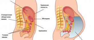
The outer surface of the organs in the abdominal cavity and the abdominal cavity itself consists of translucent cellular tissue, which was formed using two plates:
- Parietal (parietal);
- Visceral commissure (covering organs).
Parietal Visceral
These two plates gradually merge into one another and form a closed space called the abdominal cavity. All the spaces that have formed between the organs and their walls have no emptiness. All of them are filled with serous matter.
Adhesion adhesions are divided into 2 types:
- Viscero-parietal. Organs or intestines adhere to the wall of the abdominal cavity.
- Viscero-visceral. Intestinal loops or organs grow together.
What type of adhesions and what places of their localization make it possible to understand the obvious symptoms of the disease, and also provide an opportunity to recognize the impact on the functioning of other organs.
Treatment of adhesive disease of the abdominal organs
It’s worth pointing out right away that doctors take all possible preventive measures even at the stage of surgical interventions on the abdominal organs. Such actions of specialists are called preventive treatment of adhesive disease:
- During surgery, the polymer icodextrin (4% solution) is used;
- creating a jelly-like barrier around the intestines using polyethylene oxide and carboxymethylcellulose;
- the use of barrier membranes with hyaluronic acid, cellulose or other materials.
The peculiarity of the disease in question is that it can develop in the long term after surgery. If symptoms of adhesive disease of the abdominal organs appear, then under no circumstances should you hesitate! No folk methods, massages, enemas or other home remedies will help, although they may bring some relief. But in the pursuit of a quick “cure,” time is lost, adhesions rapidly develop and cause irreparable harm to organs.
Treatment of adhesive disease of the abdominal organs is carried out exclusively by surgical methods.
Symptoms
There are a number of symptoms that can be used to identify adhesions disease. These indicators are:
- Long-term constipation . With a normal and correct diet, the absence of feces is noticed. This happens for more than two days. This symptom is the most dangerous because it indicates the most dangerous complication of the adhesive process, such as intestinal obstruction.
- Dyspeptic syndrome . This symptom means a disruption in the normal functioning of the stomach. In the area of the digestive system, inconvenience, discomfort, pain occur, and difficulty in digestion appears. With such symptoms, a tendency to constipation is often noticed. The patient may feel distension in the abdomen, fullness, bloating, and heaviness.
- Painful sensations . Either nagging or short-term attacks may appear, which may intensify with physical exertion or sudden turns and changes in body position.
- Impaired intestinal motility . In case of failures, short-term constipation is observed. In a normal state, the walls contract and move feces towards the exit.
How do adhesions in the pelvis affect the body?
The abdominal cavity and pelvis are lined with parietal tissue, and the outer shell of the internal organs is covered with a visceral layer. If this part of the human body has not been subjected to negative effects in the form of mechanical injuries, surgical operations, inflammatory processes of infectious etiology, then both types of tissues have a perfectly smooth surface.
Under conditions of normal functioning of the parietal and visceral tissues, the synthesis of peritoneal fluid is performed, the main purpose of which is to ensure the movement of organs inside the abdominal cavity and directly in the pelvis. For example, the bladder cavity after filling can move slightly and move the uterus without damaging its walls.
If the internal organs of the pelvis have been subjected to pathological effects, then fibrin begins to form on the surface of the visceral tissue.
This substance should prevent the spread of the inflammatory process to neighboring tissues. Fibrin has a viscous consistency, and its excess amount can cause fusion of parietal and visceral tissues. As a result of this process, adhesions of varying sizes and severity are formed.
Most often, adhesive disease develops in the pelvic area in women, affecting the organs of the reproductive system. For example, if infectious microorganisms entered the cavity of the fallopian tubes, acute inflammation developed, and the patient did not receive adequate drug therapy, then over time the released fibrin forms an adhesive plug, blocking their abdominal opening.
Other internal organs located in the pelvis also undergo pathological changes. As the adhesions grow, a tubo-ovarian neoplasm (pyosalpinx) is formed, which covers the walls of the pelvis, intestinal walls, bladder, and appendages in women.
This type of tumor is difficult to treat surgically, disrupts the blood circulation of internal organs, and also reduces their performance. To achieve a complete cure for the patient, the doctor will need to show skill and put a lot of effort into removing adhesive tissue without disturbing healthy visceral tissue.
Diagnostics
| Diagnostic procedures | Explanation |
| Diagnostic laparoscopy | To perform this procedure, the patient is given general or local anesthesia. First, a thin tube with a lens and an eyepiece is inserted into the patient. With this method, a specialist who has discovered intestinal adhesions can safely remove them if there are only a small number of them. |
| X-ray of the intestine | This procedure uses barium sulfate contrast agent. In order for the examination to be successful, a barium solution is taken orally four hours before the procedure, or the patient is given an enema when examining the colon. |
| Ultrasound | This procedure is performed on an empty stomach to eliminate false results due to food intake. |
| Clinical blood test | During this examination, the specialist evaluates the level of platelets, leukocytes and hemoglobin in the blood. Very often, during the adhesive process, there is a large number of leukocytes in the blood and an increased erythrocyte sedimentation rate. |
Prevention measures
Prevention of acute and chronic adhesive disease consists of timely treatment of inflammatory processes in the abdominal and pelvic organs, corrective therapy for concomitant diseases, and competent surgical interventions.
Recommended:
- lead an active healthy lifestyle;
- monitor the health of the genitourinary system (plan to visit a gynecologist, urologist, promptly treat genital infections);
- exercise regularly;
- monitor the regularity of bowel movements and the type of stool.
After operations, patients are recommended to undergo various physical procedures aimed at resolving adhesions and preventing the formation of scar tissue (electrophoresis, magnetic therapy).
In case of adhesive disease, excessive loads are contraindicated. Women should not lift weights more than 5 kg, men - more than 7 kg.
Postoperative immunoenzyme therapy (taking immunomodulators with prolonged hyaluronidase activity) and the inclusion of fibrinolytic drugs that prevent fibrin deposition in rehabilitation therapy are effective.
Unfortunately, adhesive disease is a serious complication of surgical interventions, and all measures taken to prevent the formation of adhesions do not guarantee a positive result. Each subsequent operation increases the number and density of adhesions, provokes their proliferation and increases the risk of relapse of OSCN. Therefore, the main task of preventing adhesions is timely and competent treatment of the underlying disease.
Treatment
Intestinal adhesions can only be removed surgically. There are no other methods to free the intestinal loops from connective tissue. During the surgical period, a course of conservative therapy is also prescribed.
Surgical method
The operation to remove adhesions has two types .
The first is laparoscopy (this is a micro-surgery). Three small incisions are made on the anterior abdominal wall. A thin fiber-optic tube with a video camera and a diode lamp, as well as a manipulator for fixing internal organs, is inserted through them.
The operation does not involve severe injuries, so the patient can be discharged already on the 3rd day.
The second is laparotomy. A 15 centimeter incision is made on the abdominal wall . This method is used when there are a large number of intestinal adhesions.
Conservative treatment
- Special diet.
- Installation of a nasogastric tube.
- Antispasmodics;
- Hypertensive enemas;
- Intravenous injections of solutions.
Folk remedies
Pain with adhesions. Use 3 tablespoons of flax seeds, place in a bag and dip in boiling water for three minutes. Then take it out, squeeze it out, and apply the finished compress to the sore area.
General discomfort. Use 1 tablespoon of dry St. John's wort and pour a glass of boiling water. Leave to brew for about 15 minutes and strain. The finished broth is divided into three parts and drunk throughout the day.
Treatment of intestinal pathology
Formed adhesions can only be removed surgically. Fibrous fibers that tightly connect internal tissues cannot be broken using conservative therapy. Especially if the fusion process began a long time ago and the adhesions have become quite strong.
Ultrasound is a fairly informative method that allows you to identify adhesions
However, along with surgical interventions, doctors always recommend conservative treatment to patients.
Drug therapy
Medicines are prescribed immediately after the patient undergoes surgery. This makes it possible to timely, at the initial stage, prevent the transformation of fibrin into full-fledged adhesions.
In addition, drug therapy is indicated for people with progressive pathology. In this case, conservative treatment is to prepare the patient for surgery.
The choice of tactics to combat pathology is selected individually and usually includes:
- Antibiotics. Such medications are prescribed if necessary to suppress the infectious process. Usually recommended: Trimezol, Tetracycline.
- Fibrinolytic drugs. They are most in demand in the postoperative period. These products are designed to dissolve fibrin, which will eventually form adhesions. Medications used in therapy: Fibrinolysin, Urokinase.
- Painkillers. To eliminate pain, it is recommended to take No-Shpa, Spazmalgin, Tempalgin.
- Antihistamines. They help reduce inflammation, reduce pain, and relieve swelling. Doctors prescribe: Suprastin, Diphenhydramine.
- Anticoagulants. These medications thin the blood. The most commonly recommended drug is Heparin.
- Vitamin complexes. Medicines containing vitamin E are useful. Therapy usually includes: Folic acid, Tocopherol.
- Anti-inflammatory drugs. To reduce severe pain and eliminate inflammation, the patient may be recommended: Diclofenac, Ketorol, Paracetamol, Ibuprofen.
- Medicines for resolving adhesions. They help reduce inflammation and speed up the process of resorption of fibrin strands. Suppositories can be prescribed: Longidaza, Lidaza. Sometimes injections are recommended: Longidaza, Plazmol.
- Laxatives. For chronic constipation, the doctor may recommend means to ensure timely bowel movements. Rectal suppositories are usually prescribed: Glycerin, Bisacodyl, Microlax.
- Medicines to restore peristalsis. To normalize the functioning of the digestive system, they recommend: Iberogast, Hilak Forte, Bifidumbacterin.
Medicines in the treatment of adhesions - photos
Tetracycline helps suppress the infectious process
Urokinase helps dissolve fibrin
No-Spa provides antispasmodic and analgesic effects
Suprastin reduces tissue swelling
Heparin thins the blood
Vitamin E is very useful for the body during adhesions
Ibuprofen is prescribed for severe pain
Longidaza ensures resorption of adhesions
Glycerin suppositories help with constipation
Iberogast helps normalize the functioning of the digestive system
Surgical treatment: will surgery help?
If the adhesive process has led to disruption of the blood supply to the intestines, then the patient is indicated for urgent surgical intervention. It involves removing obstacles and restoring the functioning of the digestive tract.
To cut adhesions, 2 types of surgical interventions are used:
- Laparoscopy. This low-traumatic operation is performed only with a small number of adhesions. The instruments and camera are inserted into the peritoneum through small punctures. All splices are dissected, and the blood vessels are cauterized.
- Laparotomy. If there are a large number of adhesions or complications occur (for example, intestinal necrosis), dissection of the anterior wall is resorted to. A large incision (about 15 cm) allows the surgeon to gain normal access to the internal organs.
The main problem of operations is the re-development of adhesions . To reduce the risk of such consequences, surgeons try to perform interventions with minimal trauma.
To do this they use:
- Laser. This is a minimally invasive treatment, which is possible with a small area of damage and a clearly visible localization of the fusion.
- Hydraulic compression, during which a special fluid is injected under pressure into the connective tissue. This drug ensures the destruction of adhesions.
Laparoscopy allows, using small punctures, to insert instruments and a camera for cutting adhesions into the abdominal cavity.
Diet and lifestyle
For patients suffering from adhesive disease, dietary nutrition is necessarily recommended.
It is strictly forbidden to starve or overeat. This will lead to aggravation of the pathology and serious consequences.
Patients are advised to follow these dietary rules:
- Eat food at certain hours, strictly according to the schedule.
- Eat food in small doses 4-5 times a day.
- Eliminate foods that strengthen the intestines and flatulence.
- Refuse hot and cold foods. Eat only warm food (it eliminates cramps).
The changes also concern lifestyle:
- Physical exercise. It is necessary to lead an active lifestyle. But at the same time, all loads must be adequate.
- Rejection of bad habits. Smoking and alcohol abuse lead to deterioration in the functioning of all body systems.
Prohibited and healthy foods - table
| Unhealthy food | Useful products |
|
|
Useful products - gallery
Low-fat broths are beneficial
Omelettes allowed
Fermented milk products are useful
Boiled chicken meat is recommended
Nutrition in the postoperative period
After surgery, it is necessary to provide the intestines with the most gentle diet possible.
The following diet is recommended for the patient:
- First day. Eating is completely excluded. You are only allowed to drink liquids (water, diluted rosehip infusion).
- Starting from 2 days. Dietary products are introduced into the diet: weak low-fat broths, grated porridges, liquid vegetable purees. It is recommended to drink herbal teas and juices, but always in diluted form.
- For 7–8 days. You can already diversify your diet with denser foods: eggs, fish, meat, vegetable purees (beetroot, carrot, apple). It is recommended to consume fresh kefir and drinking yoghurts. But all dishes are still well chopped and pureed.
At about 2-3 weeks, the doctor will allow you to go back to your normal diet.
Folk remedies
Healer's recipes cannot replace the treatment prescribed by a doctor. But sometimes, especially in the initial stages of pathology development, they can become a good support for conservative therapy and bring significant relief.
Folk remedies can help eliminate severe pain
The following folk remedies are usually recommended:
- To improve the condition. Dry St. John's wort herb (10 g) is poured with boiling water (250 ml). The mixture is simmered for 15 minutes over low heat. Then infuse the decoction for 1 hour. It is recommended to take the strained drink three times a day, 50 ml. Treatment lasts about 1–2 months.
- To relieve pain and improve the functioning of the digestive system. Plantain seeds (15 g) are poured with boiling water (200 ml). Boil the mixture in a steam bath for 3–4 minutes. The cooled product is filtered. It is necessary to consume 20 ml half an hour before meals 3 times a day for 1–2 months.
- To soften adhesions. Milk thistle seeds (10 g) are poured with boiling water (200 ml). The composition is boiled for approximately 3–5 minutes. After cooling, the broth is filtered. It is recommended to take 1 tbsp. l. before every meal. Treatment lasts 1–2 months.
- For resolving adhesions. Chopped hogweed grass (5 tbsp) is poured with vodka (0.5 l). The mixture should be infused in a dark place for 15 days. In this case, it is necessary to shake the tincture daily. After 15 days, the mixture is filtered. It is recommended to take 40 drops before meals, twice a day. The duration of treatment is 21 days.
You can use folk remedies to treat adhesive disease only after consultation with your doctor.
Folk remedies for adhesions - gallery
St. John's wort helps improve overall health
Plantain seeds are used to relieve severe pain
Milk thistle helps soften adhesions
The boron uterus promotes the resorption of adhesions in the fallopian tubes
Physiotherapy
As an additional treatment, the patient may be prescribed physiotherapeutic procedures:
- Electrophoresis. It is usually carried out with analgesics or medications that ensure the resorption of adhesions. Using electric current, the penetration of medicinal substances into the deeper layers of the affected areas is achieved.
- Paraffin applications. The event helps to soften fibrin strands.
Physiotherapy
For adhesions, the patient is recommended to perform special exercises.
She is called upon:
- ensure a sufficient level of activity;
- improve the elasticity of internal tissues;
- activate blood circulation;
- strengthen the peritoneum.
The patient will benefit from the following set of exercises:
- Lying on your back. The legs are simultaneously bent at the knees and pulled towards the stomach. Then the limbs are returned to their original position.
- Legs are bent at the knees. Hands are placed behind the head. The head and shoulder blades are lifted off the floor. The abdominals should be tense. After holding this position for a couple of seconds, they return to the initial state.
- Lying on your back, legs bent at the knees, feet on the floor. Raise the lower back and pelvis. At the top point they linger for a few seconds and then lower to the floor.
- Bent knees and tilt to one side, then to the other. At the same time, the pelvis and back are not lifted off the floor.
- Exercise "bicycle". It is recommended to work with the legs with maximum amplitude.
- Pairs of legs and arms are torn off the floor one by one. You need to touch your left elbow to your right knee. Repeat the exercise for the other pair of limbs.
Do physical exercise smoothly, without sudden movements. Each exercise is performed 8–10 times.
Special exercises help strengthen the peritoneum and improve tissue elasticity
In addition to the complex described above, yoga classes are very useful for adhesive disease.
Disease history
Even in ancient times, people suffered from symptoms of the disease “intestinal obstruction”. History says that the Danish anatomist Bartholin was the first to diagnose it in 1654. The adhesive process was first described by Hunter in 1797.
But officially the history of the disease dates back to 1906. It was then that Biller was able to demonstrate for the first time an x-ray image of gas bubbles in the abdominal cavity. In 1911, Schwartz proposed using a contrast method for examining the intestines to determine mechanical intestinal obstruction.
In 1910, Weistermann used the method of long-term probe aspiration of gastric contents to treat paresis of the gastrointestinal tract. In 1931, Heller used gastrostomy.
Adhesions after abdominal delivery
Adhesions after cesarean section are almost an optional consequence of the operation.
- Firstly, a cesarean section is an abdominal operation and is performed with high tissue trauma.
- Secondly, massive blood loss during surgery (from 600 to 1000 ml) is also an important factor in adhesions.
- In addition, cesarean section is often performed for emergency reasons, which is one of the reasons for the development of postoperative metroendometritis and additionally contributes to the formation of adhesions.
Pathogenesis
The internal organs of the abdominal cavity are covered with thin sheets of peritoneum. Normally, they have a smooth surface and secrete a small amount of fluid to allow the abdominal organs to move freely relative to each other. Various provoking factors lead to tissue swelling and the appearance of fibrin plaque on the peritoneum. Fibrin is a sticky substance that helps connect nearby tissues. If adequate therapy is not carried out at this time, then after the pathological processes subside, adhesions will form at the site of gluing.
The process of formation of adhesions takes place in several stages: first, fibrin falls out, then after 2-3 days special cells (fibroblasts) appear on it, which secrete collagen fibers. The pronounced replacement of inflamed tissues with connective tissue begins on the 7th day and ends on the 21st. During this period, the adhesions turn into dense adhesions, capillaries and nerves grow into them.
Preventive measures
Prevention of complications during surgery is manifested in its careful implementation, timely pumping out of excess blood accumulations and avoidance of dehydration of the abdominal cavity. Additional preventive measures: introduction of dry preparations and removal of foreign bodies.
In the postoperative period, such types of physiotherapy as UHF and electrophoresis are used. Exercise therapy and diet will be helpful. If all the doctor’s instructions are scrupulously followed, the prognosis is favorable. But the possibility of relapse cannot be ruled out.
Diagnosis of adhesions
As is correct, a preliminary diagnosis is based on medical history, but for a final diagnosis you should undergo an examination, which includes:
- Study of vaginal microflora, PCR - these tests will help determine the presence or absence of infections that can cause adhesions.
- Ultrasound is effective only in half of the cases: adhesions are not visible during this examination. Their presence can be suspected only by indirect signs (for example, reduced mobility of the ovaries relative to the uterus, etc.). The doctor interprets the results obtained based on other indicators of the patient’s condition.
- Laparoscopy - with this study you can examine the uterus, fallopian tubes, ovaries and detect the pathological process. During the procedure, there is the possibility of cutting the adhesions. However, it is impossible to detect synechiae in the uterine cavity or fallopian tubes using laparoscopy.
- Hysterosalpingography is a study of the uterus and fallopian tubes to study their structure, allowing to detect adhesions, their location and number. The examination is recommended to take place on days 5-11 of the cycle.
- Hysteroscopy is an examination of the inner surface of the uterus using a hysteroscope, special endoscopic equipment. The method allows not only to detect intrauterine synechiae, but during the examination it is possible to dissect them.
Specifics of diagnostics and therapeutic therapy
Intestinal obstruction is suspected based on the existing clinical picture. The main diagnostic method is an abdominal x-ray using barium. This study helps to identify distension of intestinal loops. Ultrasound, MRI, CT, and laparoscopy are used to determine an accurate diagnosis.
Therapeutic manipulations are prescribed by the attending physician based on the following data: type and stage of pathology, period of its development, concomitant pathologies. If the stage of the disease is early, therapy begins with conservative methods. Doctors normalize the functioning of the gastrointestinal tract. In many cases, such therapy serves as a measure of preoperative preparation of the patient. The optimal duration of such treatment must be selected in order to complete the operation on time.
When primary symptoms are detected, patients are prescribed intravenous nutrition and measures are prescribed to help improve intestinal motility. Such measures prevent intoxication and also improve the general condition of the patient.
If signs of pathology appear on the third day after surgery, it is necessary to neutralize the paretic part of the obstruction. To do this, Trimecaine is injected into the epidural space. In parallel with these actions, the stomach is washed, an enema is given, and Proserin, a solution of Sodium Chloride, is administered intravenously. If there is no positive dynamics within six hours, a laparotomy is performed.
If a strangulation form of adhesive obstruction is suspected, short-term conservative treatment is allowed. After this, the operation and postoperative care of the patient are carried out. As for the late stage of the disease, surgical intervention is urgently indicated. Without proper treatment, there is a risk of intestinal necrosis.
Before proceeding with surgery, it is important to properly prepare the patient. First of all, a siphon enema is given, the stomach is thoroughly washed, and Prozerin is administered. If after a few hours the pain subsides and the symptoms fade away, anti-adhesion therapy and inpatient treatment are prescribed. If no improvement is observed, laparotomy is indicated to release adhesions, establish an anastomosis, or perform resection. When a patient is taken to a medical facility in serious condition, he is prescribed intensive therapy to eliminate the intoxication of the body and undergo surgery.
Adhesive obstruction is a dangerous disease that requires urgent treatment. If symptoms occur, you should immediately seek medical help.
Drug treatment
How to treat intestinal adhesions in a child and an adult, and with what? If no progress is observed, there are no symptoms of adhesive pathology, no special treatment is required. The patient is under constant observation with periodic medical examinations. For pain and mild dysfunction, the use of analgesics and antispasmodics is indicated to relieve spasm of the intestinal muscles. To limit the adhesive process and partial resorption, injections of vitreous body, aloe preparations, Splenin, and enzymes (trypsin, chymotrypsin, lidase) are indicated. Medicines are prescribed for mild forms of pathology or as prophylaxis in accordance with the prevailing symptoms.
Paralytic obstruction
This form of the disease can manifest itself in the form of a progressive decrease in peristalsis and intestinal tone. As a result, complete paralysis of the affected organ often develops.
Paralytic obstruction is characterized by:
- pain, uniform bloating and vomiting;
- gas and stool retention.
Pain with this disease affects the entire abdominal area. They have a bursting character and do not give anywhere.
Vomiting with paralytic intestinal obstruction visits the patient many times. At first it consists of gastric and then intestinal contents. In case of diapedetic bleeding from the walls of the intestines and stomach, as well as acute ulcers, the vomit is hemorrhagic in nature.
Severe gas formation causes chest breathing. Patients are also diagnosed with low blood pressure, tachycardia and dry mouth.
Traditional methods of treatment
There are many different folk remedies used for intestinal adhesions. However, their effectiveness and safety have not been studied in studies, so you should consult your doctor before using these methods.
Castor oil
Relieves pain and inflammation, and with long-term use can reduce scar tissue and adhesions. You need to moisten several layers of cotton or woolen cloth with castor oil and place it on the stomach over the intended adhesions. Cover this fabric with cellophane wrap and secure it in place by tying something around your waist. Then apply a hot heating pad to it. This heat will allow the castor oil to penetrate the skin. You need to keep this bandage for 2 hours, then remove it. You need to make compresses every other day.
Medicinal plants
To treat abdominal adhesions, it is recommended to use comfrey and calendula, which can be used separately or in combination with each other.
Comfrey and calendula tea:
- ½ teaspoon comfrey leaves:
- ½ teaspoon calendula flowers:
- 2 cups of water.
Boil water and add herbs. Leave for 15 minutes and strain. Add honey if needed. Drink daily.
Comfrey and Calendula Oil:
- 1 cup dried comfrey leaves:
- 1 cup dried calendula flowers;
- Castor oil;
- olive oil.
Place the herbs in a jar. Using equal parts olive and castor oils, pour them over the herbs. Place a piece of cloth on the bottom of the multicooker and place a jar of herbs and oil on it. Add water to the multicooker bowl so that it does not reach the top of the jar. Set to keep warm mode and keep the jar in it for 5 days. Every day you need to add a little water to the slow cooker. After 5 days, strain off the oil.
Gently massage this oil into your stomach twice a day. This should be done regularly, over several weeks or months.
It should be recalled that before using any traditional medicine, you should consult a doctor.
Clinical picture
The pathology has acute, critical, and chronic forms. Each is defined by characteristic symptoms. The development of the clinical picture is determined by the type of disease and signs of its development.
- Obstructive. The intestinal loops are compressed by adhesions, circulatory disturbance occurs. Attacks of acute pain, vomiting with bile and green color, bloating, impaired gas removal, constipation.
- Strangulational. Necrosis of the digestive tract, compression of the intestinal mesentery. Severe pain, pale skin, vomiting, constipation, difficulties with gas removal, signs of toxicosis (increased pulse rate, increased or decreased blood pressure, weakness).
- Conjunctive. Complications after surgery: Asymmetrical bloating, pain on palpation, lack of stool, vomiting, cramps, intoxication.
The development of pathology a month or more after surgery confirms the adhesive sign of a late stage. The patient suffers from sharp and sudden pain and vomiting.
The act of defecation and gas removal is not carried out independently. Intoxication develops quickly. A person suffers from general malaise, fatigue, weakness, dizziness.
Chronic stage of the disease
Adhesive intestinal obstruction may have signs of a chronic stage. This form of pathology is defined as abdominal adhesive disease. Women who have had an appendectomy in childhood are at risk. It occurs due to the characteristics of the female body, since the fallopian tube and ovaries are located close to the intestines.
The occurrence of adhesions is accompanied by repeated signs of intestinal obstruction. Manifested by the following factors:
- severe pain at the site of the postoperative scar;
- prolonged retention of stool or its complete absence;
- vomiting occurs rarely.
In some cases, this form of pathology can be cured using conservative methods. Enemas will help effectively, as they do not empty the intestines. Its function is restored, pain disappears, and general condition improves.
How to diagnose?
How is complete or partial intestinal obstruction detected? The symptoms of this disease are quite similar to the signs of other diseases occurring in the gastrointestinal tract. Therefore, when making a diagnosis, it is impossible to rely only on external manifestations.
The main way to diagnose this pathology is an X-ray examination of the abdominal cavity, as well as a blood test. In addition, some specialists use ultrasound as an adjunct.
Upon objective examination, the patient’s tongue is dry and covered with a white coating. The patient also experiences uneven abdominal bloating.
How to treat adhesions
Consisting of dense connective tissue, synechiae cannot resolve on their own. But treatment of adhesions is carried out in cases where they cause problems in the body (pain, reproductive dysfunction). The only available way to remove them today is separation, usually by laparoscopic method. During the operation, the synechiae are dissected, and the internal organs are freed from adhesions. Surgery using laparoscopy is performed under general anesthesia. The procedure is performed through small punctures on the anterior wall of the abdominal cavity; there is no need for stitches at the end of the operation. In addition, the advantages of this method include an excellent cosmetic effect and a short rehabilitation period.
Types of disease
The symptom of intestinal obstruction can vary. It often depends on the type of disease and the cause of its occurrence.
In medical practice, the disease in question is usually classified as follows:
- congenital;
- acquired.
Based on the causes of development and the mechanism of the disease, it should be clarified that the congenital form of intestinal obstruction is the result of intrauterine anomalies.
As for the acquired disease, it is the result of a developmental mechanism. The group of this type includes the dynamic or so-called functional type of obstruction with paralytic and spastic forms. The first is a consequence of paralysis and intestinal paresis. As a rule, it appears only in the secondary stage and may be the result of postoperative stress.
The spastic form of the disease is associated with reflex spasms of the intestines. This disease is a consequence of helminthic infestations or intoxications.
Stages of the disease
How does intestinal obstruction occur in infants? Symptoms of this disease in children and adults depend on its stage.
According to medical practice, such a disease develops in three stages:
- Initial. It lasts about 3–12 hours and is also accompanied by pain in the abdomen, increased peristalsis and flatulence.
- Intermediate. It lasts about 13–36 hours. In this case, the pain syndrome subsides and a time of imaginary well-being begins. During this period, symptoms of intoxication and dehydration increase.
- Terminal. As a rule, this stage occurs two days after the formation of the disease. The person's condition is noticeably deteriorating. In this case, there is an increase in symptoms of dehydration, damage to internal organs and the nervous system.
Diagnosis of pathology
The presence of adhesions can only be determined using laboratory and instrumental studies.
Article on the topic: What does periarteritis nodosa look like, why does it appear and how is the pathology diagnosed?
If tissue fusion is suspected, the patient will be advised to:
- Irrigography. Contrast (barium suspension) is injected into the digestive tract and X-rays are taken. They allow us to identify abnormalities of the intestinal loops.
- Colonoscopy. Using endoscopic equipment, the condition of the rectum is studied.
- Electrogastroenterography. The study characterizes the peristaltic ability of the digestive system.
- Ultrasound. The condition of the peritoneal and pelvic organs is studied. In most cases, ultrasound shows the presence of adhesions and characterizes their prevalence.
- Computed tomography (CT). This measure is prescribed only if the ultrasound does not allow the pathology to be determined.
- Laparoscopy. The most informative method. It involves inserting a camera into the peritoneum. This diagnostic method usually ends with full-fledged surgical treatment.
- Blood analysis. It is prescribed to eliminate the likelihood of inflammation developing in the body.
What types are there
Adhesive intestinal obstruction has several varieties. They differ in the time of appearance after injury (surgery) and methods of treatment. It can be acute, chronic, complete or partial. Let's look at them in more detail using the example of an operation. This:
- Adhesive-paretic (with the development of intestinal paresis) obstruction. It is detected 2-6 days after surgery.
- Early. From 5 days to 2-3 weeks after surgery.
- Late. Later 3-4 weeks after surgery. Sometimes symptoms appear months or years after the intervention.
The adhesive-paretic type of intestinal obstruction is distinguished by the fact that the cause appears against the background of “stopping” the movement of the intestines (their paresis). Under such conditions, the first postoperative days are accompanied by “gluing” of the intestinal loops to each other. This makes it difficult to promote their content. The condition is reversible, the adhesions resolve after 5-7 days, sometimes later.
Diagnosis of such obstruction is not difficult. The clinical picture is manifested by abdominal pain, severe bloating, and vomiting. Treatment of such conditions is almost always conservative (without surgery); it has several directions that eliminate the causes of intestinal paresis:
- Elimination of the symptom of intestinal bloating (gastric, intestinal tubes, gas tube are inserted).
- Complete pain relief is carried out, since the symptom of pain causes a cessation of intestinal movement. For this, epidural anesthesia is most often used for several days.
- They use medications that stimulate intestinal motility (prozerin, hypertonic sodium chloride solution, etc.), various types of enemas.
Early adhesive obstruction has similar causes of occurrence, mechanism of development, symptoms; its diagnosis is relatively simple. It differs in that adhesions do not always eventually resolve (partially or completely). They compress the outside of the intestinal lumen and may cause a mechanical obstruction to the passage of food through the intestines.
Symptoms of such obstruction may persist despite conservative treatment (it has similar principles to the treatment of adhesive-paretic obstruction). Then it is necessary to perform surgical intervention to eliminate the cause due to the severity of the patient’s condition.
Late intestinal obstruction, caused by adhesions, can be complete or incomplete, acute or chronic. Conservative treatment does not work if these adhesions cause insurmountable obstacles to the intestinal contents and cause symptoms of a serious illness. The clinical picture of acute obstruction is typical: vomiting is indomitable and does not bring relief. It causes dehydration due to the large loss of intestinal juices with vomiting (up to 6-8 liters per day).
Severe abdominal pain increases sharply when palpating the abdomen. With the development of obstruction of the large intestine, asymmetry of the abdomen is observed. Treatment with conservative methods described above is used as preparation for surgery. Continuous intravenous administration of solutions is used to replace fluid loss. To make a diagnosis and start treatment, you can make an appointment with your doctor in Moscow here: https://www.mos-clinics.ru/lechenie/
Symptoms
In the acute form of adhesive intestinal obstruction, various symptoms are observed, which are determined by the duration of the disease. Main characteristics: abrupt onset, intense pain in the abdominal area, large volumes of vomit mixed with bile. In later lines, exicosis is observed: facial features sharpen, the tongue becomes dry, the stomach increases in size, and the level of intestinal motility increases. Intestinal loops in a distended state begin to contour through the walls of the peritoneum (Wal's symptom). During a rectal examination, you can notice that the rectal cavity is empty and balloon-like inflated. Radiological symptoms become more pronounced: in the presence of low obstruction, multiple levels of intestine (or Kloiber's cup) are noticeable. At a high level, they are single and appear as darkening of the lower abdomen.
A smooth onset is also characteristic of the form of the disease that is caused by an inflammatory infiltrate. But before the disease begins to manifest itself, you may notice a weakening of the general condition of the body and an increase in body temperature. In the area of the peritoneum, a compacted accumulation of infiltrates can be felt, causing pain. Blood tests show the presence of purulent inflammation.
Strangulation obstruction lasts as a hyperacute form. The pain is cramping in nature. There is a regular urge to vomit. Vomit contains food debris and mucus. The level of manifestations of toxicosis and exicosis increases very quickly. At an early stage, the stomach does not swell. It is mild and symmetrical in shape, but with painful symptoms. Peristaltic sounds are clearly audible in the intestinal cavity. Later, the patient appears lethargic and inactive. His signs of toxicosis and peritonitis are increasing.
Characteristic signs
If a person develops adhesions in the intestines, the first symptom is the manifestation of a feeling of pain in the place where the scar is localized. If treatment is not started in time, then the painful sensation spreads throughout the lower abdomen, interfering with full functioning. In some cases, scars do not cause severe pain; the patient feels discomfort in the abdomen, which intensifies and then subsides. Over time, the number of attacks increases, the patient feels unwell, loses weight, and other disturbances in the functioning of the body develop along the way: problems with the respiratory and cardiovascular systems appear. Due to intestinal obstruction, a process of intoxication begins in the body, when decay products are not eliminated fully and naturally. The patient becomes ill, has attacks of angina pectoris and suffocation, dizziness, and loss of consciousness. The patient is plagued by the following symptoms of acute adhesive obstruction:
- nausea and frequent bouts of vomiting;
- constant constipation, due to which a large amount of gas is formed in the intestines;
- heaviness in the stomach due to the fact that food is not fully digested and stagnates in the organs of the gastrointestinal tract.
Nausea and frequent bouts of vomiting are symptoms of acute adhesive obstruction, in the presence of which you should immediately seek medical help.
If you have such symptoms and have regular pain attacks in the abdominal area, you should immediately seek medical help, since delay and untimely treatment causes serious complications that can only be eliminated through surgery. To make a final diagnosis and begin adequate treatment, the doctor refers the patient to diagnostic studies and tests.