The second pair of cranial nerves is the most important element of the visual system, because through it the connection between the retina and the brain occurs. Although other structures continue to function correctly, any deformation of the nervous tissue affects the properties of vision. Optic nerve atrophy cannot be cured without leaving a trace; nerve fibers cannot be restored to their original state, so it is better to carry out prevention in a timely manner.
Basic information on the disease
Optic nerve atrophy or optic neuropathy is a severe process of destruction of axons (nerve tissue fibers). Extensive atrophy thins the nerve column, healthy tissue is replaced by glial tissue, and small vessels (capillaries) are blocked. Each of the processes causes certain symptoms: visual acuity decreases, various defects appear in the visual field, and the shade of the optic nerve head (OND) changes. All pathologies of the optic nerves account for 2% of the statistics of eye diseases. The main danger of optical neuropathy is absolute blindness, which occurs in 20-25% of people with this diagnosis.
Optic neuropathy does not develop on its own; it is always a consequence of other diseases, so a person with atrophy is examined by different specialists. Typically, optic nerve atrophy is a complication of a missed ophthalmological disease (inflammation in the structures of the eyeball, swelling, compression, damage to the vascular or nervous network).
Functions
The main function of the nerve is to conduct signals from retinal receptors, which are processed in the occipital cortex of the brain.
A feature of the structure of the human visual analyzer is the presence of the optic chiasm - a place where the nerves from the right and left eyes are partially intertwined with their parts closest to the center.
Thus, part of the image from the nasal region of the retina is translated to the opposite region in the brain, and from the temporal region it is processed by the hemisphere of the same name. As a result of combining images, the right visual fields are processed in the visual area of the left hemisphere, and the left ones - in the right.
Damage to the optic nerves always affects the visual field
Causes of Optic Neuropathy
Despite the many causes of optic nerve atrophy known to medicine, in 20% of cases they remain unclear. Usually these are ophthalmological pathologies, central nervous system diseases, autoimmune disorders, infections, injuries, intoxications. Congenital forms of ADN are often diagnosed together with cranial defects (acrocephaly, microcephaly, macrocephaly) and hereditary syndromes.
Causes of optic nerve atrophy from the visual system:
- neuritis;
- arterial obstruction;
- myopia;
- retinal dystrophy;
- uveitis;
- retinitis;
- oncological lesion of the orbit;
- unstable eye pressure;
- local vasculitis.
Injury to nerve fibers can occur during a traumatic brain injury or even the mildest injury to the facial skeleton. Sometimes optic neuropathy is associated with the growth of meningioma, glioma, neuroma, neurofibroma and similar formations in the thickness of the brain. Optical disturbances are possible with osteosarcoma and sarcoidosis.
Causes from the central nervous system:
- neoplasms in the pituitary gland or cranial fossa;
- compression of chiasmata;
- multiple sclerosis.
Atrophic processes in the second pair of cranial nerves often develop as a result of purulent-inflammatory conditions. The main danger is brain abscesses, inflammation of its membranes.
Systemic risk factors
- diabetes;
- atherosclerosis;
- anemia;
- avitaminosis;
- hypertension;
- antiphospholipid syndrome;
- Wegener's granulomatosis;
- systemic lupus erythematosus;
- giant cell arteritis;
- multisystem vasculitis (Behcet's disease);
- nonspecific aortoarteritis (Takayasu disease).
Syphilis, tuberculosis and similar severe infections often lead to the death of optic nerve axons. Less commonly, atrophy is detected after influenza, ARVI, measles, rubella and similar viral diseases. The influence of some parasites (toxoplasmosis, toxocariasis) cannot be ruled out.
Causes of pathology
During scientific experiments, it was found that 2/3 of cases of optic nerve atrophy were bilateral. The cause is intracranial tumors, edema and disorders of the vascular system, especially in patients aged 42 - 45 years.
The causes of the disease are:
- Nerve damage. These include: chronic glaucoma, neuritis, neoplasms.
- Neuropathy (ischemic), chronic neuritis, edema is a secondary pathology.
- Hereditary neuropathy (Leber).
- Neuropathy (toxic). The disease is caused by methanol. This component is found in surrogate alcoholic drinks and medications (Disulfiram, Ethambutol).
The causes of the disease include: retinal damage, TAY-SAXS pathology, syphilis.
The development of atrophy in children is influenced by a congenital anomaly, a negative hereditary factor, and a malnutrition of the optic nerve. Pathology entails disability.
Classification of optical neuropathy
All forms of optic nerve atrophy are hereditary (congenital) and acquired. Congenital diseases are divided according to the type of inheritance; they often indicate the presence of genetic abnormalities and hereditary syndromes that require in-depth diagnosis.
Hereditary forms of ADS
- Autosomal dominant (juvenile). Predisposition to nerve destruction is transmitted in a heterogeneous manner. The disease is usually detected in children under 15 years of age; it is recognized as the most common, but weakest form of atrophy. It is always bilateral, although sometimes the symptoms appear asymmetrically. Early signs are detected by 2-3 years, and functional disorders only at 6-20 years. Possible combination with deafness, myopathy, ophthalmoplegia and distance.
- Autosomal recessive (infantile). This type of ADN is diagnosed less frequently, but much earlier: immediately after birth or during the first three years of life. The infantile form is bilateral in nature and is often detected in Kenny-Coffey syndrome, Rosenberg-Chattorian, Jensen or Wolfram disease.
- Mitochondrial (Leber's atrophy). Mitochondrial optic atrophy is the result of a mutation in mitochondrial DNA. This form is considered a symptom of Leber's disease; it occurs suddenly and resembles external neuritis in the acute phase. Most patients are men 13-28 years old.
Forms of acquired atrophy
- primary (squeezing of neurons in the peripheral layers, the optic disc does not change, the boundaries have a clear appearance);
- secondary (swelling and enlargement of the optic disc, unclear boundaries, replacement of axons by neuroglia is quite pronounced);
- glaucomatous (destruction of the cribriform plate of the sclera due to surges in local pressure).
Destruction can be ascending, when the axons of the cranial nerves are affected, and descending, involving the nerve tissue of the retina. According to symptoms, they distinguish between unilateral and bilateral ADN, according to the degree of progression - stationary (temporarily stable) and in constant development.
Types of atrophy according to the color of the optic disc:
- initial (slight blanching);
- incomplete (noticeable blanching of one segment of the optic disc);
- complete (change in shade over the entire area of the optic disc, severe thinning of the nerve pillar, narrowing of the capillaries).
Main classification of the disease
Atrophy of the optic nerve is determined by pathological and ophthalmoscopic signs.
Acquired and congenital form
The acquired form is primary or secondary in nature. Caused by the influence of etiological factors. The process occurs as a result of: inflammation, glaucoma, myopia, and metabolic disorders in the body.
Congenital form: occurs against the background of genetic pathology. There are 6 types of hereditary atrophy: infantile (from birth to 3 years), dominant (juvenile blindness from 3 to 7 years), opto-oto-diabetic (from 2 to 22 years), Beer syndrome (complicated form, appears from 1 year) , increasing (from an early age, gradually progressing), Leicester's disease (hereditary), occurs at the age of 15 - 35 years.
Primary and secondary atrophy
The primary form is localized in a healthy eyeball. Occurs when microcirculation and nutrition of nerve fibers are disrupted.
The occurrence of secondary atrophy is caused by various eye pathologies.
Descending and ascending form
Descending atrophy is characterized by an inflammatory process in the proximal zone of the axon. Retinal disc damage is observed.
In the ascending form, the retina is initially affected. Gradually, the destructive process is directed to the brain. The rate of degeneration will depend on the thickness of the axons.
Partial and full degree
Diagnosing the extent of damage:
- initial (damage to some fibers);
- partial (diameter damaged);
- incomplete (the disease progresses, but vision is not completely lost);
- complete (loss of visual functions in full).
There is unilateral and bilateral atrophy. In the first case, damage to the innervation of one eye is observed, in the second - two.
https://medglaza.ru/zabolevaniya/bolezni/atrofiya-zritelnogo-nerva.html
Symptoms of optic atrophy
The degree and nature of optical disturbances directly depends on which nerve segment is affected. Visual acuity can decrease critically very quickly. Complete destruction ends in absolute blindness, blanching of the optic disc with white or gray spots, and narrowing of the capillaries in the fundus. With incomplete ONH, vision stabilizes at a certain time and no longer deteriorates, and the pallor of the ONH is not so pronounced.
If the fibers of the papillomacular bundle are affected, the deterioration in vision will be significant, and the examination will show a pale temporal zone of the optic disc. In this case, optical disorders cannot be corrected with glasses or even contact lenses. Damage to the lateral zones of the nerve does not always affect vision, which complicates diagnosis and worsens the prognosis.
ADN is characterized by various visual field defects. The following symptoms allow one to suspect optic neuropathy: scotomas, concentric narrowing, tunnel vision effect, weak pupil reaction. In many patients, the perception of colors is distorted, although more often this symptom develops when axons die after neuritis. Often changes affect the green-red part of the spectrum, but its blue-yellow components can also be distorted.
Atrophy due to methyl alcohol poisoning
Methyl alcohol and technical alcohol mixtures can cause serious damage to vision. Pathology sometimes occurs due to methyl alcohol poisoning.
The first sign of poisoning is characterized by: migraine, dizziness, nausea, vomiting, diarrhea. The pupil becomes dilated, the clarity of vision is impaired, and light reflections cannot be distinguished. There is a sharp decrease in vision.
Therapy for this form of atrophy consists of using: alkalizing medications, calcium, B vitamins, ascorbic acid.
In patients diagnosed with methyl arthophy, the prognosis for recovery is pessimistic. Vision restoration is observed in only 15% of patients.
Optic nerve atrophy is damage to nerve fibers. When the process is prolonged, the neurons die, which leads to loss of vision.
Diagnosis of optic nerve atrophy
The expressive clinical picture, physiological changes and functional disorders greatly simplify the diagnosis of ADN. Difficulties may arise when actual vision does not match the degree of destruction. To make an accurate diagnosis, the ophthalmologist must study the patient’s medical history, establish or deny the fact of taking certain medications, contact with chemical compounds, injuries, and bad habits. Differential diagnosis is carried out for peripheral lens opacification and amblyopia.
Ophthalmoscopy
Standard ophthalmoscopy allows one to establish the presence of ADN and accurately determine the extent of its spread. This procedure is available in many regular clinics and does not cost much. The results of the study may vary, but some signs are detected in any form of neuropathy: changes in the shade and contour of the optic disc, a decrease in the number of vessels, narrowing of the arteries, and various venous defects.
Ophthalmoscopic picture of optical neuropathy:
- Primary: clear disc boundaries, optic disc sizes are normal or reduced, saucer-shaped excavation is present.
- Secondary: grayish tint, blurred disc borders, enlarged optic disc, no physiological excavation, peripapillary reflex to light sources.
Coherence tomography
Optical coherence or laser scanning tomography allows us to study the nerve disc in more detail. Additionally, the degree of mobility of the eyeballs is assessed, the reaction of the pupils and the corneal reflex are checked, visometry is performed with tables, visual field defects are examined, color vision is checked, and eye pressure is measured. Visually, the ophthalmologist determines the presence of exophthalmos.
Plain radiography of the orbit allows you to identify pathologies of the orbit. Fluorescein angiography shows dysfunction of the vasculature. To study local blood circulation, Doppler ultrasound is used. If atrophy is due to infection, laboratory tests such as enzyme-linked immunosorbent assay (ELISA) and polymerase chain reaction (PCR) are performed.
Electrophysiological tests play a key role in confirming the diagnosis. Optic nerve atrophy changes the threshold sensitivity and lability of the nervous tissue. The rapid progression of the disease increases the retino-cortical and cortical time indicators.
The level of reduction depends on the location of the neuropathy:
- when the papillomacular bundle is destroyed, sensitivity remains at a normal level;
- damage to the periphery causes a sharp increase in sensitivity;
- atrophy of the axial fascicle does not change sensitivity, but sharply reduces lability.
If necessary, check the neurological status (x-ray of the skull, CT or MRI of the brain). When a patient is diagnosed with a brain tumor or unstable intracranial pressure, a consultation with an experienced neurosurgeon is prescribed. In case of orbital tumors, it is necessary to include an ophthalmic oncologist in the course. If destruction is associated with systemic vasculitis, you need to contact a rheumatologist. Pathologies of the arteries are dealt with by an ophthalmologist or vascular surgeon.
Diagnosis of the disease
First of all, the doctor identifies other diseases and deviations from the normal functioning of the patient’s body. This will help clarify the cause of optic nerve atrophy. The nature of the patient’s lifestyle, the consumption of any medications, episodes of poisoning and intoxication of the body are also important. During the collection of anamnesis, it is imperative that the patient’s complaints about his health be clarified. Particular attention is paid to headaches, dizziness, fainting and other neurological symptoms.
When examining and palpating the eye, the ophthalmologist identifies the degree of eye mobility, the presence of exophthalmos, abnormalities or normal pupil reaction to light, as well as the corneal reflex. A standard examination of vision, color vision, and perimetry is carried out.
The most accurate picture of the condition of the optic nerve is provided by the ophthalmoscopy procedure. Optic neuropathy can have completely different causes and prerequisites, therefore the picture visualized by a specialist can vary greatly. But there are still some similarities that are inherent in each type of optic atrophy. Among them: varying degrees of pallor of the optic disc, pathological changes in the shape and shade of the nerve (from gray to pale waxy), excavation of the upper part of the disc, a drop in the number of capillaries and a decrease in the diameter of the arteries of the retina, anomalies in the structure of the veins. The specific condition of the optic nerve head is determined using laser scanning and optical tomography.
VEP as a type of electrophysiological diagnosis determines an increase in the sensitivity threshold and a decrease in the lability of nervous tissue. If optic neuropathy develops against the background of glaucoma, then tonometric measurement reveals high intraocular pressure. Tumors and other diseases of the orbit are visible on x-rays.
Vascular pathologies are detected by angiography. Blood flow parameters in the arteries of the eye area are measured using Doppler ultrasound.
If there are indications, the examination program includes:
- consultation with a neurologist;
- X-ray of the skull, area of the sella turcica;
- MRI, CT scan of the head;
- consultation with a neurosurgeon (for diagnosing tumors and other formations, as well as dropsy, increased intracranial pressure);
- consultation with a rheumatologist (if systemic vasculitis, systemic lupus erythematosus is suspected);
- consultation with an ophthalmic oncologist (if a tumor process is detected);
- consultation with an angiosurgeon (for blockage, stenosis of arteries).
If optic neuropathy develops as a result of damage to the optic nerve by infectious agents, then the type of infection is diagnosed using ELISA and PCR tests. Optic neuropathy is differentiated from amblyopia and cataracts.
How is optic atrophy treated?
The treatment regimen for each patient with optic neuropathy is always individual. The doctor needs to get all the information about the disease in order to create an effective plan. People with atrophy require immediate hospitalization, while others are able to maintain outpatient treatment. The need for surgery depends on the causes of ASD and symptoms. Any therapy will be ineffective if vision weakens to 0.01 units or below.
It is necessary to begin treatment of optic nerve atrophy by identifying and eliminating (or stopping) the root cause. If cranial nerve damage is caused by intracranial tumor growth, an aneurysm, or unstable cranial pressure, neurosurgery must be performed. Endocrine factors influence hormonal levels. Post-traumatic compression is corrected surgically by removing foreign bodies, removing chemicals, or limiting hematomas.
Conservative therapy for optic neuropathy is primarily aimed at inhibiting atrophic changes, as well as preserving and restoring vision. Drugs are indicated to expand the vasculature and small vessels, reducing capillary spasm and accelerating blood flow through the arteries. This allows all layers of the optic nerve to be supplied with sufficient nutrients and oxygen.
Vascular therapy for ADN
- intravenously 1 ml of nicotinic acid 1%, glucose for 10-15 days (or orally 0.05 g three times a day after meals);
- Nikoshpan tablet three times a day;
- intramuscularly 1-2 ml No-shpa 2% (or 0.04 g orally);
- intramuscularly 1-2 ml of Dibazol 0.5-1% daily (or orally 0.02 g);
- 0.25 g of Nihexin three times a day;
- subcutaneously 0.2-0.5-1 ml of sodium nitrate of increasing concentration 2-10% in a course of 30 injections (increase every three injections).
Decongestants are needed to reduce swelling, which helps reduce compression of the nerve and blood vessels. Anticoagulants are used to prevent thrombosis; the vasodilator and anti-inflammatory Heparin is recognized as the best. It is also possible to prescribe antiplatelet agents (prevention of thrombosis), neuroprotectors (protection of nerve cells), glucocorticosteroids (combat inflammatory processes).
Conservative treatment of ADN
- To reduce inflammation in the nervous tissue and relieve swelling, dexamethasone solution is prescribed in the eye, intravenous glucose and calcium chloride, and intramuscular diuretics (Furosemide).
- Strychnine nitrate solution 0.1% in a course of 20-25 subcutaneous injections.
- Parabulbar or retrobulbar injections of Pentoxifylline, Atropine, xanthinol nicotinate. These drugs help speed up blood flow and improve trophism of nervous tissue.
- Biogenic stimulants (FIBS, aloe preparations) in a course of 30 injections.
- Nicotinic acid, sodium iodide 10% or Eufillin intravenously.
- Vitamins orally or intramuscularly (B1, B2, B6, B12).
- Antioxidants (glutamic acid).
- Orally Cinnarizine, Riboxin, Piracetam, ATP.
- Instillation of Pilocarpine to reduce eye pressure.
- Nootropic drugs (Lipocerebrin).
- Drugs with antikinin effect (Prodectin, Parmidin) for symptoms of atherosclerosis.
In addition to medications, physical therapy is prescribed. Oxygen therapy (use of oxygen) and blood transfusion (urgent blood transfusion) are effective for ADN. During the recovery process, laser and magnetic procedures are prescribed; electrical stimulation and electrophoresis (administration of drugs using electric current) are effective. If there are no contraindications, acupuncture (use of needles on active points of the body) is possible.
Tabetic nerve atrophy
Tabes is a disease of the nervous system due to infection with syphilis. If timely treatment is not applied, the disease progresses, causing ocular trophic disorders.
Tabes optic atrophy is the only manifestation of tabes (an early symptom of neurosyphilis). The tabetic form of atrophy is characterized by bilateral vision loss.
A sign of the disease is reflex immobility of the pupils. The optic nerve papilla becomes discolored and becomes gray-white.
There is a sharp decline in vision, the pathology is difficult to treat. Therapy is prescribed by a venereologist and neurologist (treatment of the primary infection is mandatory). Initially, medications and vitamins are prescribed that stimulate metabolic processes in tissue structures.
Prescribed internally:
- vitamin A;
- ascorbic acid;
- a nicotinic acid;
- calcium (pangamate);
- riboflavin.
After three days, intramuscular injections are prescribed: vitamin B, B6, B12. The drugs are combined with aloe or vitreous extract. Treatment is carried out under the strict supervision of a specialist in a medical institution.
Surgical treatment of optic neuropathy
One of the methods of surgical treatment of the optic nerves is hemodynamic correction. The procedure can be performed under local anesthesia: a collagen sponge is placed in the sub-Tenon's space, which stimulates aseptic inflammation and dilates blood vessels. In this way, it is possible to provoke the growth of connective tissue and new vascular network. The sponge dissolves on its own after two months, but the effect lasts for a long time. The operation can be performed repeatedly, but at intervals of several months.
New branches in the vascular network help improve blood supply to nerve tissue, which stops atrophic changes. Correction of blood flow allows you to restore vision by 60% and eliminate up to 75% of visual field defects if you go to the clinic in a timely manner. If the patient has severe concomitant disorders or atrophy has developed to a late stage, even hemodynamic correction will be ineffective.
For partial atrophy of the optic nerve, the use of a collagen implant is practiced. It is impregnated with antioxidants or drugs to dilate capillaries, and then injected into the eyeball without stitches. This method is effective only when eye pressure is stable. The operation is contraindicated in patients over 75 years of age, with diabetes mellitus, severe somatic disorders and inflammation, and vision less than 0.02 diopters.
Treatment
Many people hope to cure optic nerve atrophy by looking for “miraculous” folk methods. I would like to draw attention to the fact that this condition is considered difficult to treat in official medicine. Treatment of optic nerve atrophy with folk remedies will most likely have a general strengthening and supportive effect. Decoctions of herbs, flowers, and fruits are unable to restore atrophied nerve fiber, but can be sources of vitamins, microelements, and antioxidants.
Examples of recommended remedies that require a long course of use for at least a month:
- an infusion of pine needles, rose hips and onion peels, prepared from a liter of water and plant materials in a ratio of 5:2:2.
- infusion of forest mallow and burdock with the addition of primrose, lemon balm and dolnik.
- infusion of rue herb, unripe pine cones, lemon, prepared in a sugar solution - 0.5 cups of sand per 2.5 liters of water.
Modern methods of treating this condition are based on a complex of therapeutic measures.
Drug treatment
First of all, efforts are made to restore blood circulation and nutrition to the nerve, stimulating its viable part. Vasodilators, anti-sclerotic drugs and medications that improve microcirculation, multivitamins and biostimulants are prescribed.
A breakthrough in the treatment of optic nerve atrophy is associated with the use of nanotechnology, which involves delivering a drug directly to the nerve with nanoparticles.
Traditionally, most drugs are administered as an injection under the conjunctiva or retrobulbar - A; irrigation system – B
The prognosis for treatment of partial optic nerve atrophy in children is most favorable, since the organs are still in the process of growth and development. Irrigation therapy has a good effect. A catheter is installed in the retrobulbar space, through which the drug can be administered regularly and many times without damaging the child’s psyche.
Irreversible changes in nerve fibers prevent vision from being fully restored, so achieving a reduction in the area of death is also a success.
Treatment of secondary optic nerve atrophy will bear fruit with simultaneous treatment of the underlying disease.
Physiotherapy
Along with medications, physiotherapeutic methods can also significantly improve the condition of the nerve fiber, normalize metabolic processes and blood supply.
Today, treatment methods using magnetic, electrical, and laser stimulation of the optic nerve are known; ultrasound pulses and oxygen therapy can also be used. Forced stimulation of the nerve helps to initiate normal processes of excitation and conduction, but with a large amount of atrophy, the nerve tissue is not restored.
Surgical intervention
This type of treatment can be considered in the context of removing a tumor or other formation that is compressing the optic nerve.
On the other hand, microsurgical restoration of the nerve fiber itself is gaining increasing popularity.
The newest methods include stem cell treatment. They can integrate into damaged tissue and further stimulate its repair by secreting neurotrophic and other growth factors.
Regeneration of nerve tissue occurs extremely rarely. The speed of recovery is crucial in maintaining its functionality, so it is important to seek medical help promptly if you suspect optic nerve atrophy, so as not to lose your vision.
Prognosis for optic atrophy
To prevent AD, it is necessary to regularly check the condition of those organs that regulate the functioning of the visual system (central nervous system, endocrine glands, joints, connective tissue). In severe cases of infection or intoxication, as well as severe bleeding, urgent symptomatic treatment must be carried out.
It is impossible to completely restore your vision after neuropathy even in the best clinic. A case is considered successful when the patient’s condition has stabilized, ASD does not progress for a long time, and vision has been partially restored. Many people have permanently reduced visual acuity and also have defects in lateral vision.
Some forms of atrophy continually progress even with adequate treatment. The ophthalmologist’s task is to slow down atrophic and other negative processes. Having stabilized the symptoms, it is necessary to constantly prevent ischemia and neurodegeneration. To do this, long-term maintenance therapy is prescribed, which helps improve the blood lipid profile and prevent the formation of blood clots.
The course of treatment for optic nerve atrophy must be repeated regularly. It is very important to eliminate all factors that can affect the optic nerve axons. A patient with optic neuropathy should regularly visit specialists as indicated. It is necessary to constantly prevent complications and improve lifestyle. Refusal of therapy for optical neuropathy inevitably leads to disability due to total death of nerves and irreversible blindness.
Any changes in the layers of the optic nerve negatively affect a person's ability to see. Therefore, it is necessary to undergo timely examinations for people with a predisposition and treat all diseases that contribute to optic nerve atrophy. Therapy will not help restore vision to 100% when optic neuropathy has already developed enough.
Diagnostics
As a rule, this disease is diagnosed without any difficulties. It is impossible not to notice a sharp decrease in vision, so the vast majority of patients themselves turn to an ophthalmologist, who then makes a diagnosis. An examination of the visual organs of a sick person will certainly in this case show a deformation of the optic nerve, its color paler than necessary.
But cromhexal drops are an effective remedy for conjunctivitis.
To clarify the diagnosis, additional types of examinations may be prescribed:
- measuring intraocular fluid pressure;
- visual field examination;
- X-ray;
- electrophysiological analysis.
It is important to establish the cause of the pathology, since it is the elimination of the underlying disease that leads to a qualitative cure for the patient. In some cases, surgical intervention is also required, since conservative treatment may not produce results. To accurately establish the cause of the disease, you need to take a blood test (general and biochemical), as well as tests for syphilis and berylliosis. The symptom of yellow eyes in a person indicates a serious illness.
It is unacceptable to diagnose the disease on your own, since only a qualified specialist will be able to carry out all the necessary studies and procedures in this case. In addition, the symptoms are similar to cataracts, so professional help is necessary in any case. What drops dilate the pupil can be read in our article.
Symptoms
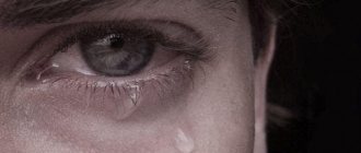
The main sign of the onset of pathology can be a steadily progressive deterioration of vision in one or both eyes, and it cannot be corrected by conventional methods.
Visual functions are gradually lost:
- Disappearance of lateral vision (narrowing of visual fields);
- The appearance of “tunnel” vision;
- Formation of scotomas (dark spots) in various parts of the visual field;
- Decreased pupillary reaction to light while maintaining the friendly reaction.
The onset of symptoms may last several days or months, depending on the severity of the lesions, but without a timely response it invariably leads to complete blindness.
Prevention
Optic atrophy is a serious disease.
To prevent it, you need to follow some rules:
- consultation with a specialist if there is the slightest doubt about the patient’s visual acuity;
- prevention of various types of intoxication;
- promptly treat infectious diseases;
- do not abuse alcohol;
- monitor blood pressure;
- prevent eye and traumatic brain injuries;
- repeated blood transfusion for profuse bleeding.
Timely diagnosis and treatment can restore vision in some cases, and slow or stop the progression of atrophy in others.
Normal ophthalmic appearance of the optic disc
When examining the fundus of the eye using ophthalmoscopy, the doctor sees the following on the retina:
- The optic disk is usually light pink in color, but with age, with glaucoma or atherosclerosis, the disc becomes pale.
- There are normally no inclusions on the optic disc. With age, small yellowish-gray disc drusen (deposits of cholesterol salts) sometimes appear.
- The contours of the optic disc are clear. Blurred disc contours may indicate increased intracranial pressure and other pathologies.
- The optic disc normally does not have pronounced protrusions or depressions; it is almost flat. Excavations are observed with high myopia, in the later stages of glaucoma and other diseases. Disc edema is observed during congestion both in the brain and in the retrobulbar tissue.
- The retina in young and healthy people is bright red, without various inclusions, and is closely adjacent to the choroid over its entire area.
- Normally, there are no bright white or yellow stripes along the vessels, as well as hemorrhages.
Causes
One of the factors that can provoke diseases of the optic nerve is multiple sclerosis. This affects the myelin covering the nerve cells of the spinal cord and brain. Damage to the brain's immune system develops. People with brain disorders are at risk. Damage to the optic nerve is caused by autoimmune diseases such as sarcoidosis and lupus erythematosus.
Neuromyelitis optica leads to the development of neuritis. This occurs because the disease is accompanied by inflammation of the spinal cord and optic nerve, but no damage to brain cells. The appearance of neuritis is also provoked by other factors:
- The presence of cranial arteritis, characterized by inflammation of the intracranial arteries. Disturbances occur in the blood circulation, blocking the supply of the required amount of oxygen to the cells of the brain and eyes. Such phenomena provoke a stroke and loss of vision in the future.
- Viral, infectious, bacterial diseases, measles, syphilis, cat scratch disease, herpes, rubella, Lyme disease, neuroretinitis lead to inflammation of the nerve, the development of chronic or purulent conjunctivitis.
- Long-term use of certain medications that can provoke the development of nerve inflammation (Ethambuton, prescribed for the treatment of tuberculosis).
- Radiation therapy. Prescribed for certain diseases that are severe.
- Various mechanical effects - severe intoxication of the body, tumors, insufficient supply of nutrients to the cornea and retina.
What can trigger the development of eye pathology? By and large, these are both congenital/inherited pathologies and common childhood trauma. There are cases when people suffered a certain inflammatory disease, after which atrophy began to develop.
We should not exclude such factors as inflammation of the eyeball and its dystrophy, swelling and stagnation, damage and subsequent compression of a certain area of the nerve, unexpected hemorrhage.
For prevention purposes
Prevention of this disease consists of maintaining a healthy lifestyle, timely treatment of concomitant diseases, and avoiding hypothermia.
To prevent optic nerve death, you must:
- prevent the development of inflammatory processes in the body, infectious diseases, stop them;
- avoid eye damage and brain injury;
- regularly visit an oncologist and conduct appropriate research for timely diagnosis of the disease and treatment;
- do not drink alcohol, stop smoking;
- monitor blood pressure readings daily;
- monitor proper nutrition;
- lead a healthy lifestyle with sufficient physical activity.
Health care
Treatment of optic nerve atrophy is a difficult task, since degenerative processes in the nerve limit its regeneration.
The treatment process must be systemic and comprehensive, taking into account the diagnostic results and the causes of the pathology.
The treatment program also depends on the duration of the disease, age and general condition of the patient. Therapy must begin with identifying the cause of optic nerve atrophy.
The therapy is long-term in nature and is aimed at obtaining the following clinical effects:
- stimulation of blood circulation in the vessels supplying the optic nerve (vasodilators and anticoagulants are used);
- improving metabolism in nerve tissue and activating the process of regeneration of damaged tissues through the use of biostimulants (aloe extract, B vitamins, enzymes, immunostimulants (eleutherococcus, ginseng), amino acids);
- stopping the inflammatory process, improving the functioning of the nervous system with the help of nootropic drugs: Cerebrolysin, Phezam, Emoxipin, Cavinton, hormonal drugs.
In the absence of contraindications, physiotherapeutic procedures are prescribed. Visually impaired patients are recommended to undergo rehabilitation courses in an inpatient setting in order to compensate for disability.
Unconventional methods of therapy using folk remedies pose a danger to the patient. This is due to the fact that when treating such a complex disease as optical neuropathy, it is important not to waste time and begin drug treatments to prevent vision loss as early as possible. For this disease, treatment with folk remedies is ineffective.
conclusions
Optic nerve atrophy is an almost incurable disease in the later stages that threatens the patient with complete blindness. However, partial atrophy can be stopped, and the main direction before developing medical tactics should be extensive diagnostics - after all, it is this that will allow us to establish the cause of the changes and try to stop them.
Therefore, try to pay special attention not only to the health of your eyes, but also to the health of your entire body. After all, everything in it is interconnected, and diseases of blood vessels or nerves can affect the quality of vision.
Also read about red spots under the eyes in the material.
What clinical picture does the disease have?
Symptoms of optic atrophy depend on the form of the disease. If this disease occurs, vision cannot be corrected with glasses. The most basic symptom is decreased visual acuity. The second symptom is a change in the fields of visual function. By this sign, the doctor can understand how deep the lesion has occurred.
The patient develops “tunnel vision,” that is, the person sees as he would see if he put a tube to his eye. Peripheral (side) vision is lost and the patient sees only those objects that are directly in front of him. In most cases, such vision is accompanied by scotomas - dark spots in any part of the visual field. Later, color vision disorder begins; the patient first ceases to distinguish between green and then red.
When nerve fibers concentrated as close as possible to the retina or directly in it are damaged, dark spots appear in the center of the visible image. With a deeper lesion, half the image on the side of the nose or temple may disappear, depending on which side the lesion occurred on. With secondary atrophy caused by any ophthalmological disease, the following symptoms occur:
- the veins of the eyes dilate;
- blood vessels narrow;
- the boundaries of the optic nerve area become smoothed;
- the retinal disc becomes pale.
Important! If even slight clouding appears in the eye (or both eyes), you should visit an ophthalmologist as soon as possible. Only by detecting the disease in time can you stop it at the stage of partial atrophy and restore vision without allowing complete atrophy.
What are the features of pathology in children
With the congenital form of the disease, it can be determined that the baby’s pupils do not react well to light. As the child grows up, parents may notice that he does not respond to an object brought to him from a certain direction.
Important! A child under two or three years old cannot report that he sees poorly, and older children, whose problem is congenital, may not realize that they can see differently. That is why it is necessary for the child to be examined annually by an ophthalmologist, even if there are no symptoms visible to the parent.
Parents should take their child to the doctor if he rubs his eyes or unconsciously tilts his head to one side while trying to look at something. The forced tilt of the head to some extent compensates for the function of the affected nerve and slightly sharpens vision. The basic clinical picture of optic nerve atrophy in a child is the same as in an adult.
If diagnosis and treatment are carried out in a timely manner, provided that the disease is not genetic, during which the nerve fibers are completely replaced by fibrous tissue during the period of intrauterine development, then the prognosis for the restoration of the optic nerve in children is more favorable than in adult patients.