Disturbances in the functioning of small blood vessels often lead to the development of hemangioma. The disease refers to tumor-like neoplasms of a benign nature, more often diagnosed in newborns, less often in adults.
The formation can be localized on any part of the skin and affect internal organs. In the practice of doctors, hemangioma often occurs on the hand or fingers. This formation does not pose a particular danger to human life, but with frequent injury or active growth of a skin growth, the risk of complications increases significantly.
What is hemangioma
Hemangioma is a benign vascular formation of soft tissue, which can be cutaneous, subcutaneous and intramuscular. It accounts for up to 1% of all benign tumors and occurs in people over 30 years of age. There are two types of tumor:
- superficial (capillary);
- deep (cavernous hemangioma).
Intramuscular tumors often occur in the tissues of the lower extremities. Most often they are presented as isolated formations. The exception is the rare diffuse form, which usually affects one arm or leg. The disease is caused by disturbances in the formation of segments of the vascular system. Hormones can influence tissue modulation. In adults, hemangiomas appear in 20% of cases after trauma, and in infants - as a result of difficult childbirth.
Reasons for the development of hemangioma on the hands
Despite advances in modern medicine, the exact causes that trigger the development of hemangioma are unknown. Many doctors believe that tumor growth is facilitated by disorders or anomalies of the vascular system during fetal development, therefore hemangiomas are classified as congenital pathologies.
The following factors can be a trigger for the development of hemangioma on the arm in adults or children:
- vascular diseases or pathologies of the circulatory system;
- systematic or prolonged exposure to ultraviolet radiation;
- frequent hypothermia of the body;
- bad ecology;
- heredity;
- frequent stress, depression;
- viral diseases (flu, measles, ARVI);
- long-term use of potent drugs;
- smoking, drinking alcoholic beverages.
If we talk about congenital hemangioma, which appeared immediately after the birth of a child, then the cause of the disease is intrauterine disorders, illnesses of the mother in the first trimester, when the formation of the fetal circulatory system occurs.
When a hemangioma appears on the finger of an adult, and there were no previous signs of the disease, it is necessary to consult a doctor and undergo a series of studies that will help determine the nature of the formation and eliminate possible risks.
Hemangiomas in newborns
Infantile hemangiomas (IGs) are benign tumors that develop from the division of cells lining blood vessels. Approximately 5% of children have some form of vascular formation. The following patterns of tumors were revealed:
- They are more common in premature babies with low birth weight - in approximately 20% of the category of newborns.
- They appear more often in women, in the head and neck area, less often on the body and limbs.
The anomalies are so common that the diagnosis can be made by a neonatologist without consulting a surgeon. If there is doubt about tumor differentiation, a biopsy is performed.
Complications and prevention
Capillary hemangioma is an insidious disease. A small spot can grow to a huge size. If neoplasms are detected, you should immediately consult a doctor to prevent complications.
If the hemangioma is not removed in time, then active growth begins not only on the surface, but also inside the body, affecting the bones, muscles and spine. Germination into the spinal column is dangerous due to compression of the organ, which leads to paralysis. A tumor on internal organs causes frequent bleeding and, if ruptured, can lead to blood poisoning.
Thrombosis becomes a frequent companion of large neoplasms; when a blood clot breaks off, it enters the circulatory system and leads to blockage of the vessel.
Medicine has not fully identified the causes of the tumor and preventive measures are considered general:
- Prematurity is recognized as one of the factors for capillary hemangioma, so you should consult a doctor if you have the slightest ailment during pregnancy.
- Chronic and primary diseases of the expectant mother - you should beware of colds and, before planning, undergo a full medical examination for the presence of hormonal imbalances.
- Lead a healthy lifestyle, excluding smoking, alcohol and taking medications without a doctor’s prescription.
Factors that cannot be influenced:
- Rh conflict between mother and fetus;
- heredity.
Capillary hemangioma appears only in a benign manner. When a disease is detected, do not panic and attribute it to cancer. The tendency to grow is a rare phenomenon of this type of tumor, and often takes the form of a mole. You should also not underestimate the disease; if it is visible or causing discomfort, you should immediately contact an oncologist.
Today, a large number of neoplasms on human skin are known. They have a different nature of origin and can be benign or malignant. If you detect any formation on your body, it is better to consult a dermatologist. He will conduct a thorough examination and decide whether to remove or treat the tumor.
Today, hemangiomas—vascular formations on the skin—are very common. What are they and what harm can they cause to the human body?
Causes of tumors
Scientists have not yet identified the factors responsible for the formation of vascular tumors. Studies confirm the influence of estrogen signaling on the formation of pathology. In 2007, it was discovered that elevated levels of the female sex hormone can stimulate tissue proliferation. Another theory suggests the influence of placental circulatory insufficiency on the vascular supply of the fetal dermis. Genetic studies refute the influence of the maternal organism on the tendency to infantile hemangiomas.
Classification of the disease
Capillary hemangioma is divided by doctors into two types according to the degree of damage:
- Segmental. Differs in large sizes. Capable of growing quickly. It doesn't look very nice from the outside.
- Localized. Affects only one part of the body. Reaches small sizes.
This formation is also classified by location (localization).
- Superficial. Color – dark red. It affects only the upper layers of the skin.
- Deep. It can be localized not only in skin cells and dermis, but also spread to internal organs and mucous membranes.
- Mixed. The patient simultaneously develops both superficial and deep hemangiomas.
What does a hemangioma look like?
Signs of hemangioma are varied and depend on the type, location and age of appearance. Most often, the tumor is not visible at birth, but changes are visible in its place:
- vascular network;
- the skin is distinguished by a red mark resembling a bruise.
Development begins 2 weeks after birth, and the fastest period occurs in the first 3–6 months of life. Pediatricians and parents are concerned that tissue proliferation can affect adjacent structures. Tumor formation slows down after 6 months, but more often they increase up to a year. After 12 months, the tumors enter the involution phase - compression and fading.
The tumor cells are replaced by fat and scar tissue. First, the infantile hemangioma turns from a bright pink shade to gray, softens, and takes on the appearance of a skin fold. Involution continues for years and ends around 7–10 years. In approximately 50% of children, no trace is found at the site of the anomaly. After a hemangioma, there may be thinned or thickened skin, a hanging fold, residual fat mass, or deformation of the structures under the tumor. Sometimes the cartilage located under the tumor in the nasal or ear cavity is damaged.
Major complications for newborns include:
- Development of ulcers due to destruction and opening of the tumor. The skin is damaged and weakened due to impaired blood supply and thinning. Minor trauma and moisture opens the skin. Often, ulcers form in the diaper area. Using cream and powder helps avoid injury to the hemangioma. You should definitely contact your surgeon for advice.
- Anatomical problems are associated with the possibility of deformation of fine structures in the head and neck area. Cavernous hemangioma on the eyelids, nose, lip, and ears can permanently damage cartilage, pigmentation, or skin quality, resulting in deformation of these structures. A large tumor may interfere with vision. Babies' brains will not receive the full image from the eye and will ignore the information later on, leading to amblyopia. Therefore, early treatment is recommended in some cases. Compression of the eyeball can cause astigmatism. Location in the chin and neck area can block the airways.
Approximately 20% of infants have more than one hemangioma on their body, but it is extremely rare that their number exceeds five. The concern is that tumors can be found on internal organs. Most often it affects the liver, and its growth provokes tension in neighboring tissues - the stomach, heart.
PNACE syndrome means a combination of developmental anomalies of the posterior part of the brain (P), hemangioma (H), cerebral vascular pathology (A), heart defects (C) and coarctation of the aorta. Almost 9 out of 10 patients with the syndrome are female. Infants with the combined anomaly have an increased risk of stroke.
Clinical manifestations
A simple hemangioma is a red, bumpy neoplasm that rises above the surface of the surrounding healthy skin ( Fig. 1 ). Cavernous hemangioma is located in the subcutaneous fatty tissue and looks like a bluish tumor ( Fig. 2 ). Combined hemangioma is located both on the surface and inside the skin. There are also mixed hemangiomas , which are combined with lipomas, fibromas, keratomas and other neoplasms.
An important differential feature of hemangiomas is blanching when pressed - this distinguishes them from port-wine stains.
Rice. 1. Simple capillary hemangioma
https://www.danderm-pdv.is.kkh.dk/atlas/7-65.html
Rice. 2. Cavernous hemangioma
https://www.danderm-pdv.is.kkh.dk/atlas/7-69-2.html
Depending on the location, hemangioma can cause various complications. Thus, a periobrital tumor in 43–60% of cases provokes amblyopia - “switching off” the affected eye from the visual process. Located in the larynx, the tumor can cause stridor and airway obstruction. In general, despite the benign nature of hemangiomas, about 10% of them are destructive in nature, causing serious aesthetic defects.
Main types of hemangiomas
Based on histological characteristics, the following types of tumors are distinguished:
- cavernous - consists of numerous dilated vessels;
- capillary - consists of small vessels resembling capillaries;
- arteriovenous - consists of dysplastically degenerated arteries and veins, has the appearance of a bunch.
Mixed types of tumors are common. Cavernous hemangioma, unlike capillary hemangioma, is located in the layers of subcutaneous tissue and can grow into muscles and organs.
Microscopy shows a cluster of thin-walled vessels with an endothelial membrane. Blood-filled vessels are separated by thin connective tissue, and blood clots form in the lumens. For differential diagnosis with malignant tumors, the histochemical marker Glut1 is used.
A benign tumor is classified according to the number of elements, where the hemangioma is located, and the age of the patient. There are independent types of vascular tumors:
- Superficial is excessive growth and thickening of skin cells. There are single and multiple lesions. In some cases, surgical treatment is required.
- Neonatal hemangiomatosis is the presence of several capillary hemangiomas at birth or formation during the first few weeks of life. If only the skin is affected, the pathology is called benign eruptive neonatal hemangiomatosis - the formations go away on their own over time. If organs are affected (liver, brain, eyes), then this is diffuse neonatal hemangiomatosis. The outcome depends on the location of the tumor. A newborn with multiple hemangiomas on the skin should be carefully examined for damage to internal organs.
- Ulcerogenic hemangiomatosis is a rare disorder associated with multiple lesions that form ulcers and lead to severe tissue damage.
- Acquired multiple hemangiomatosis - a large number of tumors appear in childhood or adulthood on the skin and internal organs, especially on the bones, brain and liver. Tumors persist for a long time, but have no symptoms.
Kasabach-Merritt syndrome (hemangioma-thrombocytopenia) is a rare complication that causes rapid growth of vascular lesions. Infantile hemangiomas rarely cause it.
Appearance and symptoms of pathology
Regardless of the location of the tumor process, the clinical picture is almost the same: on the affected area of the skin of the hand or fingers there is a growth of any size - from tiny to large. This formation does not have clear boundaries, rises slightly above the skin, has a red color of different shades - the color of the hemangioma depends on the number of arteries in it. With increased stress, blood flow increases, so at this moment the color of the growth becomes brighter.
A hemangioma can be soft or hard to the touch. Soft tumors tend to grow, while dense tumors do not tend to grow. In addition to the growth itself, which causes aesthetic discomfort, pain can often appear, which occurs when surrounding tissues are compressed or nerve endings are damaged.
Internal hemangiomas
People with multiple hemangiomas on the skin should be checked for internal tumors. Most patients with internal masses have no external signs because they appear in adulthood.
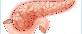
Most often, a vascular tumor is found in the liver, but a small hemangioma does not cause symptoms. Formation more than 4 cm in diameter leads to discomfort and a feeling of fullness in the stomach. Less commonly, weight loss, nausea, and dyspepsia are provoked.
Vascular neoplasms rarely occur in the brain:
- Hemangioblastoma accounts for 2% of all intracranial tumors. More often they are formed in the lower sections: the trunk and cerebellum. These are slow-growing tumors that form from the cells that make up the inner layer of blood vessels.
- Hemangiopericytes are less common and form in the membranes of the brain or spinal cord.
In practice, doctors are faced with hemangiomas of the liver and brain, which is why information about diagnosis and treatment is most widespread. Tumors are detected during random MRI examinations. Formations on the esophagus are extremely rare: among 20 thousand autopsies, only 3 such cases were found.
Most organ hemangiomas discovered incidentally do not require intervention. Tumors are surgically removed if they compress neighboring organs and tissues.
Special forms of hemangiomas
Rapidly involuting hemangiomas (RICHs) were not distinguished from infantile immature tumors for a long time. There are now clear criteria based on differential clinical, histological and immunophenotypic features. Round or oval, solitary, superficial or infiltrating vascular tumors are often located near joints on the extremities, on the scalp, forehead or around the ear.
Tumors develop in utero and do not grow postnatally. Complete involution occurs after about a year. Hemangiomas of this type on the scalp originate from the subperiosteal region and can provoke bone-related defects. The surface of the tumor is soft or telangiectatic, and shades vary from pink to purple. The skin around the tumor becomes poor, and a linear scar forms in the center. After regression, two types of complications occur: lipoatrophy with a pale bluish tint to the skin or telangiectatic plaque.
Color Doppler ultrasound is used for diagnosis. If clinical and radiological findings are equivocal, a biopsy is performed in newborns. Pathological studies demonstrate large and dense cell lobules, areas of fibrosis, and areas of irregularly shaped vessels. Differential diagnosis with angiomas, infantile myofibromatosis, and rhabdosarcomas is required.
Antenatal diagnosis of RICH during prenatal ultrasound monitoring is possible in the 3rd trimester, and sometimes from the 20th week of pregnancy. Genetic counseling is not provided.
Often the tumor is left to observe the end of spontaneous involution. For residual effects, lipofilling and surgical resection are used. Corticosteroid therapy is sometimes used.
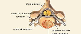
Spinal hemangiomas
It is believed that hemangioma is a childhood disease. However, vascular tumors in the bones develop after age 50. Most often they appear in the middle or lower back - the thoracic and lumbar regions. Only 5% of patients with spinal hemangiomas have symptoms. Tumors are accidentally discovered during X-rays and MRIs of the spine for other issues: back pain that is felt on the skin. Symptoms include numbness and weakness.
To accurately diagnose a spinal cord mass, an MRI or CT scan is required to detect damage to the nerves, spine, or spinal canal, as well as compression of the spinal cord itself.
Treatment is carried out if the tumor causes pain and affects movement. In rare cases, the tumor can cause paralysis. Treatment of spinal hemangiomas includes:
- embolization - a minimally invasive procedure that blocks blood flow to the tumor;
- ethanol injections promote blood clotting in the tumor to prevent bleeding;
- radiation therapy is aimed at destroying tumor cells;
- a surgical procedure to remove vertebrae (vertebractomy) or vertebral bones (laminectomy) is used when the segment is completely damaged.
Prognosis and prevention
Most hemangiomas have a favorable prognosis. They are not capable of degenerating into a malignant tumor. Some of them never increase in size, others quickly regress. If the formation does not cause discomfort and does not grow, then treatment is not prescribed to the patient. The hemangioma is monitored.
There is no specific prevention, but the disease can be avoided if you follow these recommendations:
- Avoid stressful situations.
- Prevent hormonal imbalances.
- Maintain good hygiene so that the pores on your skin do not become clogged.
- Limit consumption of fatty and high-calorie foods and sweets.
- Avoid prolonged exposure to direct sunlight. In the summer, you should use protective equipment.
- Strengthen your immune system with multivitamins.
If a person lives in an ecologically unfavorable area, then it is better to change his habitat. Prevention rules will not be able to completely protect against the appearance of such formations, but they will significantly reduce the risk of their development.
Capillary hemangioma is a benign formation on the skin or internal organs, consisting of a large number of capillaries, and the cavity is filled with blood. In the international classification (ICD-10) it has code D18. Pathanatomy defines the disease as a continuous mass of vessels covered with endothelium and connective tissue.
A characteristic feature of the tumor is that it heals on its own, and this can happen unexpectedly. The appearance of the tumor varies from red to purple, with a rough and raised shape. It is formed in the prenatal period, but its appearance is detected after the birth of the child; it is also possible for hemangioma to appear in the first months of life. During the first six months, the formation actively grows, reaching a size that can significantly interfere with normal life activities. Statistics show that usually after a year the tumor stops activity and a cure occurs.
If there is no regression or the tumor becomes large and causes discomfort to the child, you should consult a doctor and conduct a full diagnosis to determine treatment options.
Capillary hemangiomas in internal organs (liver, spleen, etc.) are highly dangerous.
Treatment options for hemangiomas
A single small hemangioma usually does not require treatment, as it goes away on its own. Treatment of vascular tumors that cause lesions or ulcers is required. There are several treatment options:
- Corticosteroid drugs are injected into the hemangioma to reduce its growth and relieve inflammation.
- Beta blocker creams are applied several times daily for 6 to 12 months to small superficial hemangiomas. The drug has no contraindications. Beta-adrenergic blockers in tablet form are also used in childhood.
- Laser therapy is used to remove hemangioma. In difficult cases, the surgeon uses a laser on the vessels to reduce redness and speed up the process of tissue regeneration.
- Becaplermin gel is used to heal ulcers on superficial cutaneous hemangiomas, but does not affect the tumor. The product is not used often due to the increased risk of cell degeneration.
- Surgery is necessary for large lesions in sensitive areas such as the eyes, nose, and throat.
Internal hemangiomas require treatment if they are large enough to impair organ function or cause pain. Surgical removal of a tumor, an organ as a whole or a section thereof, or embolization of an artery that supplies blood to the tumor is used.
Beta blockers in the treatment of childhood hemangiomas constrict blood vessels, suppress their growth, stimulate the death of tumor cells, and at the site of their new formation, creams should be used in the proliferative phase. The drugs act on beta-adrenergic receptors, which are expressed precisely during the period of tumor growth.
In domestic practice, Propranolol is used among beta blockers. Children with vascular manifestations are prescribed doses of 1–3 mg per kilogram of body weight per day. Side effects of a beta blocker include sleep disturbances, decreased blood pressure, and weakness in the child.
Medicinal methods
Depending on whether the patient has a particular complication, treatment tactics will be different - for example, topical corticosteroids may be prescribed. At the same time, the clinical response to even the most powerful drugs develops quite slowly—over several weeks. Therefore, this method is not suitable for vision-threatening conditions.
Injectable corticosteroids provide a faster effect: blanching of hemangiomas is observed already on days 2–3, and involution becomes noticeable after 2–4 weeks. The success rate of this therapy is about 75%.
Systemic corticosteroids are used for amblyogenic life-threatening lesions. A marked response is usually observed in 30% of patients, a moderate response in 40%, and no response in 30%.
Alpha-2a interferon is prescribed when hemangioma is resistant to corticosteroid therapy. Despite its good effectiveness, its use is associated with undesirable side effects: fever, arthralgia, retinal angiopathy.
Propranolol can be used to treat hemangiomas , although the evidence base is relatively weak and mainly consists of anecdotal cases. However, some studies indicate that propranolol may prevent loss of visual acuity in the case of periocular hemangioma.
For some superficial (and in some cases deeply located) neoplasms of a limited area, timolol .
Symptoms
Hemangioma is detected in most cases in the first days and weeks of a child’s life. The most rapid growth is observed in the first 6 months, then it usually slows down.
Traditional locations for hemangiomas in children are: scalp, face (nose, eyelids, cheeks), genitals, oral cavity, bones, internal organs, arms and legs, upper torso.
Superficial hemangiomas in children are formations rising above the skin ranging in size from several millimeters to tens of centimeters. Their shape and color may vary. To the touch, the area of the vascular formation is perceived to be hotter than the surrounding soft tissue (a symptom of temperature asymmetry).
Hemangioma in a child on the face, head, arm and other open areas of the body is also a cosmetic defect that causes significant psychological discomfort to both the child and his parents.
As the hemangioma grows, it begins to compress the surrounding tissues, thereby causing disruption of their functions. It is easily traumatized and bleeds. Hemangiomas on a child’s hand are especially often injured.
Hemangioma on a child’s hand is often traumatized and bleeds
Capillary hemangiomas in children can resolve spontaneously. In the process of such spontaneous regression, the stages of early and late involution can be distinguished. Regression of the vascular formation begins with the formation of a blanching focus in its central part, which very slowly increases. Often, complete disappearance of hemangioma occurs only at the end of puberty.
Risk factors
Although the reasons are not exactly clear, statistics allow us to roughly estimate in which case the risk of a tumor in a child is greatest. Risk groups include:
- children born as a result of multiple pregnancy;
- presence of birth injuries;
- persistent hypertension in a pregnant woman;
- maternal intoxication;
- smoking;
- the age of the expectant mother is over 40 years old;
- birth of children before 36 weeks.
If the expectant mother monitors her lifestyle and habits during pregnancy, the risk of developing pathologies in the baby can be significantly reduced.
Diagnostics
Usually, a visual examination of the patient is sufficient to diagnose a simple hemangioma. Most often, neoplasms are detected already in the neonatal period. If you suspect the possible presence of internal tumors and to accurately determine the structure of skin formations, the doctor may insist on additional studies - ultrasound or CT. Such diagnostic methods make it possible to find out the features of the localization of hemangiomas (including proximity to large vessels, the possibility of germination into the dermis, etc.), as well as their structural features. The detection of deep or extensive tumors may be a reason to perform other studies, in particular angiography.
Conservative and surgical treatment methods
At the initial stage of the formation of skin hemangiomas on the legs, especially when they are small in size, experts prefer to take a wait-and-see approach. If the defect does not have a pronounced cosmetic significance and does not bother you in any way, then the patient is given general recommendations for improving the health of the body. The immune system copes with the problem on its own.
Drug therapy has a beneficial effect on the course of the tumor:
- products based on propranolol/timolol - for example, "Propranobene" or "Timaderm";
- cytostatics - suppress the growth of tumor cells directly in the lesion, for example, Vincristine;
- corticosteroid medications - based on diprospan or prednisolone.
Stages of pathology development
There are 3 stages in the development of this benign tumor:
- progression;
- growth arrest;
- regression.
At the stage of active growth, the neoplasm has a bright red color and intensively increases in both area and height. In severe cases, the area of the hemangioma can increase to 2-4 mm per day.
Around the end of the first year of life, growth processes slow down, and by the age of 5, in most cases, they stop completely.
The process of reverse development occurs in 2% of cases of all pathologies. The tumor shrinks within 10 years. The redness at the site of formation is replaced either by scar tissue or healthy skin.
General information
Hemangioma is a vascular tumor that most often appears in children.
Hemangioma is a reddish spot that gradually appears on different parts of the body, as well as on the face. According to existing statistics, a child diagnosed with this disease is born once in a hundred cases. The reddened area continues to grow as the child grows, but the hemangioma does not cause any pain. But you should know that such a tumor is especially destructive due to its active growth inside the body. During its development, the functioning of the organ around which the redness has formed may be disrupted. For example, a hemangioma that is located in the ear area can eventually damage the eardrum , which can lead to hearing loss in the patient. Therefore, this disease should be treated to get rid of it as early as possible.
Vascular tumor on the skin
A true capillary tumor is located exclusively on the surface of the skin. Usually it rises above the skin, its nutrition is provided by the vessels of the dermis. At its core, such a neoplasm is a network of capillaries, closely intertwined and filled with blood. In this case, the vessels that make up the hemangioma can grow together, forming a ball of vascular tissue.
This neoplasm is considered the most common among all types of hemangiomas; it does not have a tendency to malingization (malignancy processes), but its development can be accompanied by growth into the skin and even ulceration. But this development of events does not indicate cancer.
To date, the exact reasons for the formation of capillary hemangiomas are not yet known to scientists. In newborns, such tumors appear due to malformations of the vascular system and can be provoked by disorders during pregnancy.