The world learned about mononucleosis back in 1887, when N.F. Filatov discovered this disease. Today we will talk about what mononucleosis in children is. Mononucleosis occurs in almost 90% of children under 10 years of age. This disease is caused by herpes type 4, called Epstein-Barr virus. Let's take a closer look at how infectious mononucleosis progresses in children's bodies, what symptoms it produces, and what to do if you find signs of mononucleosis in a child.
Causes
As a rule, children are often in large closed groups, such as a kindergarten, school, theater, public transport - in places of mass public gathering. In such public places, mononucleosis in a child can occur through transmission of infection from a sick person. There are several lines for obtaining the Epstein-Barr herpetic virus, these are:
- Close contact. When kissing, which is primarily associated with predominant infection by saliva. The virus penetrates from a sick person into the body of a healthy child through the mucous membrane of the larynx, mouth and nose - the respiratory tract. Infectious mononucleosis in children can occur as a result of blood transfusion from an infected donor.
- Airborne transmission of the virus. Despite the fact that the virus usually dies quickly in the environment, even in this situation the infection can enter the body.
- Household transmission route. General use of household items - cup, spoon, glass, plate, bottled water, towel, toothbrush, etc.
The incubation period usually ranges from 5 to 14 days - an average of a week. In some cases, according to statistics, mononucleosis in children can last from one and a half to two months. The reasons for this phenomenon are not well known.
Viral mononucleosis is possible when the following forms of infectious disease occur:
- Atypical. The characteristic symptoms of mononucleosis in both children and adults are associated with an incredibly stronger severity than usual. For example, children can have a fever when they are sick, or they can get sick without a fever. Atypical mononucleosis initially has a predisposition to cause serious complications and serious consequences.
- Chronic. It is considered as a catastrophic result of deterioration in the activity of the immune system of the child’s body.
Infectious mononucleosis in children with symptoms and treatment of any plan can vary significantly. This may depend absolutely on the subjective characteristics of the child’s body. First of all, this is the work of the immune system.
What is infectious mononucleosis
For reference. Infectious mononucleosis is an acute infectious pathology caused primarily by herpetic viruses.
Clinically, the infectious process manifests itself:
- specific inflammation of lymphoid tissues,
- intoxication and febrile symptoms,
- development of hepatosplenomegaly,
- the appearance of AM (atypical mononuclear cells) in blood tests.
The code for infectious mononucleosis according to ICD10 is B27. Depending on the causative agent of the infectious process, an additional code is indicated after the main code:
- 0 – for a disease caused by EPB (Epstein-Barr viruses);
- 1 – for MI caused by cytomegalovirus infection;
- 8 – for other forms of MI caused by a specified pathogen;
- 9 – for unspecified infectious mononucleosis (B27.9).
Symptoms
Because today there is practically no prevention against mass infection of mononucleosis in children. In cases where a child comes into contact with sick children, his health should be closely monitored. If somatic signs of mononucleosis do not appear, then the child either did not become infected, or the immune system of the child’s body coped with the infection and the disease passed without danger.
There are many infectious diseases. To understand what kind of disease this is, you need to understand the symptoms:
- Somatic manifestations of a prodromal nature are detected. Catarrhal symptoms - health gradually but noticeably worsens; the temperature remains at a low-grade point; there is a persistent soreness in the throat; when the nose is stuffy, breathing becomes very difficult; pathological swelling of the tonsils occurs.
- Signs of general intoxication appear - rashes on the body; severe chills; a sharp increase in temperature; physical weakness; significant enlargement of lymph nodes.
- When suddenly infected with mononucleosis, the symptoms in children are more pronounced. Under the current circumstances of such a plan, fever is not excluded - the temperature rises from 38 to 39 degrees and lasts for several days; in rare cases within a month. Excessive sweating, severe chills, excessive drowsiness, general weakness. Characteristic signs of intoxication are headache, sore throat when swallowing, aches throughout the body or in the muscles.
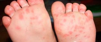
- Then usually comes the culmination of somatic infectious mononucleosis in children. The main characteristic features of the clinical picture of the disease are clearly expressed. Sore throat - the posterior wall of the pharyngeal mucosa appears crumbly, hemorrhages in the mucosa, and follicular hyperplasia may occur. Hepatosplenomegaly is also observed - a sharp enlargement of the spleen and a significant enlargement of the liver. Lymphadenopathy is a significant enlargement of the lymph nodes. The appearance of rashes over a large part of the body.
With infectious mononucleosis, rashes most often appear simultaneously with fever, a condition of enlarged nodes of the lymphatic system. The rash can be quite intensely localized in the legs, torso (back, arms or stomach) and face in the form of tiny spots of red and sometimes pale pink.
Such rashes do not require treatment, and the use of ointments is not recommended in any case. The rash self-destructs due to the increased fight of the immune system against the virus. If the rash begins to itch while taking antibiotics, this indicates an allergic reaction to these medications, because with mononucleosis the rashes do not itch.
How dangerous is the disease?
As a rule, the disease occurs in a benign form, complications rarely develop.
In isolated cases, the disease may be complicated by:
- autoimmune hemolytic anemias,
- thrombocytopenia,
- granulocytopenia,
- neurological pathologies (encephalitis, cranial nerve palsies, paralysis of facial muscles, etc.),
- meningoencephalitis,
- polyneuritis,
- transverse myelitis,
- psychoses,
- pericarditis,
- myocarditis,
- pneumonia,
- splenic ruptures.
Polyadenitis
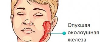
But still, the most significant somatic sign of infectious mononucleosis is usually considered to be polyadenitis - a joint group inflammatory process of the lymph nodes. Usually results from hyperplasia of lymphoid tissue. In the vast majority of cases, multiple island deposits in the form of gray and whitish-yellowish shades are formed on the tonsils. These loose and lumpy formations can be removed without much difficulty.
In addition to all this, the lymph nodes of the nervous system enlarge. The virus is actively retained in them. The lymph nodes on the back of the neck are especially enlarged. When you turn your head, the lymph nodes become very noticeable. Since the lymph nodes located nearby are interconnected, their damage is bilateral.
In some cases, the lymph nodes become enlarged even in the abdominal cavity. They compress the nerve endings, which provokes the possible occurrence of symptoms of an acute abdomen, which can lead to an incorrect diagnosis.
Infectious mononucleosis in children is characterized by hepatosplenomegaly - simultaneous enlargement of the liver and spleen. These are one of the most sensitive organs to the disease, so significant changes occur already in the initial stage of infection. The spleen can become so enlarged that it ruptures because the tissue cannot withstand the pressure.
Over the course of a month, there may be a continuous increase in the size of these organs. Sometimes it lasts even after the child recovers. When body temperature is restored, the condition of the liver and spleen is normalized.
Palpation of the lymph nodes is not so painful due to mobility and loose contact with the skin.
Complications
Complications from infection can be avoided if proper treatment is carried out, but in some cases the disease is confused with a cold and the wrong medications are initially prescribed.
- Complications from internal organs include: retinal hemorrhage, development of kidney inflammation, problems with the respiratory system, the appearance of purulent wounds, mumps, pancreatitis, thyroid diseases, liver dysfunction.
- When the spleen ruptures, the patient feels a sharp pain inside the abdomen, along with which bleeding occurs. Such an incident could be fatal. In this case, emergency hospitalization is performed.
- Due to liver damage, improper treatment can result in hepatitis. As a result of the destruction of liver cells, jaundice occurs.
- Meningitis occurs in a number of complications. It is characterized by inflammation of the lining of the brain. It is a serious disease, the treatment of which is a long and complex process.
- The appearance of anemia.
- The presence of hallucinations, frequent depression, mental disorders, Bell's palsy, encephalitis, inflammation of the brain.
When treating infectious mononucleosis, the main factor is timely consultation with a doctor, as well as compliance with the recommendations. With the right approach, the disease does not have serious and dangerous consequences. Drug therapy includes the removal of all inflammatory processes and symptoms, as well as restoration of the functioning of the affected organs.
It should be remembered that this virus is a type of herpes virus, which is why it cannot be completely cured. That is why repeated relapses in adults are possible after a certain time.
To avoid recurrent illness, it is necessary to support the immune system and lead a healthy lifestyle.
Article design: Mila Friedan
Diagnostics
For mononucleosis in children, treatment can be carried out only after visiting the clinic. Specialists, if the differential diagnosis is correctly established, will prescribe appropriate treatment after confirmation of special tests. Analyzes are examined in special laboratories.
To detect the Epstein-Barr virus, you need to take several tests to study them:
- EBV DNA is examined using the scientific PCR method;
- IgMk antibodies to EBV capsid antigen;
- antibodies of the IgM, IgG type to viruses by ELISA;
- IgGk antibodies to EBV nuclear antigen;
- IgGk antibodies to the capsid antigen.
Typically, such a diagnosis proceeds without any particular difficulties. Laboratory examinations are widely used. All these medical examinations clearly reveal the possible presence of infection over time. The stage of the disease is clearly revealed: acute or chronic.
Diagnosis of the disease
There is no specific test for mononucleosis; the main diagnostic method is a clinical blood test; in the presence of infection, it shows an increased level of atypical mononuclear cells, which appear 15-20 days after infection.
Additionally, there is a high content of leukocytes, lymphocytes, monocytes, and ESR in the blood; all indicators exceed the permissible age norms by 1.5 times.
What other tests need to be taken:
- biochemical blood test - allows you to determine the presence of malfunctions in the functioning of internal organs;
- HIV test;
- general urine test - shows the functioning of the urinary system;
- ELISA - analysis shows the presence of antibodies to pathogens in the blood;
- PCR – shows the presence of DNA of pathogenic microbes in the body.
In case of severe pathology, the doctor will prescribe an ultrasound or CT scan to determine the extent of damage to internal organs by pathogenic microorganisms.
Treatment
Treatment for mononucleosis in children inevitably involves the complete removal of somatic signs of the disease.
- To regulate a child's body temperature, antipyretic medications are recommended: Paracetamol for children is an excellent medication for reducing fever and relieving any painful signs of the disease. Analogues - Panadol, Efferalgan, Calpol.
- To eliminate the symptoms of sore throat and to remove plaque, it is advisable to prescribe throat sprays - Kameton and Ingalipt. Recommendations for gargling are saline solution, furatsilin and chamomile flowers.
- In case of a particularly depressing hypertoxic painful condition, a course of Prednisone is prescribed.
- If it is necessary to strengthen the immune system, immunomodulators are prescribed - children's Anaferon, Cycloferon, Imudon, Viferon, vitamins B, C, P.
- For a secondary viral infection, the attending physician prescribes antibiotics.
Diagnosis of Epstein-Barr viral infection
Diagnosis is based on a set of leading clinical symptoms (fever, lymphadenopathy, enlarged liver and spleen, changes in peripheral blood).
In addition to examining the blood picture, diagnosis is based on the detection of heterophilic antibodies and specific antibodies to EBV.
Heterogeneous antibodies. Modified heterohemagglutination reactions are used: the Paul–Bunnell reaction (sheep red blood cell agglutination reaction) is currently not recommended due to low specificity. Hoff-Bauer reaction - agglutination of formalinized horse erythrocytes (4% suspension) with the patient's blood serum, the reaction is carried out on glass, the results are taken into account after 2 minutes; Can be used for express diagnostics. Heterophilic antibody titers reach a maximum at 4–5 weeks from the onset of the disease, then decrease and can persist for 6–12 months. However, this reaction can also produce false positive and false negative results.
The most specific and sensitive methods are based on the determination of antibody markers of EBV antigens (NRIF, ELISA), which make it possible to determine the form of infection.
Table 18-27. Diagnostic value of antibodies to Epstein–Barr virus
Antibodies (IgM) to the capsid antigen in infectious mononucleosis are detected from the end of the incubation period; they are detected no more than 2–3 months. IgI to the capsid antigen appears in the acute period of infection and persists for life. Antibodies to early antigens (IgM) appear at the height of the disease in 70–80% of patients and quickly disappear, while antibodies to IgI persist for a long time. An increase in the titer of antibodies to early antigens is typical for the reactivation of EBV infection and for tumors caused by this virus. Antibodies against nuclear antigen appear 6 months after infection and remain in low titers for life.
An additional confirmation of Epstein-Barr viral infection can be a test to detect viral DNA in blood or saliva using the PCR method. Its use is effective for detecting EBV infection in newborns, when the determination of serological markers is ineffective due to an immature immune system, as well as in difficult and doubtful cases when diagnosing EBV in adults.
Differential diagnosis of infectious mononucleosis
It is necessary to differentiate with febrile diseases occurring with lymphadenopathy and hepatolienal syndrome; occurring with acute tonsillitis syndrome and occurring with the presence of atypical mononuclear cells in the blood.
Table 18-28. Differential diagnosis of infectious mononucleosis
Indications for consultation with other specialists
All patients diagnosed with infectious mononucleosis and suspected of having it must be examined for HIV infection in the acute period of the disease, after 1, 3 and 6 months during the period of convalescence.
If hematological changes persist, consultation and examination with a hematologist is indicated; if abdominal pain occurs, consultation with a surgeon and ultrasound of the abdominal organs.
If neurological symptoms appear, consultation with a neurologist is necessary.
An example of a diagnosis formulation
B27.0. Infectious mononucleosis. Moderate course.
Complication: rash after taking ampicillin.
Indications for hospitalization
Patients are hospitalized according to clinical indications. The main indications for hospitalization and treatment of a patient in a hospital are: prolonged high fever, jaundice, complications, diagnostic difficulties.
Home treatment
Treatment of infectious mononucleosis in children can also be carried out at home, combining the use of medications with herbal medicine. To prepare the decoction, you need to take herbs in equal proportions - chamomile flowers, coltsfoot, string, immortelle, calendula flowers, yarrow. Pour four tablespoons of dry herbs into one liter of boiling water. Leave in a thermos for about 10-12 hours. Then strain and drink half a faceted glass half an hour before meals.
Children are mainly treated for infectious mononucleosis at home. But in some cases, for certain reasons, treatment is carried out inpatiently. Children are hospitalized if quite severe laryngeal edema occurs (if breathing is difficult, pulmonary ventilation and tracheotomy are performed). If the spleen and liver are enlarged, it is possible to perform an operation - splenectomy.
Treatment of mononucleosis in children
Mononucleosis is a viral disease, so the use of antibiotics against it is pointless. There is no single medicine for the treatment of mononucleosis; various antiviral agents are used in therapy (Acyclovir, Isoprinosine, etc.). However, the main forces to fight the virus come from the body’s natural immunity, and the higher it is initially, the greater the chance of a quick recovery without complications.
☝☝☝Children's doctor Komarovsky says that acute mononucleosis is a disease that in most cases is treated on an outpatient basis, i.e. at home, subject to regular visits to the doctor.
However, in severe cases (especially for infants), hospitalization of the child in the hospital is indicated. The admission criteria are as follows:
- Temperature above 39.5 C;
- Development of complications;
- Severe signs of intoxication of the body - vomiting, nausea, prolonged fever, etc.;
- Severe breathing difficulties, risk of suffocation.
➡Mononucleosis can be treated using various means. As mentioned earlier, the first method of therapy is symptomatic, designed to eliminate the manifestations of the disease while the body’s immune system independently fights the virus. The drugs used for this purpose are mainly antipyretics.
Prednisolone for complex myocardial infarction
In the event that mononucleosis causes a complication in the form of a sore throat, local antiseptics are used, and immunomodulatory nonspecific drugs are also prescribed to maintain the body's defenses. Antibiotics are prescribed orally or by injection only if a bacterial infection is attached and it is detected in tests.
Often, treatment of mononucleosis is accompanied by the prescription of general strengthening vitamins, because The body loses many useful substances while fighting the disease. Hepatoprotectors and other drugs are also used to improve liver function. To avoid allergic reactions, antihistamines are prescribed in response to decreased immunity.
In the case of a severe course of the disease with clear signs of toxicosis, a short-term course of prednisolone is prescribed in a hospital setting. The drug is also used when there is a high risk of asphyxia. Also, if there is swelling of the larynx and severe breathing difficulties, a tracheostomy is installed, and the child is transferred to artificial ventilation.
Another dangerous complication of mononucleosis is splenic rupture. To avoid this, ultrasound monitoring of the organ’s condition is carried out regularly, and in case of rupture, surgery is necessary.
☝You can often meet people who recommend treating mononucleosis with homeopathy. In particular, you can meet people who give positive feedback about such treatment. The popular rumor about the benefits of homeopathy is explained by the fact that the remedies themselves do not make the body any better or worse, and mononucleosis is sometimes cured on its own provided the child has a strong immune system.
⚠However, with such treatment, complications can easily develop, which in turn can lead to consequences including death.
Children's diet
Strict and absolutely correct baby nutrition is recommended in the form of a mandatory gentle diet for mononucleosis in children. By adhering to these rules, you can count on a quick recovery and subsequent recovery.
- Exclude from the baby food diet: fried and fatty foods; sweets, pickles, preserves, smoked meats, onions, garlic, beans, peas and similar products. Reduce the consumption of sour cream; cheeses; fat cottage cheese; fat milk; oils - both butter and vegetable.
- Include in the baby's diet: milk porridge; all low-fat dairy and fish, as well as boiled meat products; fresh fruits and vegetables.
- Complex vitamins for children are a must.
Such a diet takes a lot of pressure off the liver in children, which was significantly damaged during the period of herpetic disease.
Additionally
To get the most objective picture of the disease, you need to take blood tests several times, and without fail - after recovery. Reliable results are ensured by compliance with the rules of preliminary preparation for analysis.
The patient needs:
- within 2-3 days, eliminate fatty foods, fried foods, and alcoholic beverages from the diet;
- stop taking medications;
- on the eve of the procedure, limit sports and other physical activities;
- observe a fasting regime of 8-12 hours (you need to donate blood for all tests strictly on an empty stomach).
- Stop nicotine at least an hour before blood sampling.
You can get acquainted with the results of biochemistry and OKA the next day. To perform special studies, a weekly execution interval is provided.
Recovery
After suffering herpetic mononucleosis in children, a recovery stage is established, which can last up to a whole year.
- Children who have recovered from the disease still feel tired, sleepy, overwhelmed, and apathetic for quite a long time.
- Most often, children have a poor appetite, which is why it is necessary to follow light, tasty diets. Drinking (natural juices, fruit drinks made from natural berries, warm herbal teas) should be plentiful.
- Do not overload children with homework or sports under any circumstances. Children should avoid both hypothermia and overheating. Children need walks in the fresh air. Spend more time in nature, at the dacha or in the village.
To summarize, it should be noted that during the entire recovery stage, children should be observed by their attending physician. Mononucleosis is not always a dangerous disease, especially if the child’s immune system works very well and fights the virus, but in any case, an integrated approach to treatment, correct diagnosis and good recovery are necessary.
Hematological parameters
OKA is carried out using capillary blood (from a finger). A general clinical analysis allows us to identify disturbances in biochemical processes characteristic of monocytic tonsillitis (another name for mononucleosis). Indicators of the leukogram, which are made up of white blood cells - leukocytes (in the study form are designated WBC), are important in diagnosing the disease.
They are responsible for protecting the body from bacteria, viruses, parasites and allergens. Subgroups of leukocytes:
- granulocytes: neutrophils - NEU (band and segmented), eosinophils - EOS, basophils - BAS.
- agranulocytes: monocytes - MON and lymphocytes - LYM.
When deciphering the results of an analysis for infectious mononucleosis, increased attention is paid to the following parameters:
- the presence of leukocytosis or leukopenia (increased or decreased values of leukocytes);
- shift in leukocyte formula (leukogram).
- presence of atypical mononuclear cells;
- shift in the values of monocytes and lymphocytes;
- hemoglobin concentration;
- change in erythrocyte sedimentation rate - red blood cells (ESR);
- the level of platelets (blood platelets that reflect the degree of blood clotting) and red blood cells.
The marker of Filatov's disease is atypical mononuclear cells (otherwise, virocytes or monolymphocytes) - young mononuclear cells from the group of agranulocytes. In a general analysis of healthy biofluid (blood), a scanty amount of these cells is detected, or they are not detected at all.
Characteristics of the disease
Infectious mononucleosis is an infectious viral .
Has an acute form of flow. The causative agent of the pathology is the Epstein-Barr virus, but it is believed that the herpes virus can also provoke the development of the disease.
Manifestations of pathology depend on the age of the child. In young children general signs of malaise ; recovery, as a rule, occurs 2-3 weeks after infection.
If we talk about teenagers, then everything is more complicated here, the clinical manifestations of the disease are more expressive and vivid, and the healing process takes a very long period of time (up to several months).
The infection is transmitted by airborne droplets and spreads gradually throughout the child’s body.
Initially, the areas of the upper respiratory tract , then, entering the bloodstream, it spreads throughout the body, primarily affecting nearby lymph nodes.
Infectious mononucleosis (Epstein Barr virus): symptoms, diagnosis, treatment
Etiology
Epstein-Barr virus (EBV), also known as human herpes virus 4, is the etiological agent in approximately 80-90% of cases of infectious mononucleosis.
In the remaining cases, mononucleosis syndrome not associated with Epstein-Barr virus can also be caused by human herpes virus 6 (9%), CMV (5-7%), HSV-1 (6%) and rarely streptococcal pyogenes, Toxoplasma gondii, HIV-1, adenovirus, as well as Corynebacterium diptheriae, Francisella tularensis, hepatitis A and B viruses, rubella or enteroviruses. This syndrome can also be caused by connective tissue diseases, malignancies, and drug reactions. The etiology of most cases of infectious mononucleosis not associated with Epstein-Barr virus remains unknown. Infection of people occurs mainly through saliva, hence the name of the disease - “kissing disease”. In one study, all patients with MI caused by EBV recovered virus from the oropharynx within 6 months of onset.
In a prospective study, 22 of 24 healthy individuals with a past history of EBV infection shed virus in their saliva over a 15-month period. There is no evidence of sexual transmission of the EB virus. In young women, the risk of EBV seroconversion has been shown to increase with the number of sexual partners. In one study, the risk of EBV infection was lower among university students who always used a condom than among those who had sex without condoms. Since EBV levels are significantly lower in genital secretions compared to saliva, sexual intercourse is probably not the most important route of transmission. However, both saliva and genital secretions are considered ineffective modes of transmission.
Rare cases of EBV transmission through blood products, organ transplantation, and intrauterine transmission have been reported. The risk of acquiring EBV infection through blood transfusion is extremely low. Humans, and possibly primates, are the only known reservoir of EBV. Determining clear risk factors is not possible due to the high prevalence of infection (over 90% of infected people are adults).
Pathophysiology
Epstein-Barr virus (EBV) has lytic and latent life cycles. Early primary infection (lytic) is likely to occur in oropharyngeal B cells when EBV directly contacts these cells through the tonsillar crypts. Circulating B cells then carry the infection to the liver, spleen, and peripheral lymph nodes, which triggers a humoral and cellular immune response to the virus. Antibodies produced in response to infection are directed against EBV structural proteins, such as viral capsid antigens (VCA), early antigens (EAs), and EBV nuclear antigen (EBNA); these antibodies are used for serological diagnosis of EBV infection. A rapid cellular response by T cells is critical for suppression of primary EBV infection and determines the clinical expression of EBV infection.
Symptomatic primary infection caused by the EB virus is usually followed by a latent stage. Latent infection is associated with self-replicating extrachromosomal nucleic acid, episomes.
During latent EBV infection, the virus prolongs the lifespan of infected lymphocytes; that is, it converts normal human lymphocytes with a limited lifespan in vitro into continuous cell lines. In a healthy EBV seropositive adult, approximately 0.005% of circulating B cells are infected with EBV. It is likely that during latency there are low levels of ongoing viral replication and B cell infection in tonsillar and lymphoid tissues controlled by residual populations of EBV-specific T cells. An observational study in patients with asymptomatic primary infection suggests that symptoms may be due to an overactive immune response rather than the viral infection itself.
Key diagnostic factors
- Cervical or generalized lymphadenopathy: The nodes are usually painful.
- They become most noticeable after the second week of illness.
- Observed in 94% of cases.
- Often resembles pharyngitis caused by Streptococcus pyogenes.
- Varies from 37.8 °C to 41.1 °C; usually <38.9°C.
- Splenomegaly (Increase begins in the first week. Lasts 3-4 weeks)
- Adults are less likely to have sore throat and lymphadenopathy, but are more likely to have hepatomegaly and jaundice (due to hepatitis). Liver involvement in acute EBV infection manifests as mild hepatitis with predominantly cholestatic features, but does not always manifest as clinically significant jaundice. The overall incidence of jaundice in adolescents and adults is about 9%.
Diagnostics
Infectious mononucleosis can develop in people with a primary Epstein-Barr virus (EBV) infection, but this does not occur in all cases and depends on the age of infection and other host factors.
History and clinical examination
In developed countries, suspected infectious mononucleosis in patients 10–30 years of age presents with fever, fatigue, malaise, pharyngitis, and cervical or generalized lymphadenopathy.
The disease typically develops gradually, but some patients may have an abrupt onset. Young children may have symptoms similar to those in adults, but more often the infection in children is subclinical or mild with nonspecific symptoms. Symptoms of mononucleosis syndrome not associated with EBV are usually less severe. Mild prodromal symptoms that last for several days include a feeling of general malaise, fatigue, and sometimes fever, and progress to an acute phase.
The clinical picture of infectious mononucleosis in children and adults with weakened immune systems may be similar to the picture of the disease in patients with healthy immune systems. Symptoms of infectious mononucleosis may resolve within a few days or may persist for 3 to 4 weeks (up to 8 weeks in some patients). In some cases, a biphasic disease may occur with worsening symptoms after initial improvement. Some symptoms of infectious mononucleosis, such as fatigue, may take several months to completely resolve.
People with MI should be monitored long-term for possible complications, including airway obstruction, hemolytic anemia, and thrombocytopenia.
Diagnostic tests
| Study | Result |
Detailed blood test
|
|
Detection of heterophilic antibodies
|
|
EBV-specific antibodies
|
|
Real-time PCR
|
|
Ultrasound of the abdominal organs
|
|
Differential diagnosis
| Disease | Differential signs/symptoms | Differential examinations |
| Pharyngitis caused by group A streptococci |
|
|
| Hepatitis A |
|
|
| Acute HIV infection |
|
|
| Adenoviruses |
|
|
| Human herpes virus-6 |
|
|
| CMV infection |
|
|
| Herpes simplex virus-1 |
|
|
Treatment
MI is usually a self-limiting condition that does not require any special treatment. The basis of therapy is supportive care. Supportive treatment includes good hydration, antipyretics and pain relievers such as paracetamol and non-steroidal anti-inflammatory drugs. Aspirin should not be prescribed to children due to the possibility of developing Reye's syndrome.
Rest is a common recommendation, but its benefit is unknown in the treatment of MI. In case of infectious mononucleosis, in order to avoid possible rupture of the spleen, it is advisable to refrain from active sports (including contact sports) in the first 3-4 weeks of the disease.
Acyclovir temporarily reduces viral proliferation in the oropharynx but does not help resolve symptoms or reduce complications. Therefore, acyclovir and similar antiviral drugs are not prescribed.
Serious disease
Patients with severe systemic manifestations of MI and complications should be referred to the hospital. Systemic corticosteroids should be used in patients with severe airway obstruction, severe thrombocytopenia (<20,000 platelets/mm3) and hemolytic anemia. These short-term complications are rare but require surgical treatment.
Severe obstruction, observed in 1-5% of patients, is caused by enlargement of the tonsils and lymph nodes in the oropharynx. Systemic corticosteroids may improve symptoms of obstruction, but intubation, tracheostomy, or, in extreme cases, tonsillectomy may be required.
Severe thrombocytopenia occurs in less than 1% of cases, although moderate thrombocytopenia is more common. In most patients it resolves within 4 to 6 weeks and is likely due to platelet destruction in the spleen or antiplatelet antibodies. There is no clear interaction between clinical severity of MI and platelet count. Systemic corticosteroids may be helpful, and intravenous immunoglobulin (IVIG) may be successful for immune thrombocytopenia.
Hemolytic anemia occurs in 1-3% of cases and most likely results from the production of antibodies against red blood cells. This usually occurs in the second and third weeks of illness and resolves within 2 weeks. Systemic corticosteroids may hasten resolution of symptoms.
Although corticosteroids are usually reserved for severe complications, some practitioners treat patients with MI with corticosteroids to relieve symptoms. In one study, treatment with corticosteroids provided relief from sore throat in the short term (more than 12 hours), but this benefit was lost after 24, 48, and 72 hours. Overall, there is currently insufficient evidence to recommend corticosteroid therapy for symptom control in MI. Studies on the effectiveness of corticosteroid treatment are rare, heterogeneous and of poor quality. There is also a lack of research into side effects, potential side effects or complications, especially in the long term.
Post-infectious chronic fatigue syndrome
The reported incidence of this syndrome in patients with a history of MI varies among sources, but in general it is not considered very common. Several small studies have raised the possibility that Epstein-Barr virus (EBV) may play a role in the pathogenesis of chronic fatigue syndrome in subgroups of these patients. There is still no reliable data to routinely consider the EB virus to be the etiological agent of this widespread syndrome. Rather, new evidence suggests that post-infectious chronic fatigue syndrome is a common and patterned result of several viral and non-viral infections.
In a longitudinal cohort study, half of young patients with chronic fatigue syndrome after myocardial infarction with long-term disability showed significant improvement, including full-time and part-time employment, thereby confirming better results than expected.
Serological diagnostic method
In the 20th century, the Paul-Bunnell test began to be performed to detect heterophilic antibodies in blood serum. If in the analysis their titer exceeds the ratio of 1:224, this confirms the development of mononucleosis. However, the test result in the first week from the onset of clinical manifestations of infection is negative in 40% of cases, therefore, for reliability, a repeat test is required 7 days and 14 days after the first test.
Today, the Paul-Bunnell test is considered less effective for confirming mononucleosis, since the accuracy of the reaction is not high, and the results do not indicate the stage and severity of the disease. With this method, it is also not clear whether the infection is primary or whether it is reactivated. Often, a blood test shows a decrease in heterophilic antibodies, and then their level increases again. This phenomenon also occurs with other viral infections.
Mechanism of disease development
The Epstein-Barr virus enters the oral mucosa with saliva or its droplets and attaches to its cells - epithelial cells. From here, viral particles penetrate the salivary glands, immune cells - lymphocytes, macrophages, neutrophils and begin to actively multiply. There is a gradual accumulation of the pathogen and infection of more and more new cells. When the mass of viral particles reaches a certain value, their presence in the body activates the immune response mechanisms. A special type of immune cells - T-killers - destroy infected lymphocytes, and therefore a large amount of biological active substances and viral particles are released into the blood. Their circulation in the blood leads to an increase in body temperature and toxic damage to the liver - at this moment the first signs of the disease appear.
A special feature of the Epstein-Barr virus is its ability to accelerate the growth and reproduction of B lymphocytes - they proliferate and subsequently transform into plasma cells. The latter actively synthesize and release immunoglobulin proteins into the blood, which, in turn, causes the activation of another series of immune cells - T-suppressor cells. They produce substances designed to suppress excessive proliferation of B lymphocytes. The process of their maturation and transition to mature forms is disrupted, and therefore the number of mononuclear cells in the blood - mononuclear cells with a narrow rim of cytoplasm - sharply increases. In fact, they are immature B lymphocytes and serve as the most reliable sign of infectious mononucleosis.
The pathological process leads to an increase in the size of the lymph nodes, since it is in them that the synthesis and further growth of lymphocytes occurs. A powerful inflammatory reaction develops in the palatine tonsils, outwardly indistinguishable from a sore throat. Depending on the depth of damage to the mucous membrane, its changes vary from friability to deep ulcers and plaque. The Epstein-Barr virus suppresses the immune response due to certain proteins, the synthesis of which occurs under the influence of its DNA. On the other hand, infected mucosal epithelial cells actively release substances that initiate an inflammatory reaction. In this regard, the amount of antibodies to the virus and a specific antiviral substance, interferon, gradually increases.
Most of the viral particles are eliminated from the body, but B-lymphocytes with embedded viral DNA remain in the human body for life, which they pass on to daughter cells. The pathogen changes the amount of immunoglobulins synthesized by the lymphocyte, and therefore can lead to complications in the form of autoimmune processes and atopic reactions. Chronic mononucleosis with a relapsing course is formed as a result of an insufficient immune response in the acute phase, due to which the virus escapes aggression and remains in sufficient quantities for exacerbations of the disease.
Symptoms and first signs
The incubation period (from the moment of introduction to the first clinical manifestations) lasts 4-7 weeks. During this period, the virus penetrates through the mucous membranes (oropharynx, salivary glands, cervix, gastrointestinal tract). Afterwards, the virus begins to contact B-lymphocytes, infecting them, replacing their genetic information with its own, this causes further disorganization of the infected cells - in addition to foreign DNA, they also receive “cellular immortality” - practically uncontrolled division, and this is very bad, because they no longer perform a protective function, but are simply carriers of the virus.
The main clinical manifestations of benign lymphoblastosis are:
- increased fatigue;
- lymphadenopathy (enlargement of regional lymph nodes);
- hyperthermia;
- a sore throat.
The following clinical manifestations (alone or in various combinations) may also occur:
- myalgia;
- arthralgia (joint pain due to lymph stagnation);
- headaches (including migraines);
- catarrhal tracheitis;
- catarrhal bronchitis;
- decreased general immunity.
As a rule, the first symptom of infectious mononucleosis is general malaise without any other manifestations of pathology. The initial period lasts on average about a week. As the disease develops, enlargement (up to 2-3 cm) and tenderness of the cervical lymph nodes and an increase in general temperature to febrile values (38-39°C) occur.
Infectious mononucleosis is accompanied by liver damage, and therefore symptoms such as a feeling of heaviness in the right hypochondrium and a change in the color of urine (it becomes dark) are often noted. The spleen is also involved in the pathological process, so the patient has splenomegaly (an increase in size of this organ).
The total duration of the disease is on average 1-2 weeks, after which a period of convalescence begins. The patient's condition gradually improves, but general weakness and enlargement of the cervical nodes may be observed for another 3 weeks.
Forecast
Patients who have recovered from viral mononucleosis are usually given a favorable prognosis.
It is worth noting that the main condition for the absence of complications and adverse consequences is the timely detection of leukemia and constant monitoring of changes in blood parameters. It is also extremely important to monitor the well-being of patients until they fully recover. Scientific research revealed:
- body temperature above 37.5 degrees persists for approximately several weeks;
- symptoms of sore throat and sore throat persist for 1-2 weeks;
- the condition of the lymph nodes is normalized within 4 weeks from the moment of manifestation of the disease;
- Complaints of drowsiness, fatigue, weakness can be detected for another 6 months.
Adults and children who have recovered from the disease need regular medical examinations for six months to a year, with mandatory regular blood tests.
Polymerase chain reaction for mononucleosis
The most sensitive method is PCR.
It is used to diagnose mononucleosis in newborns, with questionable results of previous tests, as well as for people if their clinical infection is complicated. To carry out PCR, a person needs to donate blood from a vein or saliva, urine, or amniotic fluid. Before collecting biomaterial, you should not eat for at least 8 hours, and the day before you should eat fatty foods. PCR diagnostics allows you to identify the type of virus and detect its DNA in cells at the earliest stages. A positive value in the test indicates the presence of HHV-4 and thereby confirms the cause of the infection.
Prevention
Currently, medical science has not yet created a specific vaccine against mononucleosis. Therefore, disease prevention is extremely important. Preventing infectious disease includes:
- following strict rules of personal hygiene;
- use of individual cutlery;
- using a personal toothbrush;
- scrupulous examination of donor blood for the presence of the virus.
In addition, you should not forget about strengthening the immune system:
- do hardening;
- do exercises;
- exercise;
- be in the fresh air more often;
- take vitamins comprehensively.
Considering that a person has already had mononucleosis in childhood or adolescence, the possibility of a dangerous relapse in adults is unlikely.
Symptoms of the disease
The incubation period ranges from several days to one and a half months. Signs of mononucleosis in adults are as follows:
- The oral cavity and pharynx are affected. The palatine tonsils become enlarged, which leads to difficulty breathing and hoarseness. In the first days of the disease, the tonsils become covered with a thick white coating. Nasal mucus is not always present, but there is congestion in the nasal passages.
- Enlarged lymph nodes. They become inflamed in the neck, back of the head, elbows and intestines, but they remain mobile and do not connect with the underlying tissues.
- Temperature. There is a sharp rise to 39-40 degrees.
- Enlarged spleen and liver. A week after the onset of the disease, the organs reach their maximum size. In this case, sometimes yellowness of the skin and sclera of the eyes is observed. Organ enlargement lasts up to three months.
- Skin rashes. With the active development of the disease, a rash similar to measles or scarlet fever appears on the skin. There are pinpoint hemorrhages in the oral cavity and palatal area.
- Cardiovascular system disorders. Possible tachycardia, systolic murmurs and decreased heart sounds.
When treating mononucleosis in adults, symptoms disappear after two to three weeks, but atypical mononuclear cells are still detected in the blood for a long time.
Pharmacotherapy of infectious disease
For mild forms of the disease, treatment is carried out on an outpatient basis, and for severe forms - in the infectious diseases departments of the hospital. During the acute period, the patient must remain in bed; in addition, he is recommended to drink plenty of fluids: fruit juice, compote, tea and light diet meals. The following medications are used to treat the symptoms of mononucleosis in adults:
- Antipyretics - to normalize body temperature: Nimesulide, Ibuprofen.
- To support the immune system - Interferon-alpha.
- Antiviral – activate the body’s resistance to viruses: “Cycloferon”, “Tiloron”.
- Antibiotics - used if necessary to prevent bacterial infections: Azithromycin, Ceftriaxone.
- Glucocorticoids - prescribed when problems with the respiratory system occur: Dexamethasone, Prednisolone.
- Solutions for intravenous administration - reduce intoxication, make the patient feel better: “Dextrose”, saline solution.
- Vitamin and mineral complexes - to restore the body.
The average duration of treatment is from two weeks to a month. After this, the patient is registered at the dispensary for a year, undergoing laboratory monitoring of blood counts every three months.
Clinical picture of the chronic course of the disease
Unlike the acute form, the disease is sluggish, and all symptoms are mild:
- The patient feels weakness, drowsiness, slight malaise, and headaches.
- The temperature stays between 37.2–37.5 degrees.
- There is a weak, aching and painful sensation in the throat. Purulent plugs come out of the lacunae with an unpleasant odor.
- The cervical and sublingual nodes are inflamed, a nagging pain is felt when talking or turning the neck.
- Skin rashes in chronic mononucleosis in adults are minor and may be present on the neck, chest, arms and face.
- The nasal passages are blocked, the mucus secretion is small.
- Slight enlargement of the liver and spleen is also present.
There are no signs of damage to the gastrointestinal tract or lungs. After about a week, the symptoms of the disease disappear on their own, but the disease is not cured. Once in the body, the Epstein-Barr virus remains in it for life. At the same time, it makes itself felt as soon as the immune system weakens, and each time it manifests itself differently.