Amebiasis is an infection by amoebas, microorganisms that parasitize the large intestine. You can get sick at any age. For a long time a person does not even suspect an invasion, because... no symptoms appear. Clinical signs are diagnosed when there is a large accumulation of worms in the intestine (parasites damage the mucous membrane of the organ). Treatment is medicinal.
Amebiasis is an infection by amoebas, microorganisms that parasitize the large intestine.
Prevalence statistics
The maximum prevalence is observed in Mexico and India, with a high rate in South Asia and Africa. In the post-Soviet space, trouble remains in Tajikistan, Kyrgyzstan, and Turkmenistan.
Middle-aged men are more likely to get sick. Every tenth person who encounters the pathogen is susceptible to intestinal amoebiasis. According to WHO, there are about 480 million carriers of dysenteric amoeba in the world. Every year there are 50 million new cases of the disease. In this case, death occurs in 2%.
Sanitary conditions in Bangladesh present the best conditions for the spread of amoebiasis
Violation of sanitary well-being accompanies natural disasters, migration of people in search of work, and social problems. Therefore, intestinal amebiasis remains a problem for developed countries. We will look in detail at what amoebiasis is, starting with a description of the pathogen and its properties.
Symptoms of intestinal amoebiasis
Patients may not experience this pathology for many years.
At the moment when the disease enters the invasive stage, characteristic symptoms begin to appear:
- Initially, necrotic areas will begin to form on the organ mucosa. Over time, they transform into ulcerative lesions, which can increase not only in depth, but also diagonally.
- There is a foul odor from the mouth.
- Due to the progression of necrotic processes, ulcerative lesions will begin to perforate, leading to the development of peritonitis.
- If ulcerative lesions are located in the rectosigmoid part of the intestine, then patients will develop dysentery syndrome. Impurities of mucus, blood and purulent masses may be detected.
- In the case where the cecum was affected, the patient will complain of pain in the lower abdomen and constipation.
If the pathology occurs in an acute form, then patients may exhibit the following symptoms:
- diarrhea begins;
- pain of a cramping nature occurs;
- feverish conditions appear;
- vomiting occurs, etc.
With the development of the necrotizing form of the disease, the following clinical picture is observed:
- signs of intoxication;
- internal bleeding;
- perforation of ulcerative lesions;
- development of peritonitis.
With prolonged intestinal amebiasis, patients experience the following symptoms:
- defecation processes are disrupted;
- motor dysfunction;
- general weakness develops;
- pain occurs in the lower abdomen;
- loss of appetite;
- nausea appears, etc.
Characteristics of the pathogen
The causative agent of amoebic dysentery is the simplest single-celled organism from the amoeba family. Of all the representatives of the class, dysenteric amoeba (histolytica) is considered pathogenic for humans, others do not pose a danger.
Intestinal amoeba has characteristic structural features that distinguish it by microscopy from other parasitic microorganisms:
- the form is constantly changing;
- core large, transparent;
- there are pseudopods (pseudopodia), with the help of which the amoeba moves;
- the outer shell is thin;
- intracellular fluid (cytoplasm) is colorless.
Movement is carried out by pouring contents into outgrowths (pseudopods). The favorite conditions for the parasite to live are created in the human colon. This sign determines the main symptoms and methods of treating the disease. The size of the amoeba and internal inclusions depend on the stage of development.
Dysenteric amoeba goes through 2 stages of development (vegetative and cystic), while retaining its viability in each.
During the growing season it takes 4 forms:
- tissue - cell size on average up to 25 microns, detected only during acute inflammation directly in the affected tissue, and not in the feces;
- large histolytic - occurs when the amoeba develops the ability to phagocytose erythrocytes with the release of special enzymes, sizes reach up to 40 microns, extends up to 80 microns in length, the pathogen penetrates the mucous and submucosal layers of the intestine, causes ulceration and necrosis, is detected in stool analysis, characteristic for the acute course of the disease;
- luminal - dimensions up to 25 microns, movements are sluggish, which is associated with adaptation to other microorganisms, the formation of connections, detected in persons who have suffered acute amebiasis, suffering from a recurrent chronic form, with an asymptomatic course;
- precystic – ensures the transition of the amoeba from the luminal stage to the cyst, size 10–18 microns.
Laboratory technicians compare the appearance of this simple organism to “broken glass”
It is important that dysentery amoebas cannot exist for a long time in the external environment in the form of luminal and large histolytic, and are quickly destroyed. A cyst is a state of rest and “waiting for the right opportunity” to manifest infectious properties. The microorganism is protected as much as possible from the external environment to maintain viability. Characteristic:
- roundness of shape;
- dense shell;
- in a ripe state - four kernels;
- the presence of “reserves” of glycogen, protein in the cytoplasm;
- localization in the sigmoid and rectum.
It is found in the feces of persons who have had amoebic dysentery, during the recovery period, and in carriers of the infection. Activation of the cyst occurs after the dissolution of the outer shell. It turns into an intermediate form and contains 4 nuclei. Each nucleus undergoes fission in half. The process continues until 8 new amoebas are formed in the luminal form, each containing one nucleus. Once in the large intestine, they are transformed into the most pathogenic form (tissue and large vegetative).
Clinical picture and types
There are two types of pathology:
- intestinal amebiasis, in which parasites affect only the intestines;
- extraintestinal amebiasis, in which the pathogenic agent can be found in other organs, usually the liver.
Intestinal amoebiasis
It can be asymptomatic for many years.
However, at any time a transition to invasive amoebiasis is possible, in which signs of the disease appear. First, small necrotic foci appear in the mucous membrane of the colon, which over time can grow and ulcers form. At the same time, not only new areas of the intestinal mucosa, but also deeper tissues are involved in the pathological process. Ulcers form throughout the intestine. They can cause perforation of the intestinal wall and the development of peritonitis.
If the ulcers are localized in the rectum and sigmoid colon, dysentery syndrome develops, and in some patients impurities of pus, blood and mucus can be detected in the stool.
If mainly the cecum is affected, the patient experiences constipation and pain in the lower abdomen on the right. These symptoms resemble those of appendicitis, which often occurs against the background of amoebiasis.
Damage to the ileum in amebiasis is rare.
Depending on the course of the infection, there are:
- acute form of amoebiasis;
- fulminant (fulminant) colitis;
- prolonged or primary chronic amoebiasis.
Acute form
In the acute form, a characteristic symptom is loose stools. Other symptoms of amoebiasis develop less frequently:
- amoebic dysentery syndrome, in which there is an acute onset, spastic pain, bloody stools with mucus;
- temperature;
- vomiting and dehydration, which occurs quickly in young children.
One of the signs of amebiasis may be abdominal pain
Fulminant colitis
This development of the disease is more often diagnosed in women expecting a child or immediately after childbirth. This is a necrotizing form, which is characterized by a severe course and often leads to the death of the patient.
The following symptoms are characteristic of fulminant colitis:
- toxic syndrome;
- involvement of deep layers of the colon mucosa in the pathological process;
- bleeding;
- rupture of the intestinal wall;
- inflammation of the peritoneum.
Fulminant colitis may develop after treatment with corticosteroid hormones.
Protracted amoebiasis
With this development of the disease, the following signs are observed:
- impaired intestinal motor function;
- diarrhea;
- difficult defecation (observed in 50% of patients);
- loose stools followed by constipation;
- asthenia;
- nausea;
- abdominal pain;
- loss of appetite.
Intestinal amebiasis can lead to the following complications:
- perforation of the intestinal wall, which can cause peritonitis and abscess of the abdominal cavity;
- the amoebic structure, which is formed by granular tissue, can cause constant constipation and local intestinal obstruction
- appendicitis;
- massive bleeding from the intestines;
- ameboma is a neoplasm in the wall of the colon.
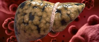
Extraintestinal amoebiasis
Extraintestinal amebiasis, depending on the location of development of the pathological process, comes in several forms.
Liver abscess. It is more often diagnosed in adult men. Basically, the right lobe of the liver is involved in the pathological process.
This course of the disease is characterized by the appearance of the following symptoms:
- night fever, accompanied by severe sweating and chills;
- hepatomegaly;
- pain in the right hypochondrium;
- increased leukocyte levels;
- jaundice, when it appears the prognosis is unfavorable.
Due to the latent course of amebic abscess, adequate therapy is difficult.
A liver abscess can burst, causing inflammation of the peritoneum and damage to the organs of the chest cavity.
The pleuropulmonary form develops as a result of rupture of an amoebic liver abscess and entry of pathogenic agents into the lungs. In rare cases, microorganisms may enter the bloodstream.
With this course of the disease, the following signs are observed:
- dyspnea;
- moist cough;
- chest pain;
- traces of blood and pus in the sputum;
- fever accompanied by chills;
- increase in the number of leukocytes.
Amoebic pericarditis develops as a result of rupture of a liver abscess into the serosa of the heart. This is a very dangerous condition and can cause cardiac tamponade and death.
The cerebral form has an acute onset, rapidly progresses and ends in the death of the patient. With this course of amebiasis, abscesses can form in any part of the brain.
The cutaneous form usually develops in weakened and malnourished patients. As a rule, ulcers are localized around the anus.
There are no specific signs of intestinal and extraintestinal amebiasis, and a diagnosis cannot be made solely on the basis of the patient’s complaints. Therefore, before prescribing certain medications, the doctor must carry out diagnostic measures.
How does infection occur?
It was found that up to 400 million amoeba cysts are excreted in the feces of a sick person or carrier per day. They are able to remain in the external environment for a long time, on objects that an infected person has touched with dirty hands.
Most often, the routes of transmission of infection are provided by:
- dirty human hands;
- with insufficient food processing;
- through unwashed vegetables or fruits;
- through the soil when working in garden plots and vegetable gardens;
- common dishes, household items;
- bed linen and underwear;
- flies and cockroaches.
Drinking water from lakes, rivers, swimming pools contaminated with sewage water is one of the ways of infection
Infection with dysentery of amoebic etiology is possible only when cysts of the pathogen enter the oral cavity. This mechanism of transmission is called “fecal-oral”. With particles of feces, cysts are swallowed and reach the intestines. Here they begin to actively reproduce.
Infection often occurs through contact with a sick patient or carrier, or shaking hands. It is classified as contact-household. Direct contact makes infection likely for lovers of oral-anal sex. Summer swimming in polluted waters contributes to the spread of the disease if water is swallowed.
Routes of entry into the body
This disease is classified as an anthroponotic infection, as it can only be transmitted from person to person. Amoebas can remain in the intestines for a long time without any signs, feeding on bacteria there and without causing damage to the human body, while their carrier becomes an active source of infection for other people. Infection with these bacteria can occur in two ways: fecal-oral or contact-household.
Dysenteric amoeba cysts can enter the body along with unwashed vegetables, berries and fruits, if water gets into the mouth while swimming in a pond. Also, linen or dishes can become a source of infection if they are used by an infected person. At risk are women during pregnancy and people with reduced immunity, since when dysenteric amoeba penetrates into a weakened body, it immediately begins its harmful activity and causes the development of intestinal amoebiasis.
Development mechanism
Pancreatic enzymes act on cysts that enter the small intestine and cause the dissolution of the membrane. The released amoeba reproduces by division. Passes into the large intestine. The luminal form may not cause disease for a long time. For the symptoms of intestinal amebiasis to appear, the pathogen must transform into a tissue form.
The conditions for activating the transition to tissue forms are:
- massive intestinal infection;
- different types of damage to the mucous membrane (inflamed areas, microtrauma);
- violation of intestinal peristalsis (muscle contractions);
- altered balance of intestinal microflora;
- the presence of other parasitic diseases (giardiasis, helminthiasis);
- fasting, low-calorie diet without sufficient intake of proteins and vitamins;
- immunodeficiency state of the body;
- disrupted hormonal levels (a current condition for infection of amebiasis in women during pregnancy);
- stress and physical overload.
For sufficient reproduction of the amoeba, an incubation period of two weeks to three months is required. This explains the fact that the initial symptoms of amebiasis appear 3-4 months after the alleged infection. The pathogenic form then “attacks” the intestinal mucosa.
Having secured itself on the intestinal wall, the microorganism secretes cytolysin and proteolytic enzymes. These substances have the ability to destroy cellular epithelial proteins and disrupt the structure of their membrane. Dead cells enable the amoeba to penetrate into the deeper submucosal layer of the intestinal wall. Active reproduction continues here and primary foci are formed. They open inward and form ulcers.
Part of the mucous membrane independently recovers and becomes covered with scars. The affected area appears as a combination of ulcers and healed areas. This is the pathogenesis of the disease.
On the surface, the ulcers look like isolated structures, but at the level of the submucosal layer a network of “tunnels” appears for increased movement of amoebas
Cause of illness
Dysenteric amoeba is a sarcode protozoan that lives in the human intestine. There are 2 stages in the amoeba development cycle: vegetative and cystic. In turn, the vegetative stage consists of 4 forms: large vegetative, small vegetative, tissue and precystic. The transition from one form to another depends on the conditions created in the intestines.
Small vegetative form (luminal form)
A unicellular small organism of irregular shape with pseudopodia, up to 12-20 microns in size. It is located in the upper part of the large intestine, where it feeds mainly on its contents and bacteria. This form is unstable in the environment and therefore dies quickly. With increased intestinal peristalsis, it is excreted from the body through feces. If the peristaltic waves are weak, then the small form further enters the lower part of the large intestine and turns into the pre-cystic form. A small form of amoeba can be detected in persons with acute amoebiasis, in carriers and in chronic amoebiasis.
Precystic form
It is an intermediate form between the luminal form and the cyst. It is inactive, round in shape, with a diameter of up to 10-15 microns. Covered with a single-layer shell, contains 1-2 cores. Under unfavorable conditions, the precystic form quickly turns into a mature cyst.
Cyst
The cyst of the dysentery amoeba is quadruple, 10-12 microns in size, immobile. The outside is covered with a two-layer shell. Chromatin grains and chromatoid bodies are located in the cytoplasm. The cyst is resistant to environmental conditions. It can be stored in feces for more than 1 week at an air temperature of +26-+30 degrees. Survives even longer in moist soil and water. The double-circuit shell of the cyst allows it to withstand dry weather well. The cyst dies quickly when boiled or using disinfectants (except for solutions containing chlorine). This form of amoeba is found in the stool of convalescents and cyst carriers.
Large vegetative form
Under favorable conditions it turns from small. Its dimensions vary from 40 to 60-80 microns. This form is active and mobile due to pseudopods. Cytoplasm is represented by ectoplasm and endoplasm, which contains 1 nucleus. The larger form is capable of producing the enzyme hyaluronidase, which damages the intestinal mucosa and capillary walls. This form of amoeba is also called erythrophage, because it feeds on red blood cells. The capture of red blood cells is carried out by pseudopodia, then the red blood cells end up in the cytoplasm, where they are digested. Under poor living conditions, the large form is not able to transform back into the small form or form a cyst. Therefore, she quickly dies. This form of amoeba can only be isolated from the feces of patients with acute amoebiasis.
Fabric form
It is a type of large vegetative form, only smaller in size (up to 20-25 microns). It also has ecto- and endoplasm, is motile, produces hyaluronidase, and contains a specific protein lectin-N-acetylgalactosamine. The tissue form can only be detected during an acute process in the affected organ, because it is embedded in the intestinal wall and does not exit into its lumen. Very rarely, when ulcers in the wall of the large intestine disintegrate, the tissue form can be released into the feces.
Classification of clinical forms
If the ulceration affects the blood vessels, the pathogens penetrate the blood and spread to different organs and tissues. Thus, extraintestinal forms of the disease develop. The most sensitive to amoebas are:
- liver;
- brain;
- respiratory organs (lungs and pleura);
- leather.
According to the area of the lesion, the clinic distinguishes:
- intestinal form (amebic colitis);
- extraintestinal.
And depending on the severity of symptoms, there are:
- asymptomatic - typical pathogens and cysts are detected in stool by chance when the patient is feeling well;
- manifest forms - all types of disease that occur with clear typical symptoms.
Asymptomatic course is equivalent to carriage
Treatment
The main goals of therapy for intestinal amoebiasis are the elimination of dysenteric amoeba, both tissue and luminal forms, as well as the restoration of impaired metabolic links.
Specific therapy
It involves the use of drugs that destroy the dysenteric amoeba. Therapy begins with drugs that kill tissue forms of dysenteric amoeba:
- ornidazole;
- metronidazole;
- Tinidazole
The duration of the course of treatment is selected individually depending on the severity of the process and the presence/absence of extraintestinal lesions. After this, the effect is exerted on the luminal forms in order to exclude the re-development of severe clinical symptoms. For this purpose are intended:
- paromomycin;
- etofamide;
- diloxacin fuorate.
When foci of extraintestinal amebiasis form, surgical removal of the amoebic abscess is practiced.
Nonspecific therapy
Typically, the patient is recommended to have increased nutrition to restore metabolic processes. Only during periods of severe colitis are mechanically gentle foods prescribed to reduce pain (with less fiber).
We recommend reading:
Symptoms of parasites in the intestines and methods of getting rid of them
For severe anemia, iron supplements are prescribed.
Signs and features of intestinal amebiasis
The intestinal form of amebiasis affects the large intestine (dysenteric amoebic colitis). It is the most common localization of specific changes. Has an acute or chronic course. A typical clinic develops gradually. Initial signs may be:
- unknown malaise;
- loss of appetite;
- intermittent abdominal pain.
Severe symptoms of amoebiasis appear:
- diarrhea - the frequency of diarrhea in the first day is 5-6 times, subsequently it reaches 20 times, there is a lot of mucus in the stool, blood clots, impurities of pus;
- strong thirst - indicates intoxication of the body;
- weakness and drowsiness;
- cramping pain in the left or right half of the abdomen;
- tenesmus (false urge) to defecate - caused by local damage to the sigmoid and rectum.
The temperature remains normal or increases slightly. Palpation reveals a soft abdomen, spastic contraction and pain along the intestine.
In case of severe cases, the typical symptoms of intoxication are:
- heat;
- headache;
- aches all over the body.
With an amoebic process in the cecum and appendix, the symptoms are very similar to acute appendicitis. Palpation of the abdomen is accompanied by local muscle tension.
The acute form of the disease lasts 4–6 weeks, independent recovery and improvement of well-being are not excluded, but there is no guarantee that the person will not become a carrier of the infection. In the absence of timely therapy, the acute process becomes chronic with alternating periods of exacerbation and remission (remission).
Chronic amoebic dysentery occurs in a relapsing form or is accompanied by a continuous, unremitting process. Chronic amoebiasis is characterized by:
- the patient complains of a constant unpleasant taste in the mouth;
- lack of appetite;
- dehydration (loss of fluid), so facial features look sharp;
- pain along the intestines;
- burning or pain in the tongue area;
- coating of the tongue with a dense grayish coating;
- prolonged stool disorder (diarrhea alternating with constipation, unstable blood clots and mucus in the stool);
- low performance;
- constant feeling of fatigue;
- palpitations, arrhythmias.
During the period of remission, pain becomes rare, and rumbling in the stomach and bloating are disturbing. Against the background of a continuous course, the condition does not improve, but new symptoms are added:
- significant weight loss;
- insomnia;
- mental disorders (irritability or apathy, tearfulness;
- memory loss;
- stabbing pain and a feeling of arrhythmia in the heart;
- unstable blood pressure.
Forms of amoebiasis
Pathomorphological changes and symptoms of the disease make it possible to distinguish invasive and non-invasive amebiasis. The first form is accompanied by pathological changes in the patient's body. Its characteristic features:
- symptoms of infection;
- endoscopic examination reveals characteristic changes in the intestinal mucosa;
- serological tests show the presence of specific antibodies;
- presence of parasites in feces.
The non-invasive form (passive) is defined as the “carriage” of amoebic cysts. Peculiarities:
- lack of a characteristic clinic;
- absence of antibodies and pathological changes in the intestines;
- absence of hematophagous trophozoites in feces.
The first form of amebiasis is accompanied by pathological changes in the patient's body. Endoscopic examination reveals characteristic changes in the intestinal mucosa.
In 90% of infected people, the non-invasive form is present. These individuals are asymptomatic carriers.
The clinical picture of invasive amebiasis has a wide range of symptoms, from mild manifestations of invasion to amoebic liver abscess.
Endoscopic picture of the disease
Modern endoscopic technology allows for examination of the mucous membrane of the large intestine to use not only sigmoidoscopy, which provides access to the lower sections, but also to use fibrocolonoscopy in the diagnosis of pathology of the upper sections. In 40% of patients with intestinal amebiasis, inflammation in the area of the rectum and sigmoid colon is confirmed already in the initial period:
- on days 2–3, foci of hyperemia appear;
- 4–5 - small ulcers measuring 5 mm;
- by 6–10 days, the size of the ulcers reaches 2 cm, undermined edges and necrosis in the center are visible.
In a chronic course, the following forms on the mucous membrane:
- large ulcers;
- cysts;
- polypous growths;
- amoebomas (in 2% of patients).
The intestinal lumen is narrowed by protrusion of the amoeba
Ameboma is a tumor-like formation consisting of a large inflammatory infiltrate with the proliferation of granulations and cells from fibrous tissue. Most often located in the cecum and ascending colon. It has clear boundaries and can reach significant sizes. Protrudes into the intestinal lumen in the form of a tumor.
Possible complications and prognosis
Severe forms of amoebiasis, as well as its complications, pose a serious danger to human life. The most common ones include:
- intestinal perforation, resulting in the formation of an abdominal abscess or peritonitis;
- specific appendicitis;
- intestinal bleeding of varying degrees of intensity;
- ameboma (tumor-like formation in the wall of the intestine or other organ containing a large number of amoebas).
The luminal form of amoeba is characterized by a long and benign course. Extraintestinal and complicated forms of amebiasis without adequate treatment end in the death of the patient. With timely treatment, including surgery, the outcome of the disease is favorable.
We recommend reading:
Giardiasis: how are parasites manifested and treated in humans?
Features of the course in children
Symptoms of amoebic dysentery in childhood most often develop according to the intestinal scenario, but are characterized by severe intoxication. The child has:
- heat;
- lethargy, drowsiness;
- nausea, frequent vomiting;
- liquid or pasty bowel movements 10–15 times a day;
- an admixture of mucus and blood in the stool.
Children are more sensitive to dehydration (loss of fluid) than adults. Blood pressure decreases, rapid heartbeat and arrhythmias occur. In the treatment of amebiasis in children, it is necessary, as a matter of priority, to replenish the balance of water in the body.
Preventive actions
The list of measures to prevent amoebiasis includes:
- Early detection of sick people and carriers of amebiasis, their treatment.
- Dispensary observation of those who have recovered from the disease.
- Sanitary and hygienic measures: food processing, water purification, instilling in people the rules of personal hygiene.
You will receive more complete information about amebiasis and amoebic dysentery by watching this video review:
Lotin Alexander, radiologist
20, total, today
( 172 votes, average: 4.56 out of 5)
Cavernous pulmonary tuberculosis: causes, symptoms, treatment
Papilloma in the mouth: causes, symptoms and treatment
Related Posts
Extraintestinal forms
Some authors consider the appearance of extraintestinal forms to be a sign of a complicated course of intestinal amebiasis. Others present them as separate types of organ damage. The hepatic variant of the pathology is most often observed. Activated cysts are carried into the liver tissue through the venous bloodstream. Changes in the organ develop according to the following type:
- amoebic hepatitis;
- liver abscess.
The clinical course is acute, subacute or becomes chronic. Signs may appear simultaneously with acute colitis, or several months and years after it. The symptoms are:
- pain in the right hypochondrium;
- moderate increase in temperature;
- enlarged liver (sometimes to a large size), dense edge on palpation;
- yellowness of the skin and mucous membranes.
For amoebic abscess:
- intense pain;
- temperature is constantly high or with a sharp drop, chills;
- severe sweating at night.
In 1/5 patients, an atypical course of the abscess is detected. Symptoms include:
- signs of cholecystitis;
- only slight fever;
- unclear jaundice.
The danger is a breakthrough of an abscess into the abdominal cavity or through the diaphragm into the chest. Damage to the pleura and lungs in amebiasis is most often associated with the breakthrough of an abscess from the liver, but a hematogenous route of penetration of the pathogen is also possible.
Pleural empyema, abscess formation of lung tissue, and suppuration of the hepatobronchial duct (fistula) develop. Patients develop:
- chest pain when breathing;
- dyspnea;
- cough;
- blood mixed with pus is found in the sputum;
- the temperature rises to high levels, accompanied by chills.
The cerebral form is caused by an infection entering the brain through the blood. Single or multiple abscesses form in the brain tissue. The left hemisphere is most often affected. Usually the course of the disease is lightning fast and is rarely diagnosed during life.
Pericarditis is one of the possible complications
Amoebic pericarditis accompanies the rupture of an abscess in the left lobe of the liver through the diaphragm. Inflammatory fluid accumulates in the heart sac, which can lead to tamponade and death.
Weakened, exhausted patients with extremely low immunity are more susceptible to cutaneous amebiasis. Erosions and ulcers are most often located in the area around the anus, on the buttocks.
There are known cases of genitourinary amoebiasis. The pathology is associated with the entry of the pathogen from rectal ulcers directly to the genitals. For women, the risk of subsequent development of cervical cancer has been established. Homosexual relationships are characterized by ulcerations in the anus and external genitalia.
Complications
In some cases, amoebic liver damage develops in the form of hepatitis, hepatocholecystitis and abscesses. Sometimes these complications occur in the absence of intestinal symptoms. Liver damage due to amebiasis often occurs at elevated temperatures. With liver abscesses, in most cases the right lobe is affected; Multiple abscesses are possible. Abscesses can open into the peritoneal cavity with the subsequent development of peritonitis. Sometimes there are lung abscesses, as well as ulcerative skin lesions (most often in the perineal area). Secondary anemia and suppression of gastric secretion are often observed.
Diagnostics
During the preliminary diagnosis, the possible source of infection, patient complaints, and examination results are taken into account. Laboratory diagnostics are performed to confirm. The tests include examination of stool for coprogram, occult blood, preparation of a smear and its microscopy.
In a smear under a microscope, you can see living forms of an amoeba or cyst. The study rule includes the mandatory viewing of at least four unstained smears. Mobile luminal and tissue forms are revealed. The study can be effective with microscopy within 30 minutes after defecation. During long-term storage, amoebas die.
Staining with iodine promotes good visibility of the nuclei of unicellular organisms. The diagnosis is considered confirmed when large vegetative forms of the pathogen are detected. The study includes differential diagnosis with dysentery caused by shigellosis.
Microorganisms differ in structure, this is dysenteric Shigella
To diagnose liver abscess the following is used:
- Ultrasound;
- computed and magnetic resonance imaging;
- radioisotope scanning of the liver.
Diagnosis of amebiasis and forms of transmission of infection
Intestinal amoeba. Infection with amoebas occurs through the oral-fecal route, often through water, vegetables, and fruits contaminated with cysts.
Cysts in water live up to several months and are resistant to chlorine-containing drugs. Amoebic dysentery, like giardiasis, is considered a disease of dirty hands.
To make a correct diagnosis, it is necessary to diagnose the disease:
- general blood and urine analysis;
- coprogram (stool test for worms);
- biochemical tests: bilirubin, thymol test, total protein, transaminasins (AST, ALT);
- serological diagnostic method: immunofluorescence reaction (RIF), indirect hemagglutination reaction (IRHA), enzyme-labeled antibody reaction (ERMA);
- specific diagnostic method: examination of the sigmoid and rectum with an endoscope; detection of worms in smears taken during sigmoidoscopy;
- additional diagnostic methods: radiography, ultrasound of the abdominal cavity, sigmoidoscopy, computed tomography of the abdominal cavity.
Treatment of amoebiasis
To treat amoebiasis dysentery, drugs are needed that destroy the pathogen in various forms, including cysts. In practice, three groups are used according to their effect on amoebas:
- on those pathogens that are located in the intestinal lumen (Yatren, Diiodoquine, tetracycline antibiotics);
- located in the mucous membrane of the intestinal wall and in other organs (Emetin, Hingamine, Chloroquine);
- all possible forms (Metronidazole, Tinidazole, Ornidazole).
During the recovery stage, patients are prescribed:
- pro- and prebiotics to restore intestinal flora;
- vitamins (especially A, E, D, B and K);
- immunostimulants.
Surgical treatment is aimed at removing a limited liver abscess and preventing its breakthrough.
Pathogenesis and pathological anatomy
For the occurrence of amebiasis, infection with Entamoeba histolytica alone is not enough; additional factors are required that contribute to a decrease in the body's resistance. These factors include the bacterial intestinal flora, which changes in humans under the influence of dietary disturbances, climatic conditions, overwork and other diseases accompanied by damage to the gastrointestinal tract.
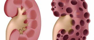
An amoebic cyst swallowed by a person excyses in the large intestine. One quadruple amoeba emerges from the cyst, which, through division, gives rise to the reproduction of amoebas. Being in the lumen of the colon, on the surface of the mucous membrane and in the crypts, amoebas crawl to the bottom of the liberkühn glands and continue to multiply there, secreting a proteolytic enzyme that causes necrosis of the cells of the mucous membrane. The mucus and products of cellular necrosis released as a result of irritation clog the lumen of the glands, which is why a yellowish nodule appears - a microabscess, which, increasing, connects with another abscess. Such abscesses deepen into the thickness of the mucous membrane and can reach the muscle layer. The rupture of an abscess leads to the release of pus into the intestinal lumen and the formation, as a rule, of deep ulcers. From small abscesses formed in the upper part of the large intestine, amoebas and other flora can penetrate through the blood vessels of the portal vein into the liver and cause secondary formation of abscesses there; it is most often observed in the upper quadrant of the right lobe of the liver.
Pathoanatomical changes in amebiasis are localized in the cecum, S-shaped, and much less often in the rectum. In the intestinal mucosa there are usually many round, suppurating ulcers with juicy, undermined red edges. The bottom of the ulcers is “greasy”, yellowish-green in color (necrotic intestinal tissue). The ulcers are surrounded by an inflammatory ridge protruding above the surface of the intestine and have undermined edges.
The discharge from the ulcers contains amoebas. The intestinal wall appears edematous and flabby. Among the ulcers, scar formations are found - traces of healed ulcers. Such scars sometimes lead to a narrowing of the intestine. Sections often reveal liver abscesses with thick mucus-like pus.
Prevention
Prevention of infection requires compliance by a person with hygiene rules in all conditions. In areas with a significant level of morbidity, “reminders” are distributed to the population and conversations are held about when to wash your hands, how to handle food, and what kind of water to drink. Utilities are involved in solving the problem. They are obliged to provide people with normal toilets, dry closets, and special wipes for hand cleaning.
Epidemiological institutions monitor drinking water and the condition of natural sources. If contamination is detected, a sign prohibiting use is posted. Treatment of acute amoebiasis must be carried out under the guidance of an infectious disease specialist. A full course of therapy will avoid complications and transition to a chronic course.
Prevention of amebiasis
To prevent amebiasis, the following preventive measures must be observed:
- observe the rules of personal hygiene, wash your hands, vegetables and fruits before eating;
- do not drink water or eat in questionable places;
- Avoid contact with possible carriers of amoebiasis.
To prevent the spread of amoebiasis it is necessary:
- isolate a patient with amoebiasis until complete recovery;
- carry out regular disinfection of the room where the patient is located;
- the patient must be under medical supervision for up to a year after treatment.
Amebiasis is a parasitic disease caused by the amoeba Entamoeba histolytica. In addition to the presence of this type of parasite in the body, other conditions are necessary for the development of pathology - weakened immunity, disturbed intestinal microflora. If amebiasis is left untreated, serious complications and damage to internal organs may develop.
Etiology
The causative agent of amoebiasis - Entamoeba histolytica - belongs to the genus Entamoeba, family Entamoebidae, phylum of protozoa - Protozoa. The life cycle of the causative agent of amoebiasis consists of two stages - vegetative and dormant, or cyst. At the vegetative stage, there are three main forms of amoeba: 1) large vegetative (tissue) 2) small vegetative (educational, commensal), 3) pre-cystic. The causative agent - Entamoeba histolytica - belongs to the type of protozoa; in the human body it can be found in the form of vegetative forms (small and large) and cysts. Its vegetative forms are very sensitive to adverse effects: disinfectant solutions and boiling destroy them instantly. Cysts are relatively stable, remaining in water for up to 1 month.
The causative agent is the protozoan Entavoeba histolytica of the genus Entavoeba of the class Sarcodina, which exists in the form of cysts and vegetative forms - precystic, luminal, large vegetative and tissue. Mature cysts are four-nucleated; vegetative forms have one nucleus.
There are pathogenic and non-pathogenic strains. Recently, non-pathogenic strains, morphologically indistinguishable from pathogenic amoebae, have been identified as a separate species E.
dispar. The luminal form, 10-20 microns in size, lives in the lumen of the large intestine without causing harm to the host.
The precystic form differs from the luminal form by low mobility and homogeneity of the cytoplasm. As the amoebas move through the colon, encystment occurs.
Vegetative forms outside the human body quickly die, while cysts in the external environment are quite stable: they can persist in feces for up to 4 weeks. in water - up to 8 months, which is of significant epidemiological significance.
Drying them out has a detrimental effect.
The causative agent is the ciliate Balantidium coli of the genus Balantidium of the order Trichostotida of the class Infusoria. The microorganism exists in the form of vegetative and cystic forms. In the external environment, vegetative forms remain viable for several hours, cysts for 3-4 weeks. Drying out has a detrimental effect on both forms.
The causative agent is the protozoan toxoplasma gondshi. An intracellular parasite 4-7 microns in size, morphologically resembles an orange slice or an onion with a stretched bowstring (Greek.
toxon - onion). It exists in the form of a vegetative form and cysts. When stained according to Romanovsky-Giemsa, the vegetative form appears as a crescent with blue cytoplasm and a ruby-red nucleus.
It is unstable to the effects of thermal and chemical factors: heating, 2% chloramine solution, 1% phenol solution, 50 o alcohol, etc. Toxoplasma cysts found in meat and meat products can remain viable at a temperature of 2-5 oC for up to a month, but they quickly die when heat treated and frozen to -20 oC.
The causative agent of amoebiasis is Entamoeba histolytica, dysenteric amoeba (synonym: Amoeba coli, Entamoeba dysenteriae, Entamoeba tetragena). Entamoeba histolytica belongs to the family Entamoebidae, order Amoebida, subclass Rhizopoda, class Sarcodina, phylum Protozoa.
The causative agent of amebiasis was described in 1875 by the Russian scientist F.A.
Leshem. Lesh experimentally proved the pathogenicity of the amoeba found in a patient with bloody diarrhea.
In the development cycle of E. histolytica there are two stages: the vegetative and the resting stage, or cysts. In the vegetative stage, the following forms are distinguished: tissue, large vegetative, luminal and precystic. Tissue and large vegetative forms are found in acute amoebiasis, luminal and precystic forms - during the period of convalescence and in cyst excretors.
Therapy
To treat amoebiasis, medications from 3 different groups are prescribed, which affect different forms of amoebas:
- Luminal or direct amoebicides are drugs that are destructive to luminal forms of amoebae, i.e. pathogens living in the intestinal lumen. They are prescribed for the treatment of amebiasis in carriers, in patients with a chronic course of the disease, in recovered people in order to prevent relapses. This group of medications includes, for example, tetracyclines, Etofamide, Paromomycin.
- Tissue amoebocides are drugs that have a detrimental effect on parasites localized in tissues and mucous membranes. Medicines in this group are prescribed for the acute course of the disease, as well as for the treatment of extraintestinal forms of amebiasis.
- Universal amoebocides that have a detrimental effect on all forms of parasites. Medicines in this group disrupt the protein structure of amoebas, as a result their reproduction is inhibited. In addition, under the influence of these drugs, free radicals are formed, which have a detrimental effect on parasites. This group includes Tinidazole, Trichopolum.
In addition, medications are prescribed that restore the intestinal microflora: Acipol, Linex.
Also, depending on the clinical picture, medications that normalize the functioning of the cardiovascular system, drugs that enhance immunity, and hepatoprotectors may be prescribed.
With the development of a severe form of amoebic dysentery, antimicrobial agents are additionally prescribed.
If a large abscess is detected, there is a high probability of its rupture, there is severe pain, and there is no effect of drug therapy, aspiration is performed. When a rupture has already occurred or closed drainage cannot be done, open surgery is indicated.
All dosages of medications and the duration of their use should be selected by the attending physician.
Classification
I. Asymptomatic amoebiasis.II. Manifest amoebiasis
Intestinal (amoebic dysentery, or amoebic dysentery
Extraintestinal:
- hepatic: acute amoebic hepatitis;
- liver abscess.
Cutaneous (this form is more common than other extraintestinal types of amebiasis, and is classified as a separate group).
Domestic medicine considers extraintestinal and cutaneous forms to be complications of intestinal amebiasis.
The International Classification of Diseases identifies the following clinical forms of infection:
- acute amoebic dysentery;
- chronic intestinal amoebiasis;
- amoebic colitis is not dysenteric;
- intestinal ameboma;
- liver abscess;
- lung abscess;
- brain abscess;
- cutaneous amoebiasis.
Classification of amoebiasis according to WHO recommendations, 1970:
- Intestinal amoebiasis;
- Extraintestinal amoebiasis;
- Cutaneous amoebiasis.
In domestic practice, extraintestinal and cutaneous amebiasis is regarded as a complication of the intestinal form.
Intestinal amebiasis can occur in the form of acute, chronic recurrent and chronic continuous variants in forms of varying severity.
The causative agent is amoeba. The life cycle consists of two stages - the vegetative stage and the resting stage (cyst), which replace each other depending on environmental conditions.
There are two forms of existence of amoeba - tissue and luminal. The main role in infecting humans and spreading amoebiasis belongs to amoeba cysts.
Amoebiasis is often registered as a mixed infection (together with other intestinal infectious diseases).
Clinical forms:
- asymptomatic amoebiasis (luminal amoebiasis, asymptomatic amoebiasis);
- amoebic dysentery (intestinal amebiasis);
- extraintestinal amebiasis (amoebic abscess (cavity filled with pus) of the liver, lungs, brain);
According to the severity of clinical manifestations:
- manifest form;
- subclinical form.
According to the duration of amoebiasis:
- acute (up to 3 months);
- subacute (up to 6 months);
- chronic (more than 6 months).
According to the nature of the flow, they are distinguished:
- amoebiasis continuously progressing;
- recurrent.
According to severity they are distinguished:
- mild course;
- moderate severity;
- severe course of amebiasis.
Epidemiology
Source and reservoir of infection
The source of infection in amebiasis is a person who secretes E. histolytica cysts. Patients with acute manifestations of intestinal amebiasis excrete predominantly vegetative forms of the parasite that are unstable in the external environment and do not pose a significant epidemiological danger. Epidemiologically, the most dangerous carriers of amoebae, as well as convalescents of acute intestinal amebiasis and patients with a chronic form of amebiasis. The release of cysts by infected individuals can continue for many years, and in one day one amoeba carrier is capable of excreting 300 million cysts or more in feces. Cysts are released unevenly. In the external environment, cysts die within a certain time after release.
Mechanism and factors of transmission of infection
The mechanism of infection is fecal-oral. The main factors in the transmission of histolytic amoeba cysts are food products, and less commonly, water contaminated with cysts. The mechanical carrier is flies. Recently, infections due to oral-anal sexual intercourse have been described.
Susceptible contingent and immunity
Living conditions, water supply, sanitary conditions of populated areas and the general cultural level of the population can determine different degrees of infection. The city population is infected to a lesser extent than the rural population. Just like carriage, the incidence of acute amoebiasis is recorded throughout the globe, but, unlike carriage, a clear connection of the disease with zones with a hot and dry climate is observed. The peculiarities of the epidemic process in amebiasis may, to some extent, be associated with a weakening of the body's resistance, which occurs more easily in hot climates, and a change in the normal basis of intestinal functioning in these climatic conditions.
The disease is recorded year-round, with a maximum in the hot months. Amoebiasis is sporadic. The incidence of acute amebiasis, as a rule, does not have the character of epidemic outbreaks; almost always, individual diseases arise without any visible connection with each other. Epidemic outbreaks develop as a result of sewage entering the water. Recently, epidemic outbreaks have been described in closed groups, in particular among prisoners in maximum security colonies.
All age groups of the population of both sexes suffer from amebiasis, but mainly men aged 20-58 years. Women in the third trimester of pregnancy and the postpartum period are especially susceptible to amebiasis, which is apparently due to the characteristics of the cellular immune response in pregnant women, as well as persons receiving immunosuppressive therapy. In general, the immunity acquired as a result of the disease in amoebiasis is unstable and non-sterile, and does not protect patients from relapses or reinfection.
Treatment of amoebic dysentery
Treatment of amoebic dysentery is carried out with specific drugs (it is better to combine or alternate them) together with a general strengthening regimen, nutritious food rich in proteins, vitamins, blood treatment, etc. under the supervision of repeated studies of stool for amoebas and repeated sigmoidoscopy;
treatment is resumed 1-2 times a year and more often until complete clinical, anatomical and parasitological cure. For acute phenomena, the most powerful remedy is emetine, an alkaloid of ipecac, which has a vigorous effect on vegetative forms. It is used only in the form of injections, preferably subcutaneously. The recommended average dosage is 0.06 per day for 12 days, i.e. a total of no more than 0.72 per course with three four-day breaks after every 3 days of treatment. In case of overdose, less often with individual intolerance, weakness, vomiting, muscle pain, polyneuritis and damage to the heart muscle occur, requiring immediate cessation of treatment, administration of vitamin B, nicotinic acid, glucose. Repeated courses of treatment with emetine, due to its cumulative properties, are permissible with an interval of at least 3-4 weeks. Yatren is a non-toxic drug, but it often causes a weakening of the intestines; applied orally at 1.5-2.0 and even 3.0 per day for 3-4 days in a row (treatment with emetine is recommended during breaks), for a total of 12-15 days or more, also with breaks. At the same time, Yatren can be administered per rectum every other day, 200 ml of a 1-2% solution after a cleansing enema. Aminarson is an organic arsenic preparation, close to osarsol and also toxic to the kidneys and liver, although to a slightly lesser extent, used for chronic recurrent forms of amebiasis and also acts on cysts. A 10-day course is carried out in persistent cases, repeated after a 10-day treatment break. Less commonly, aminarsone is prescribed in the form of enemas of 2.0 per 200 ml of 2% soda solution every other day. In cases of intolerance (abdominal pain, diarrhea, dermatitis, swelling of the eyelids, neuritis, fever), aminarsone is immediately discontinued. For amoebic dysentery, enemas of rivanol (1.0 per 200 ml of water) and many other remedies were also recommended. A good effect, as shown by the observations of Soviet authors (Baldin), is the use of gramicidin enemas (100 ml of 0.08% solution) simultaneously with the main treatment with emetine or aminarsone and from the very first days of this treatment, administered every second day after the cleansing enema through a rubber catheter to a depth of 40 cm in the patient’s knee-elbow position; a total of 6-12 enemas. Treatment of liver abscesses: in the early stages, emetine subcutaneously, preferably in combination with repeated removal of pus by liver puncture with a needle; in the late (bacterial) stage, the addition of penicillin and usually surgical opening of the cavity are necessary. Prevention is carried out on the same basis as for bacillary dysentery, according to the rules of personal and communal hygiene, by controlling flies, prohibiting cyst carriers from working in public catering establishments, etc.
Cutaneous amebiasis: what are the symptoms?
This form of amebiasis can develop as a complication. It is often experienced by patients with reduced immunity. Body ulcers mainly appear in areas where the skin comes into most contact with the toilet seat and human waste.
They look like erosions with dark edges and smell unpleasant.
Diagnosis of amoebiasis
The diagnosis can only be made by a doctor after a complete examination of the person. At the same time, reports on the sanitary and epidemiological situation of the area or region where the person lives are also taken into account.
After a long study, the doctor will prescribe the correct treatment and necessary medications, their dosage and frequency of use.
Amebiasis in adults and children is recommended to be treated only with medications, and in severe cases - with surgery.
How is amoebiasis treated in the hospital?
Patients with severe disease or in later stages are sent for treatment to an infectious diseases hospital.
For effectiveness and speed of recovery, doctors advise using medications. They can be recognized when diagnosing amoebiasis. The doctor will prescribe the necessary medications for you and select the correct dosage.
Also, if there are ulcers or cysts on the internal organs, surgery may be used. For skin amebiasis, special ointments are often prescribed.
How to treat amoebiasis using folk remedies?
Treatment of amebiasis with folk remedies at home is possible only in mild or initial stages of the disease. Even in ancient times, when there were no medicines, patients with amoebiasis were treated with decoctions of sea buckthorn, hawthorn or bird cherry. They also used garlic - they mixed one hundred milliliters of vodka and forty grams of crushed and dried garlic, infused for two weeks and took ten drops before meals with a dairy or fermented milk product.
When using folk remedies for treatment, remember that medications are much more effective and cope better with amebiasis. However, you can use them together to achieve the best effect.
general description
is an infectious disease caused by a parasitic unicellular microorganism (amoeba), and characterized by damage to the colon.
Amebiasis (amoebic dysentery) is an infectious disease accompanied by ulcerative lesions of the colon, a protracted course and the possible formation of abscesses of the liver, lungs and other organs. Classification (amoebiasis (According to WHO recommendations, 1970): Intestinal amebiasis Extraintestinal amebiasis Cutaneous amebiasis
The causative agent of amebiasis is a histological or dysenteric amoeba, the habitat of which is the large intestine. It is noteworthy that in addition to this type of amoeba, a non-pathogenic type of amoeba is also often detected in this area.
The life cycle of a histological amoeba includes the vegetative (or trophozoite) as well as the cystic stage. Dysenteric amoeba, unlike other types of amoebas, has four forms of the vegetative stage in its life cycle. These include the tissue form, E. histolytica magna, the luminal form and the precystic form. Let's look briefly at each of them.
Amebiasis (amebic dysentery) is an invasive human disease characterized by ulcerative-inflammatory lesions of the colon and the development of inflammatory foci in the liver and other organs.
Dysenteric amoeba
It is known that 6 types of amoebas can parasitize humans, 5 of them are non-pathogenic and feed on intestinal bacteria, and 1 - Entamoeba histolytica - is pathogenic and causes severe intestinal symptoms.
Dysenteric amoeba in the intestines
Like any parasite, the dysentery amoeba has 2 life forms - the trophozoite (vegetative stage) and the cyst (resting stage). The trophozoite also goes through several stages and can remain in one of them for a long time:
• tissue form (found only in acute amoebiasis in affected organs and rarely in feces); • large vegetative form (this form already lives in the intestines and is found in the coprogram; it absorbs red blood cells); • luminal form (found in chronic amoebiasis or in the stage of reconvolution after taking a laxative); • precystic form (detected under the same conditions as the luminal form).
All this is important for determining the source and methods of combating its parasites.
Cysts are found in patients with chronic recurrent amebiasis in remission and in carriers of amoebae.
The resistance of the trophozoite and all its varieties is very low; in the external environment it dies within 30 minutes. Cysts are the most resistant, for example:
- at 17-20ºС they are stored for a month, in darkened and moist soil - up to 8 days;
- in chilled food products, fruits, vegetables and household items – up to 5 days on average;
- At sub-zero temperatures they can be stored for several months.
Drying and high temperature destroy the amoeba almost instantly. Of the disinfectants, only cresol and emitin have a detrimental effect on them, and even chloramine does not have a negative effect on them.
Disease prognosis
With intestinal amebiasis, the prognosis is favorable: timely diagnosis and properly selected treatment ensure the patient a complete recovery in a few months.
The prognosis for extraintestinal forms of amebiasis is much more serious, especially if abscesses of the liver and other organs are detected late. Without treatment, or if treatment is started late, a fatal outcome (death of the patient) is possible.
If you suspect that you have amoebiasis, immediately consult an infectious disease specialist or parasitologist.
Intestinal amebiasis is an infectious disease caused by amoebas, microscopic single-celled microorganisms that parasitize the large intestine.
Infection can occur at any age. A person may not even know that he is sick for a long time, since the disease can be asymptomatic. Clinical manifestations can appear only when a lot of worms accumulate in the intestines, in which case they damage the intestinal mucosa.
pathogenesis
Stage of colon damage
The name E.histolytica is directly related to the lytic effect of the dysenteric amoeba on tissue. Its attachment (adhesion) to the intestinal mucosa occurs due to galactose-inhibiting-adhesive lectin, a peptide that forms pores in the mucosa and thus damages the intestines. In addition to this factor, many hemolysins were found in E. histolytica, which are capable of neutralizing red blood cells, leukocytes and proteases, and hyaluronidase was found, which facilitates the penetration of amoebas into tissues. When E. histolytica enters the submucosal layer, intensive multiplication of parasites occurs, and primary foci are formed in the form of microabscesses, which open into the intestinal cavity. Thus, a small ulcer appears first. In this case, amoebas multiply intensively, spreading along the periphery of the ulcer and penetrate inside, reaching the muscular layer of the intestinal wall. Small ulcers located close to each other can merge, resulting in the formation of large ones, several centimeters in diameter. On the intestinal mucosa during acute amoebiasis, ulcers of various sizes and shapes can simultaneously appear: from very small, barely noticeable to gigantic. The bottom of the ulcers is covered with necrotic masses. In uncomplicated amebiasis, the mucous membrane between the ulcers, without inflammatory changes, retains its normal appearance, which is an important diagnostic manifestation of this disease. Most often, the ulcerative process involves the cecum and the adjacent ascending part of the colon, the vermiform appendix. In second place in terms of frequency of localization are the sigmoid and rectum. Long-term, chronic intestinal amebiasis can cause the formation of cysts, polyps and amoebae.
Stage of extraintestinal lesions
When the blood vessels of the colon mucosa are eroded, E. histolytica trophozoites enter the portal venous system, causing generalization of the infection. Hematogenous dissemination of amoebae leads to the development of extraintestinal amebiasis with the formation of foci (often abscesses) in the liver, lungs, brain and other organs. In limiting the prevalence of the pathological process in invasive amoebiasis, an important role is played by the cellular mechanisms of immune defense. However, as it was found, with the development of acute invasive amoebiasis, the activity of the T-cell component of the immune defense decreases due to the production of a specific serum factor induced by E. histolytica. Nevertheless, cellular immunity has a certain importance in containing amoebic infection, which can indirectly be evidenced by the severe course of amoebic infection during treatment with corticosteroids, which suppress the immune system. The same situation can be supported by cases of fulminant amebiasis in premature infants and pregnant women suffering, first of all, from cellular immunodeficiency.
Pathogen information
First, it’s worth understanding what amoebiasis is and why it appears. This is a disease caused by a certain type of protozoa. Quite often this term is associated with infections caused by other types of amoebas, but this definition is incorrect. The causative agent of this disease is dysenteric amoeba or Entamoeba histolytica.
The following protozoa of this type can also live in the human body:
- Dientamoeba fragilis (the causative agent of dientamoebiasis).
- Entamoeba dispar (non-pathogenic amoeba living in the intestinal lumen).
- Entamoeba hartmanni (another non-pathogenic inhabitant of the gastrointestinal tract).
- Entamoeba coli (commensal intestinal amoeba).
- Endolimax nana (dwarf amoeba that does not harm the owner).
- Iodamoeba butschlii.
These types of amoebas are called opportunistic because human infection is not necessarily part of their life cycle. Among them are the following creatures:
- Entamoeba moshkovskii (manifestations are largely reminiscent of intestinal amoebiasis).
- Naegleria fowleri (Naegleria fowler, which causes primary amoebic meningoencephalitis - a rare and fatal disease).
- Acanthamoeba (the causative agent of acanthamoeba keratitis).
- Balamuthia mandrillaris (leads to the development of granulomatous amoebic encephalitis).
- Sappinia diploidea (associated with amebic encephalitis).
https://youtube.com/watch?v=x7OCTKkEv5g
These free-living protozoa typically live in warm water (Naegleria fowlera can even live in hot springs) or moist soil. Bacteria serve as food for these amoebas.
Method of infection
Cysts, an infectious form of dysenteric amoeba, usually enter the human body through the fecal-oral route. Another transmission option is through contact with dirty hands or objects.
The infection spreads through the ingestion and subsequent release of parasite cysts. It is this inactive and solid structure that is found in feces during the diagnosis of amoebiasis. Cysts are able to remain viable for a long time without a host under favorable conditions. Another form of amoeba, the trophozoite, dies immediately once outside its body. They can also be found in feces, but they serve as a source of new infection in very rare cases.
Since amoebiasis is transmitted through contaminated water and food, regions with an insufficiently developed sanitation system are considered endemic areas. People can become infected when sewage discharges into fresh water sources or when untreated human feces are used as fertilizer.
The term "amebic dysentery" is sometimes mistakenly used in connection with traveler's diarrhea. The confusion probably stems from the fact that both diseases have a high prevalence in developing countries with poor sanitation conditions. But in fact, most cases of traveler's diarrhea are bacterial or viral in origin. So the dysentery-causing amoeba has nothing to do with it.
Infection Prevention
Prevention of amebiasis is aimed primarily at observing personal hygiene measures and improving sanitary conditions. To prevent infection and spread of infection, simply follow these simple rules:
Wash your hands thoroughly with hot water and soap for at least 10 seconds after using the toilet, changing a diaper, and before handling food. Clean your bathtub and toilet frequently, paying special attention to the toilet seat and faucets. Try to use separate towels for your face and body. Do not eat raw vegetables when in endemic areas. Drink only boiled water. Avoid street food.
Compliance with sanitary standards and responsibility when disposing or treating wastewater are measures necessary to prevent intestinal infection. Amoeba cysts are resistant to chlorination, so sedimentation and filtration of water is required to reduce the prevalence of the pathogen.
Symptoms of liver amoebiasis
According to medical statistics, this form of pathology occurs in no more than 10% of patients.
Patients may experience characteristic symptoms:
- pain and heaviness appear in the right hypochondrium;
- hepatomegaly develops;
- seals appear in the organ that can be felt by palpation;
- patients have low-grade fever;
- in rare cases, jaundice appears;
- when conducting blood tests, specialists detect leukocytosis, which is moderately expressed;
- with the development of an abscess in this organ, febrile conditions develop;
- Over time, pain may radiate to the area of the right shoulder or collarbone;
- pseudocholecystitis may develop;
- Over time, an abscess breaks through, in which the contents provoke the development of peritonitis, etc.