Acute cerebrovascular accident is considered a pathological process in which blood circulation in the vessels of the head deteriorates. If characteristic symptoms appear, it is important to immediately consult a doctor to avoid complications. When stroke appears, there is a high risk of death, as well as irreversible changes.
If the disorder becomes chronic, then the person may experience blood clots as well as hemorrhage, which can also lead to death. When a patient notices how his health has worsened, he should immediately visit the hospital and begin proper treatment.
Mechanism of formation and types of stroke
Acute circulatory disorders in the brain develop in several ways. Among the key ones, three can be named.
The first is atherosclerosis. The essence of the disorder is the narrowing or blockage of an artery located in the brain. In 95% of cases, the culprit is a cholesterol plaque.
Slightly less common is a blood clot that has broken away from the site of its own formation (usually the legs or arms, sometimes the heart).
Blocking the lumen leads to the inability of blood to move further or to a decrease in the efficiency of fluid tissue circulation. For the most part, this ends with a decrease in the quality of trophism (nutrition) of nerve fibers.
In the case of predominant blockage, especially if there are problems with the vessels, a rupture with the formation of a hematoma and massive bleeding cannot be avoided.
The condition is called hemorrhagic stroke, which also refers to an acute disorder of brain nutrition.
Atherosclerosis is also represented by narrowing (spasm) of the vessel. Usually the problem is observed in smokers, patients with diabetes, pregnant women, patients with congenital anomalies of the structure of the arteries, people who have suffered from inflammatory vascular diseases and others.
The second possibility is persistent, long-term hypertension. With a prolonged increase in pressure, the thickness of the walls of blood vessels increases, they lose elasticity, and the lumen narrows.
Hence the high probability of ischemia (cell death as a result of insufficient blood supply).
A spontaneous surge in pressure with the same outcome is also possible, and this option occurs in people without hypertension and generally without health problems. In case of inadequate physical activity, taking certain medications.
Finally, there is a third mechanism. It is associated with past inflammatory diseases (for example, vasculitis) affecting the choroid from the inside (intima or endothelium). Scarring and mechanical closure of the lumen of the tubular structure occurs.
Thus, despite the “cosmetic” differences, the essence is the same: disruption of blood flow, increased pressure in the vessel and further tissue death as a result of the impossibility of blood movement or rupture.
The etiology is identified to prevent an emergency.
A transient ischemic attack develops according to the same laws, but with it there is no rupture of blood vessels, and the death of neurons is not observed.
Therefore, these pathogenetic factors are milder in nature. Otherwise everything is identical.
The symptoms are similar, but the duration of the disorder is a maximum of a few hours, it goes away without a trace and does not leave a neurological deficit.
Death also does not occur, with one exception: involvement of the brain stem in the process.
Hemorrhagic stroke
Brain hemorrhage occurs suddenly. With a hemorrhagic stroke, the patient's chances of survival are significantly lower than with an ischemic stroke. The disease can overtake a person during the most active period of life: at work, at a party, during physical activity and stressful conditions.
Hemorrhagic stroke is a non-traumatic cerebral hemorrhage and occurs in 20% of patients.
The walls of damaged vessels rupture under the influence of unfavorable factors, which leads to general cerebral symptoms of the disease.
Symptoms
There are several types of hemorrhage in brain tissue, each of which has its own symptoms and is diagnosed and treated differently.
- Blood flows between the membranes of the brain and the bones of the skull (subarachnoid space). The patient suffers from an attack of headache, nausea and vomiting. Photophobia develops, relief does not occur, and the patient may suddenly lose consciousness.
- Hemorrhage in brain tissue. Characterized by the formation of a hematoma in the brain itself. Neurological symptoms for this type of lesion depend on the location of the pathology:
- in the frontal lobe of the brain - leads to foolishness, speech impairment and unsteady gait. The patient may spontaneously extend the lips with a tube;
- in the temporal - provokes visual and hearing impairment (the patient sees part of the visual picture and does not understand his native speech). The body cramps;
- in the parietal - deprives one half of the body of sensitivity (temperature, pain);
- in the occipital region - causes loss of vision in one or both eyes;
- in the cerebellum - leads to impaired coordination of movements: unsteady gait, eyes running from side to side and decreased muscle tone. The patient may have difficulty breathing, hyperemia (redness) of the facial skin and convulsive seizures until loss of consciousness are observed.
Causes
In most cases, the causes of cerebral hemorrhage are chronic diseases and bad habits of a person.
- Arterial hypertension.
- Aneurysms and pathologies of cerebral vessels.
- Vasculitis, angiopathy, hemophilia and thrombocytopenia.
- Taking drugs with fibrinolytic properties and anticoagulants (Heparin, Aspirin, etc.).
- Smoking, alcoholism, drug addiction.
A patient with a hemorrhagic stroke exhibits a number of symptoms that suggest the location of the hemorrhage
Among the risk factors, doctors note the following:
- age over 50 years (both men and women);
- obesity;
- diabetes;
- hereditary predisposition.
Diagnostics
Upon admission to the hospital, the patient is prescribed an emergency CT or MRI. Studies help establish the correct diagnosis for stroke and plan drug therapy; with their help, doctors determine:
- type of stroke and location of the hematoma;
- presence and degree of cerebral edema;
- volume of hemorrhage and evolution of hematoma;
- data on venticular hemorrhage (presence, prevalence), etc.
Emergency care and treatment
At the first suspicion of a cerebral hemorrhage, you need to place the patient on a flat surface, slightly raising his head. It is important to ensure that the person does not choke on vomit.
Transportation of a patient with hemorrhagic stroke is carried out with the utmost care. The tremors can trigger new cerebral hemorrhages.
Emergency measures are divided into conservative and surgical and include:
- Normalization of high blood pressure.
- Elimination or reduction of cerebral edema.
- Intubation and connection to a ventilator (for problems with the respiratory system).
- Prevention of blood clots in blood vessels.
- Surgery is prescribed according to indications (to save the patient’s life) in the first few hours after a cerebral hemorrhage.
Rehabilitation
Rehabilitation of patients who have suffered a cerebral hemorrhage depends on the degree of damage to the organ tissue. As a rule, these people become bedridden invalids, their chances of living a full life are extremely small. Relatives must provide maximum care for a seriously ill patient and alleviate his condition.
Patients eat pureed or crushed food. In some cases, a feeding tube may need to be used. The patient needs constant supervision by the attending physician and strict adherence to all prescriptions.
The main problem for bedridden patients is the formation of bedsores and the development of pneumonia. Recommendations for care and treatment are described in detail by doctors.
Causes and risk factors
The moments were partially named. If we systematize all the possible ones, the following picture is created:
- Arterial hypertension. At the first stage, the likelihood of developing a stroke or transient ischemic attack is minimal. Then the risks increase. By stage 3 hypertension, when blood pressure is constantly high and the patient does not feel symptoms, the probability of a negative outcome is maximum and ranges from 15 to 25% during the year. Constantly.
- Atherosclerosis. Narrowing of the vessel or blockage of the structure by cholesterol plaque. Both options are dangerous. Requires urgent treatment.
- Endocrine diseases. Diabetes mellitus and under or overproduction of thyroid hormones are just some of the possible conditions that can trigger a stroke. Indirectly or directly.
- Osteochondrosis or hernia of the cervical spine. It provokes stenosis of the vertebral and basilar arteries, with disruption of the nutrition of the occipital lobe of the brain.
- Pathologies of the cardiovascular system. With low contractility of the myocardium, there is not enough blood to nourish the cerebral structures.
With a gradual increase in cardiac dysfunction, microstrokes often develop and only then full-fledged necrosis of nerve tissue.
Among the risk factors:
- Bad habits. Alcohol abuse, drug addiction, smoking.
- Physical activity is inadequate to the body's capabilities. It provokes excessive activity of the heart, and from a certain point the contractility decreases, and the organ becomes unable to provide for the brain. A common variant of deviation due to this reason is ischemic stroke. Hemorrhage is slightly less common.
- Stressful situations. One-time or permanent. They cause the production of hormones such as cortisol, adrenaline, followed by angiotensin and aldosterone. The substances artificially constrict blood vessels and cause secondary atherosclerosis.
- Poor nutrition, excessive consumption of animal fat.
- Lack of normal sleep.
- Presence of pulmonary diseases (if the respiratory system is affected).
- Taking certain medications: non-steroidal anti-inflammatory drugs, hormones, oral contraceptives, psychotropics (neuroleptics, antidepressants, tranquilizers).
Causes are assessed in the system to determine the likely outcome, diagnosis and treatment pathways for patients.
The basis of treatment is the elimination of the main provoking factor. Only by identifying the root cause is it possible to radically influence the situation and prevent a relapse.
Acute cerebrovascular accidents (ACI)
Acute cerebrovascular accident causes persistent disorders of brain activity. It comes in two types: hemorrhagic (bleeding) and ischemic (also called cerebral infarction).
Hemorrhagic
Etiology
Hemorrhage (hemorrhagic disturbance of blood flow) can be caused by various arterial hypertension, vascular aneurysms, congenital angiomas, etc.
Pathogenesis
As a result of an increase in blood pressure, plasma and the proteins contained in it are released, which leads to plasma saturation of the walls of blood vessels, causing their destruction. A peculiar hyaline-like specific substance (a protein whose structure resembles cartilage) is deposited on the vascular walls, which leads to the development of hyalinosis. The vessels resemble glass tubes and lose their elasticity and ability to hold blood pressure. In addition, the permeability of the vascular wall increases and blood can freely pass through it, soaking the nerve fibers (diapedetic bleeding). The result of such transformations can be the formation of microaneurysms and rupture of the vessel with hemorrhage and blood entering the white medulla. Thus, hemorrhage occurs as a result of:
- Plasmic impregnation of the walls of blood vessels of the white medulla or visual thalamus;
- Diapedetic bleeding;
- Microaneurysm formations.
Hemorrhage in the acute period is characterized by the development of hematomas due to wedging and deformation of the brain stem into the tentorial foramen. In this case, the brain swells and extensive edema develops. Secondary hemorrhages occur, smaller ones.
Clinical manifestations
Usually occurs during the day, during physical activity. Suddenly your head starts to hurt and you feel nauseous. Consciousness is confused, the person breathes quickly and whistling, tachycardia occurs, accompanied by hemiplegia (one-sided paralysis of the limbs) or hemiparesis (weakened motor functions). Basic reflexes are lost. The gaze becomes motionless (paresis), anisocoria (pupils of different sizes) or divergent strabismus occurs.
Treatment
Treatment of cerebrovascular accidents of this type includes intensive therapy, the main goal of which is to reduce blood pressure, restore vital (automatic perception of the outside world) functions, stop bleeding and eliminate cerebral edema. The following medications are used:
- Blood pressure-lowering drugs - ganlioblockers (Arfonad, Benzohexanium, Pentamin).
- To reduce the permeability of vascular walls and increase blood clotting - Dicinone, vitamin C, Vikasol, Calcium gluconate.
- To increase blood rheology (fluidity) - Trental, Vinkaton, Cavinton, Eufillin, Cinnarizine.
- Inhibiting fibrinolytic activity - ACC (aminocaproic acid).
- Decongestant - Lasix.
- Sedatives.
- To reduce intracranial pressure, a spinal puncture is prescribed.
- All drugs are administered by injection.
Ischemic
Etiology
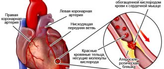
ischemic cerebrovascular accident due to atherosclerotic plaque
Ischemic circulatory disorders are most often caused by atherosclerosis. Its development can be provoked by severe anxiety (stress, etc.) or excessive physical activity. May occur during night sleep or immediately upon awakening. Often accompanies a pre-infarction condition or myocardial infarction.
Symptoms
They may appear suddenly or grow gradually. They manifest themselves in the form of headaches, hemiparesis on the side opposite to the lesion. Impaired motor coordination, as well as visual and speech disorders.
Pathogenesis
An ischemic disorder occurs when insufficient blood flows to a particular area of the brain. In this case, a focus of hypoxia arises, in which necrotic formations develop. This process is accompanied by disruption of basic brain functions.
Therapy
Treatment uses injections of medications to restore normal functioning of the cardiovascular system. These include: Korglykon, Strophanthin, Sulphocamphocaine, Reopoliklyukin, Cardiamin. Intracranial pressure is reduced by Mannitol or Lasix.
Signs of transient ischemic attack
General signs of stroke of a transient, transient nature are represented by classic manifestations of stroke, but differ in a number of ways:
- Insignificant in duration. The condition lasts from a couple of minutes to several hours.
- Complete spontaneous regression of symptoms. No medical intervention.
- No persistent neurological deficit after. The person returns to the “original position” the same as he was before.
Read more about the symptoms and treatment of transient ischemic attack here.
A stroke in itself is much more dangerous, because everything is exactly the opposite. Symptoms of acute cerebrovascular accident are divided into general and focal.
Symptoms
Each form and stage of development has its own signs of cerebrovascular accident. The general clinical picture includes the following symptoms:
- headaches for no apparent reason;
- nausea, which rarely ends with vomiting;
- memory impairment;
- decreased visual and hearing acuity;
- dizziness;
- impaired coordination of movements.
Transient cerebrovascular accidents are characterized by the following additional symptoms:
- numbness of the half of the body, which is opposite to the focus of the pathology;
- weakness of arms and legs;
- speech impairment – the patient has difficulty pronouncing individual words or sounds;
- photopsia syndrome - the visibility of luminous dots, dark spots, colored circles and similar visual hallucinations;
- drowsiness;
- stuffy ears;
- increased sweating.
Symptoms of cerebrovascular accident
Since there are symptoms such as speech impairment and weakness in the limbs, the clinical picture is often confused with a stroke. It should be noted that in the case of PNMK, acute symptoms disappear within a day, which is not the case with a stroke.
In the first stage of the chronic form, the following symptoms of cerebrovascular accident may be observed:
- frequent headaches;
- drowsiness;
- increased fatigue – a person feels tired even after a long rest;
- sudden mood swings, short temper;
- absent-mindedness;
- memory impairment, which manifests itself in frequent forgetfulness.
When moving to the second stage of development of the pathological process, the following may be observed:
- slight impairment of motor function, a person’s gait may be unsteady, as if intoxicated;
- concentration deteriorates, the patient finds it difficult to perceive information;
- frequent mood changes;
- irritability, attacks of aggression;
- almost constantly dizzy;
- low social adaptation;
- drowsiness;
- performance practically disappears.
The third stage of chronic cerebrovascular accident has the following symptoms:
- dementia;
- hand tremors;
- stiffness of movements;
- speech disorder;
- almost complete loss of memory;
- a person is unable to remember information.
At this stage of development of the pathological process, symptoms of almost complete degradation are observed; a person is not able to exist without outside help. In this case, we can talk about an irreversible pathological process. This is due to the fact that brain neurons begin to die already at the initial stages, which entails serious consequences if this process is not stopped in a timely manner.
Common stroke symptoms
The first are represented by nonspecific manifestations:
- Headache. Strong, unbearable. It is localized in the occipital region, crown, temples, and can cover the entire skull, spreading diffusely and radiating to the eyes and neck.
- Vertigo. Inability to navigate. The world is literally spinning, it’s impossible to even walk normally. Often a person takes a forced lying position.
- Nausea, vomiting. Short-term symptoms. Indomitable reflex gastric emptying almost never occurs. There is no relief after the act, because a false defense mechanism is triggered.
- Weakness, lethargy, drowsiness. Asthenic moments.
For a detailed description of stroke symptoms, read this article.
Symptoms of PNMK
The symptoms of short-term disruption of cerebral blood supply can be very different. This significantly complicates the definition of the disease and its detection in the early stages. Typically, the disease develops acutely, occurs suddenly, and symptoms disappear quickly.
Symptomatic manifestations are divided into several groups:
- General cerebral manifestations of the disease may be as follows:
- headaches of various nature and localization;
- nausea and vomiting;
- weakness throughout the body;
- feeling of lack of air;
- blurred vision;
- vasomotor reactions;
- disorders of consciousness that quickly pass.
- Focal (regional) manifestations of the disease are reduced to the following manifestations:
- numbness or tingling sensation in the face, arms or legs;
- musculoskeletal disorders;
- paretic phenomena in the area of the hand, individual fingers or feet;
- hemiplegia;
- Jacksonian epilepsy is a rare disorder;
- blindness of one eye;
- systemic dizziness indicates PNMK in the vertebrobasilar region;
- impaired swallowing ability;
- seizures of temporal lobe epilepsy;
- memory impairments indicate ischemic disorders in the medial-basal region.
Transient disturbances in cerebral blood flow can cause a variety of symptoms, each of which requires attention. You cannot ignore the messages that come from the body. They may have good reason to worry.
Focal signs
More specific and informative. Multiple areas of the brain may be affected.
Frontal lobe
Responsible for cognition. Creativity, thinking, behavior, everything is localized here.
Typical symptoms include:
- Marked decrease in intelligence, inhibition and decline in thinking productivity.
- Goofiness, inappropriate emotional reactions. Behavioral disorders may not be noticeable at first, because the patient is unable to demonstrate personal qualities. He is apathetic, mostly lies down and is silent.
- Regression. Induced infancy. Complete immaturity. Again, this refers to a behavioral disorder.
- Epileptic seizures. Heavy. Tonic-clonic. They can be repeated repeatedly, developing not into an attack, but into a status (a long course of paroxysms continuously or one after another for 30 minutes or more).
- Paralysis of the muscles opposite the localization of the lesion to the side.
- Aphasia. Inability to speak clearly.
Parietal lobe
Responsible for intellectual activity (partially), processing of tactile information (sensory component of human activity), and also the perception of smells.
- Inability to read, write, or perform simple arithmetic operations.
- Physical hallucinations. It seems to a person that someone is touching him, something is moving under the skin.
- There may be a lack of holistic perception of one's own body.
- There is agnosia, the inability to recognize an object, even the simplest one, known to everyone, by touch with eyes closed.
- There are complex complex hallucinations of the type of oneiric clouding of consciousness.
Temporal lobes
Responsible for auditory analysis, memory, verbal abilities.
- Deafness or hearing loss.
- Inability to understand speech in one's native language.
- Lack of speech, telegraph type of process. The patient expresses himself in abrupt words with formal logic intact.
- Epileptic seizures. This time they rarely turn into a full-fledged status with a continuous long-term course of the episode. Otherwise, the clinic corresponds to people’s ideas about a similar condition (seizure).
- Memory impairment. Anterograde, retrograde. Global and partial amnesia.
Occipital lobe
Responsible for visual analysis. Hence the corresponding symptoms: the simplest hallucinations such as luminous points and geometric figures, loss of areas of visibility, blindness (temporary) and other moments.
Limbic system and cerebellum
When it is affected, there is no sense of smell. Against the background of destruction of extrapyramidal structures (cerebellum), the inability to navigate in space, muscle weakness, lack of normal coordination of movements develops, and nystagmus is provoked (rapid spontaneous movement of the eyeballs to the left and right).
When the brain stem is involved in the pathological process, catastrophic consequences are possible: disturbances in heart rhythm and breathing, even stopping one or the other, critical jumps in body temperature. Even against the backdrop of a transient attack.
There is no fundamental difference between the individual forms of the disorder.
Deviation of the ischemic type is somewhat easier, because there is no additional negative factor. Namely, a blood clot, a hematoma, which compresses the tissue.
Lesions of the hemorrhagic type are more lethal; for comparison, the ratio of deaths in the first and second forms of stroke is 20 versus 43% for the small-focal type and 70/98% against the background of extensive destruction of nerve tissue.
Treatment methods
If PNMK is detected, it is imperative to undergo treatment, which will be prescribed by the attending physician. His recommendations cannot be ignored, since the consequences of an advanced disease can be irreparable. There are several treatment methods.
Medication
There are a number of medications that are prescribed when diagnosing PNMK. Only a doctor can prescribe them; you cannot prescribe medications for yourself under any circumstances. In such cases, treatment for a transient cerebrovascular accident may not only be ineffective, but also dangerous.
- Pentoxifylline or Dextran normalizes blood rheological parameters. These drugs are administered intravenously by drip.
- Acetylsalicylic acid is prescribed for long-term use.
- Bromcamphor is prescribed to people with PMN who cannot take salicylates due to contraindications.
- Neurometabolites.
- Drugs that normalize blood pressure showed, which is important for a patient with PNMC.
- To get rid of systemic dizziness and vegetative symptoms, belladonna alkaloids, diazepam, phenobarbital or belladonna extract can be prescribed.
- Anti-anxiety medications are prescribed for sedation therapy.
Folk remedies and nutrition
You shouldn’t dismiss traditional medicine, but you shouldn’t take them as a full-fledged treatment for PNMK either. Such techniques can only complement the main treatment of the disease. We have selected the most effective and at the same time simple recipes.
- Peel 4 heads of garlic and chop 6 lemons (do not peel, but remove the seeds). Grind these two components in a meat grinder, place the resulting mixture in a 3-liter jar. Add 350 gr. honey, fill the remaining space with clean water. We send the medicine to a dark place for 10 days, then filter and take a tablespoon, after diluting it in a glass of water.
- Grind 100 grams on a grater. horseradish root, add 3 chopped lemons and 3 tablespoons of honey. Place the mixture in the refrigerator for three weeks. After infusion, you need to take a teaspoon with meals twice a day.
- Place red clover (plant heads in dry or dried form) in a liter jar so that it is half filled. Add half a liter of high-quality vodka to the jar, close the jar and leave for 3 weeks. We squeeze out the flowers, strain and take 25 drops of the medicine, first diluting them in a glass of water. The course lasts a month, no more than 4 courses can be repeated per year.
It is very important to adhere to the rules in nutrition. In this way, the effectiveness of treatment of the disease can be significantly increased and relapses can be prevented.
- complete and categorical refusal of fast food;
- reducing the consumption of fatty foods to a minimum;
- exclusion of carbohydrate foods;
- avoidance of processed foods that contain large amounts of cholesterol;
- inclusion in the daily diet of vegetables and fruits, dairy products, including fermented milk;
- transition to boiled and steamed food.
Physiotherapy
For people diagnosed with PNMK, therapeutic exercises are recommended. It should only be prescribed by a specialist. You also need to be careful when performing exercises. It is recommended to combine therapeutic exercises with massage for PNMK.
Feldenkrais gymnastics demonstrates good results, after which you feel a surge of strength. In patients with PNMK, intelligence is restored and sensitivity increases. When performing exercises, the muscles are not overstrained. The essence of gymnastics is to perform slow movements and follow breathing exercises. As a result, the blood is saturated with oxygen, and the body relaxes.
First aid
It turns out immediately, at the pre-medical stage. The main thing to do is call an ambulance.
Then the algorithm is:
- Calm and reassure the patient. Fussiness will not lead to anything good.
- Place the victim on his back in a half-sitting position so that his head and torso are slightly elevated. A cushion made from scrap materials or several pillows will do.
- Provide fresh air flow. Open the window, window. This is important because it is necessary to correct hypoxia (oxygen starvation), which inevitably manifests itself during a stroke and even a transient ischemic attack.
- Loosen the collar and remove body jewelry. If there are any.
- Closely monitor the patient's condition. Assess the heart rate (based on the pulse on the carotid artery), pressure level, and the number of respiratory movements per minute.
- If you lose consciousness, turn your head to the side to prevent choking on vomit.
- If necessary, carry out resuscitation measures: cardiac massage (palm on the other, both on the middle of the sternum, do 80-100 energetic and rhythmic passes pressing the area 5-6 cm until the heart activity is restored).
Artificial respiration is done only if you have the skill, every 10-20 massage movements. If you don't have one, it's a waste of time.
Attention:
In no case should you put your head below the level of your body, give any medications, allow the patient to move, especially walk, take a bath, shower, eat or drink a lot.
For a detailed step-by-step algorithm for providing first aid for a stroke, read this article.
Diagnostic methods
We will study methods for diagnosing the disease a little later, but for now let’s turn to the international classification system ICD10 and find the code that is assigned to PNMK. This will be G45, this is the designation for this disease accepted by international medicine, and it is indicated on the medical history.
If PNMK manifests itself in the form of transistor ischemic attacks, then the essence of diagnosis comes down to excluding stenotic lesions of extra- and intracranial arteries. This is especially true for repeated manifestations of the disease. For these purposes, the following methods are used:
- Ultrasound G;
- MR angiography;
- contrast angiography;
- study of microcirculation disorders;
- the ability of the circulatory system to clot is assessed;
- CT and MRI can exclude a hemorrhagic process.
In the presence of a hypertensive crisis, the essence of diagnosis comes down to the exclusion or confirmation of secondary hypertension. In the case of meningeal syndrome, it is necessary to exclude subarachnoid hemorrhage. It is imperative to diagnose disturbances in the functioning of other organs and systems that have arisen as a result of diseases of the cardiovascular system.
Diagnostics
It is carried out after the condition has stabilized. Acute cerebral circulatory disorders require a minimal examination: measuring blood pressure, heart rate, respiratory movements, and identifying the integrity of reflexes. Next they provide assistance.
Only after recovery does it make sense to evaluate the situation more carefully and look for the reasons.
The list of events is quite long:
- Oral questioning of the patient and collection of anamnesis.
- Daily monitoring using a programmable Holter device. It measures blood pressure and heart rate levels. Provides information about indicators over a 24-hour period.
- Electro- and echocardiography. To exclude a cardiac etiology of the problem.
- MRI of the brain. To identify the consequences of a violation.
- General blood test, biochemical (especially with detailing the lipid spectrum), for thyroid, pituitary, and adrenal hormones.
Other methods are also possible. The diagnosis of stroke is specified and established. Then it is important to carry out regular preventive examinations, as part of prevention and rehabilitation.
Rehabilitation after acute cerebrovascular accident
Drug therapy can stop the progression of the disease. But she is unable to regain the ability to move. Only special gymnastic exercises can help with this. You need to be prepared for the fact that this process is quite long and be patient. The patient’s relatives must learn to perform massage and therapeutic exercises, since they will have to do them for him for six months or more.
Kinesiotherapy is indicated as the basis for early rehabilitation after dynamic cerebrovascular accident in order to fully restore motor functions. It is especially necessary in the restoration of motor skills, as it contributes to the creation of a new model of the hierarchy of the nervous system for the physiological control of the motor functions of the body. The following techniques are used in kinesitherapy:
- Gymnastics “Balance”, aimed at restoring coordination of movements;
- Feldenkrais reflex exercise system.
- The Voight system, aimed at restoring motor activity by stimulating reflexes;
- Microkenisotherapy.
Passive gymnastics “Balance” is prescribed to every patient with cerebral circulation disorders as soon as consciousness returns to him. Usually, relatives help the patient perform it. It includes kneading the fingers and toes, flexing and straightening the limbs. Exercises begin to be performed from the lower extremities, gradually moving upward. The complex also includes kneading the head and cervical regions. Before starting the exercises and finishing the gymnastics, you should use light massaging movements. It is imperative to monitor the patient's condition. Gymnastics should not cause him to become overtired. The patient can independently perform eye exercises (squinting, rotating, fixing the gaze at one point, and some others). Gradually, with the improvement of the patient's general condition, the load is increased. An individual recovery method is selected for each patient, taking into account the characteristics of the course of the disease.
Photo: basic passive gymnastics exercises
The Feldenkrais Method is a therapy that gently affects the human nervous system. It promotes the complete restoration of mental abilities, motor activity and sensuality. It includes exercises that require smooth movement when performed. The patient must focus on their coordination, making each movement meaningfully (consciously). This technique forces one to divert attention from the existing health problem and concentrate it on new achievements. As a result, the brain begins to “remember” previous stereotypes and returns to them. The patient constantly studies his body and its capabilities. This allows you to find quick ways to get him moving.
The methodology is based on three principles:
- All exercises should be easy to learn and remember.
- Each exercise must be performed smoothly, without overstraining the muscles.
- While performing the exercise, the sick person should enjoy the movement.
But most importantly, you should never divide your achievements into high and low.
Additional rehabilitation measures
Breathing exercises are widely practiced, which not only normalizes blood circulation, but also relieves muscle tension that occurs under the influence of gymnastic and massage loads. In addition, it regulates the respiratory process after performing therapeutic exercises and gives a relaxing effect.
In case of cerebrovascular accidents, the patient is prescribed bed rest for a long time. This can lead to various complications, for example, disruption of natural ventilation of the lungs, the appearance of bedsores and contractures (mobility in the joint is limited). Prevention of bedsores involves frequent changes of position of the patient. It is recommended to turn him over on his stomach. At the same time, the feet hang down, the shins are located on soft pillows, and under the knees there are cotton wool discs covered with gauze.
To prevent the development of contractures, it is recommended:
- Place the patient's body in a special position. In the first days, he is transferred from one position to another by his relatives caring for him. This is done every two or three hours. After stabilizing blood pressure and improving the general condition of the patient, they are taught to do this on their own. Getting the patient into bed early (if well-being allows) will prevent contractures from developing.
- Perform the massage necessary to maintain normal muscle tone. The first days it includes light stroking (if muscle tone is increased) or kneading (if muscle tone is decreased) and lasts only a few minutes. Subsequently, the massage movements intensify. Rubbing is allowed. The duration of massage procedures also increases. By the end of the first half of the year, they can be completed within an hour.
- Perform physical therapy exercises, which, among other things, effectively combat synkinesis (involuntary muscle contractions).
- Vibration stimulation of paralyzed parts of the body with an oscillation frequency of 10 to 100 Hz gives a good effect. Depending on the patient's condition, the duration of this procedure can vary from 2 to 10 minutes. It is recommended to carry out no more than 15 procedures.
For cerebrovascular accidents, alternative treatment methods are also used:
- Reflexology, including:
- Treatment with odors (aromatherapy);
- classic version of acupuncture;
- acupuncture at reflex points located on the ears (auricolotherapy);
- acupuncture of biologically active points on the hands (su-Jack);
- Treatment with leeches (hirudotherapy);
- Pine baths with the addition of sea salt;
- Oxygen baths.
Video: rehabilitation after a stroke, program “Live Healthy!”
Read more about comprehensive rehabilitation after strokes and ischemic attacks by following the link.
Treatment
Conservative (medicinal) predominantly. Upon arrival at the hospital, the first stage begins. The following medications are mandatory:
- Thrombolytics. Streptokinase and others. To dissolve blood clots and normalize the flow of liquid tissue.
- Statins. Eliminate cholesterol deposits on the walls of blood vessels.
- Diuretics. Furosemide or more powerful osmotic ones. As part of the prevention of cerebral edema.
- Antiplatelet agents. Restores the rheological properties of blood. Fluidity. Aspirin Cardio, Heparin.
- When there is a critical drop in blood pressure, Epinephrine and Dopamine are used.
- Increased blood pressure levels are controlled by antihypertensive drugs. There are many options, so it makes no sense to give specific names.
Next, therapy with slightly milder drugs is prescribed. In addition to diuretics, anticoagulants, and drugs for normalizing blood pressure, the following are indicated:
- Cerebrovascular. Normalizes brain nutrition and blood flow.
- Nootropics. Glycine. To restore metabolic processes in nerve fibers.
It is possible to use other means, such as protectors that prevent the destruction of blood vessels (Anavenol) and others. At the discretion of specialists.
Operations are performed according to indications if the cause is a vascular anomaly (for example, an aneurysm), severe atherosclerosis, which cannot be corrected with drugs.
Lifestyle changes are mandatory: giving up smoking, alcohol, drugs, unauthorized taking of medications, full sleep (at least 7 hours per night), physical activity within the limits of what is reasonable and allowed (walking in the fresh air at a slow pace), limiting the amount of salt (7 grams per day). day or less), diet correction (refusal of animal fat, fried foods, fortification, treatment table No. 10 is suitable).
What are cerebrovascular accidents (CVA)
The vessels of the brain have a unique, perfect structure that ideally regulates blood flow, ensuring the stability of blood circulation. They are designed in such a way that while the flow of blood into the coronary vessels increases approximately 10 times during physical activity, the amount of circulating blood in the brain, with an increase in mental activity, remains at the same level. That is, a redistribution of blood flow occurs. Some of the blood from parts of the brain with less load is redirected to areas with increased brain activity.
However, this perfect circulatory process is disrupted if the amount of blood entering the brain does not satisfy its need for it. It should be noted that its redistribution across brain regions is necessary not only for its normal functionality. It also occurs when various pathologies occur, for example, stenosis of the lumen of the vessel (narrowing) or obstruction (closure). As a result of impaired self-regulation, the speed of blood flow slows down in certain areas of the brain and ischemicity occurs.
Rehabilitation
It is carried out immediately after stabilization of the condition. It consists of systematically taking medications (already named), activity (physical therapy, exercises), massage, kinesitherapy (the same exercise therapy, but more variable, includes passive methods when movements are made with the help of another person). Also psychotherapy, restoration of emotional background.
Rehabilitation lasts from 6 months to a year. In some cases, even more. A transient attack does not require recovery for the most part.
Read about treatment after a stroke here, and the rehabilitation process is described in this article.
How dangerous is a cerebral stroke and what can be the consequences after a stroke?
The condition of most people who have had a stroke is assessed as serious. The brain contains vital centers, and if their functioning is disrupted, a person often dies or is left with severe impairments of body functions, sometimes disabling them.
After a stroke, a period of recovery (rehabilitation after a stroke) is necessary, which is no less important than the treatment process itself, and ideally is an integral part of the entire recovery process after a stroke. What needs to be done for rehabilitation if a stroke occurs - what it is and what the rehabilitation process is, read more in the continuation of the article on recovery here.
Consequences and prognosis
The consequences of stroke and stroke are quite severe; they depend on the location and size of the affected area. Death is no exception. Complications may persist until the end of the patient's life. That is why it is important for people who have suffered a stroke to undergo a rehabilitation course. The main consequences are:
- paralysis;
- pronunciation disorders;
- impaired coordination of movements;
- visual and hearing disorders;
- disorientation in space and time.
After suffering a stroke, it is difficult for a person to move independently and take care of himself. He may remain confined to a wheelchair or even a bed. Impaired memory and memorization, the formation of dementia and depression may occur. That is why completing a rehabilitation course is considered important.
Prevention
Vascular genesis develops for various reasons, but it can be prevented. Prevent the development of diseases, avoid complications. What will help with prevention:
- compliance with dietary rules and cessation of smoking and alcoholic beverages;
- monitoring body weight; in case of obesity, the risk of developing such problems is higher;
- correction of the condition, in the presence of hypertension or diabetes mellitus, specific medications are required;
- normalization of motor activity, stabilization of emotional state, avoidance of stressful situations.
As part of preventive procedures, you will have to undergo a comprehensive examination every six months. It can be recommended that, in case of pathological signs, contact a neurologist for help, and not wait for your health to worsen.
Main causes of the disease
Scientists have identified the main risk factors that provoke cerebral circulatory failure:
- genetic inheritance;
- congenital or acquired thin and fragile blood vessels;
- vascular diseases (atherosclerosis, etc.);
- increased blood viscosity;
- disturbances in the functioning of the heart (defects, changes in its rhythm, etc.);
- high blood pressure;
- disorders in the musculoskeletal system;
- diabetes;
- overweight;
- excessive abuse of alcohol and tobacco products;
- taking a certain group of medications (hormonal contraceptives or drugs that change the rheological properties of the blood);
- nervous tension or stress;
- increased physical activity;
- duration of adherence to depleting diets.
Cerebrovascular accidents occur equally among men and women. However, in older people this pathology is diagnosed much more often. This is due to the emergence of chronic diseases that cause disruption of natural blood circulation. Vascular genesis can provoke:
- Transient disorders;
- Complete or partial blockage of blood vessels;
- Rupture of blood vessels and severe hemorrhage in the brain.
It is very important to recognize cerebrovascular accidents at an early stage, this will help reduce the risk of developing concomitant diseases and complications.
What is a stroke?
Previously, a disease of the central nervous system such as a stroke was called a “stroke” or “apoplexy,” especially in Russian medical literature. And now the most authoritative journal in the field of neurology, dedicated exclusively to stroke, is called Stroke, which means “stroke” in English. This is also the name of the disease.
What is a stroke? First of all, stroke is a clinical syndrome that manifests itself as an acute disturbance of local brain functions that lasts more than 24 hours. This local disturbance at various periods of the disease manifests itself as focal neurological symptoms, or failure. Such signs include paralysis, partial paralysis (paresis), disturbances of sensitivity, movement, speech disorders, swallowing disorders, higher cortical functions of the brain and other symptoms of the disease.
In addition to the local syndrome, during certain periods of the development of a disease such as stroke, general cerebral symptoms may be present, which are manifested by headache, impaired consciousness, and sometimes meningeal symptoms.
A stroke is not always a disease of the brain: sometimes spinal strokes also occur, with acute disruption of spinal circulation, which are characterized by certain symptoms, for example, lower paraparesis in the legs with impaired sensitivity.
Symptoms
| Occurrence (how often a symptom occurs in a given disease) | |
| Severe headache spreading over the entire head | 100% |
| Numbness of one half of the body | 100% |
| Increased blood pressure (high blood pressure, hypertension, arterial hypertension) | 90% |
| Weakness in one side of the body | 90% |
| Difficulty speaking (speech disorder, speech disorder, speech problems) | 80% |
| Transient speech impairment (absence or difficulty speaking during the day) | 50% |
| Numbness of the lower body | 40% |
| Denial of the disease | 15% |
Examination algorithm
A qualified doctor will be able to make a diagnosis immediately after examining the patient. However, even despite this, a number of examinations will also be prescribed. The following studies are considered the most informative:
- Biochemistry of blood. Particular attention is paid to blood enzymes, glucose coefficient and coagulation parameters.
- CT or MRI. Allows you to determine the location and size of the affected area.
- Angiography. The condition of the affected vessels and the exact location of the problem area are determined.
- Spinal tap. Its use is considered justified only if it is not possible to determine the type of hemorrhage using newer instrumental methods.
With the help of these examinations, it will be possible to establish the specifics of the pathology, its type, location and extent of brain damage. Diagnostics will allow you to prescribe an adequate treatment regimen.
Disease prevention
Preventive measures are based on a healthy lifestyle and proper nutrition, as well as the complete elimination of factors that have an adverse effect on blood vessels. We recommend that you read the article about cleansing and strengthening blood vessels.
You need to give up smoking, alcohol, and reduce the amount of animal fats you consume. Preventive actions include measures to control blood pressure, blood sugar levels and lipid levels. An important role is played by adequate treatment of diseases that can act as causative factors of this disease.
Compliance with precautionary measures, high-quality treatment of PNMK, regular observation by a neurologist and compliance with all his treatment recommendations guarantee a favorable outcome of the disease and a quick full recovery.
Incidence (per 100,000 people)
| Men | Women | |||||||||||||
| Age, years | 0-1 | 1-3 | 3-14 | 14-25 | 25-40 | 40-60 | 60 | 0-1 | 1-3 | 3-14 | 14-25 | 25-40 | 40-60 | 60 |
| Number of sick people | 0.01 | 0.1 | 0.5 | 61.4 | 61.4 | 184.9 | 266.4 | 0.01 | 0.1 | 0.5 | 52.3 | 52.3 | 165 | 253.1 |
Causes of impaired blood supply to the brain
Cerebrovascular accidents occur as a result of the development of any pathological processes affecting large and small vessels in the brain:
- Aorta;
- Brachiocephalic trunk;
- Common arteries;
- Carotid arteries (both internal and external);
- Subclavian arteries;
- Vertebral arteries;
- Basilar artery;
- Spinal arteries;
- Radicular arteries;
- Cerebral veins;
- Venous sinuses;
- Jugular veins.
All these lesions can be the result of a wide variety of diseases, which are accompanied by thrombosis and stenosis of blood vessels, their pathological modification (for example, kinks and the formation of loops), embolism and aneurysm (protrusion of the vessel wall as a result of its excessive stretching or thinning).
Most often, a disease such as atherosclerosis leads to impaired cerebral circulation. This disease is characterized by a chronic course, and its main symptoms are:
- Deposition on the walls of arteries (less often veins) of plaques and growths formed by cholesterol and various fats;
- Pathological compaction of the walls of blood vessels and loss of elasticity;
- Narrowing of the lumen of blood vessels.
Over time, formations on the walls of blood vessels, accumulating platelets, grow so much that they not only begin to interfere with normal blood flow. Then there are two possible scenarios:
- The plaque completely blocks the lumen of the vessel with its size;
- The plaque breaks off and enters the blood vessels of the brain through the bloodstream, where it becomes the cause of their blockage and the development of a stroke (in other words, acute cerebrovascular accident).
Other reasons for deterioration of blood supply to the brain are:
- Hypertonic disease;
- Immunopathological vascular damage resulting from diffuse connective tissue diseases (systemic lupus erythematosus, polymyositis, Sjogren's disease, etc.);
- Diseases that impair heart function;
- Chronic fatigue syndrome;
- Arterial hypertension;
- Osteochondrosis affecting the upper parts of the spine (in particular the cervical);
- Various types of traumatic brain injuries.
There are a number of reasons that lead to acute circulatory disorders. This disease often occurs against the background of other pathologies, for example, diabetes or anemia. That is why it is important to treat diseases in a timely manner if you do not want to encounter complications.
It can be noted that pathology appears in two cases: when blood vessels are blocked and when they rupture. In the first situation, a cerebral infarction occurs, and in the second, a hemorrhagic deviation occurs. For each situation, a number of reasons can be identified due to which a pathological condition may appear.
Ischemic cerebrovascular accidents can occur in the following situations:
- Atherosclerotic changes in blood vessels.
- Excessively high blood viscosity.
- Low blood pressure.
- Blockage of the passage by a blood clot.
- Stenosis of large arteries.
If stroke was caused by a hemorrhagic mechanism, then, first of all, people with hypertension can be considered at risk. In addition, pathology can be caused by anemia, lack of vitamins, toxic poisoning, blood diseases, aneurysms and other reasons.
But still, the occurrence of acute cerebral circulatory disorders most often occurs in the presence of high blood pressure, when the readings are more than 200 to 110.
ACVA, as a disease of the most complex level of danger, often develops against the background of pathologies existing in the body, to which medicine classifies the following processes:
- Hypertension, with rapid changes in blood pressure.
- Congenital or acquired pathologies of the cardiovascular system of the body.
- Atherosclerotic processes in large vessels.
- Inflammatory processes in the heart or the structure of blood vessels.
- Loss of vascular tone and elasticity.
- Thromboembolism.
- Blood diseases.
- Diabetes.
- Previously suffered a heart attack.
Medicine lists the following social and subjective circumstances as additional factors that can trigger the development of pathology in combination with the precedents described above:
- The age of the person. The natural aging process of the body affects absolutely all its areas, including blood vessels, which lose elasticity and firmness over time. People of retirement age are more prone to acute stroke, but recent precedents often affect younger people.
- Drinking alcoholic beverages, especially in excessive quantities, and smoking.
- Excess body weight, which entails or is a consequence of cardiovascular problems.
- Sedentary, inert lifestyle, low level of physical activity.
- Poor nutrition, which contributes to the development of atherosclerosis and thrombosis.
- Unfavorable environmental conditions.
- Regular presence of psycho-emotional stimuli against the background of a person’s low threshold of stress tolerance.
In a complex combination of pathological processes in the body and factors contributing to the development of stroke, the likelihood of the disease increasing several times.
There are many reasons why cerebral circulation may be impaired. Here are the main ones:
- Hypertensive crisis. This is the main factor that negatively affects the blood supply to the brain. If the pressure rises sharply to high levels (a hypertensive crisis occurs), this can lead to rupture of the vessel. In turn, blood can enter the medulla, resulting in the formation of an intracerebral hematoma.
- Aneurysm. This is a congenital pathology, which is a formation on the wall of a vessel. This formation has the shape of a pouch, which lacks a muscular frame. Due to increased pressure, the aneurysm can rupture, which leads to poor circulation. Usually the cause of rupture is physical exertion or stress. If the aneurysm is located on the wall of a cerebral vessel, this leads to subarachnoid hemorrhage.
- Arterial blockage. For example, inflammation of the heart valves can cause blockage. In turn, this provokes the formation of a blood clot in the heart. The latter can come off at any time, and the bloodstream can deliver it to the brain vessel. The latter is always smaller than the thrombus, which is why blockage occurs.
- Atherosclerosis. During the disease, plaques form, which can also act as emboli and clog blood vessels. This can cause a cerebral infarction.
- Blood clotting. Any deviation of this indicator from the norm is dangerous. An increased rate can lead to thrombosis, that is, the formation of blood clots. Reduced levels cause bleeding even with minor bruises. Low blood clotting is called hemophilia.
- Vascular spasm. A sharp contraction of the muscle layer of the vessel wall can cause excessive compression, or spasm. This is how a cerebral infarction develops.
- Changes in the walls of blood vessels. And again it’s worth remembering blood pressure. If it often increases (we are not talking about a hypertensive crisis), then over time it affects the walls of small vessels that are responsible for feeding the deep structures of the brain. These changes are characterized by narrowing and even complete closure of small vessels, as a result of which the delivery of nutrients to the deeper structures of the brain is impaired or stopped.
- Chronic fatigue. If the human body experiences excessive stress, this leads to brain fatigue. Moreover, this applies to both physical and mental stress. Over time, if a person does not change his lifestyle and does not provide the entire body and brain with proper rest, this leads to the brain depleting its resources. As a result, the blood supply to the brain may also be disrupted.
- Problems with the cervical spine. In this case, the most common cause is osteochondrosis. With this disease, the arteries that deliver nutrients to the brain are pinched. As a result, blood circulation and brain functionality are impaired.
- Injuries. With a concussion, bruises, hemorrhages, etc., a disorder of the blood supply can also occur. This is explained by compression of the brain centers, which causes difficult blood supply. In turn, this can even be fatal.
It is often the latter factor that is the reason why so many people die after car accidents, falls from heights and other similar tragedies.
Epidemiology and social significance
In approximately 30% of patients, stroke and its complications lead to death at various stages of the disease. The socio-economic significance of this problem is very great: in developed countries, acute cerebrovascular accident is in third place among the causes of death, second only to myocardial infarction and cancer.
If we take the entire population of the planet, then 0.8% of the total number of people living are patients who have suffered a disease such as stroke. Half of them permanently lose their ability to work. These people have problems taking care of themselves in everyday life, and rehabilitation specialists believe that acute cerebral circulatory disorders lead to long-term physical, cognitive, social, emotional and work disability. The consequences of stroke and their correction are the most expensive of all government expenses related not so much to treatment, but to the maintenance and care of the patient.
According to 2004 data, in the Russian Federation the number of working years lost as a result of stroke, calculated per 100 thousand people, ranges from 1,800 to 2,000. That is, the total amount of time spent on stroke in a large city like Novosibirsk is up to 25 000 years annually. Indicators in the USA and EU countries are 4-5 times less. In a large city, the number of registered cases of stroke can reach 120-130 per day. In Russia, every 1.5 minutes someone has a reliable case of stroke.
Each general practitioner, who has about 2,000 people in his area, on average, once every three months is faced with a striking case of acute cerebrovascular accident, and the number of patients with post-stroke problems can reach 25-30 people, based on this number population.
Currently, there are 140 state-of-the-art vascular centers at the regional level in the Russian Federation, which treat stroke patients. Also in our country there are more than 500 primary specialized centers, whose task is to provide emergency care and redirect for further therapy to regional vascular centers.
According to Moscow Deputy Mayor for Social Development Leonid Pechatnikov, each of these centers cost the budget between 13 and 15 million euros. Today, taking into account the remaining funding for healthcare in the Russian Federation, the maintenance of these centers, the purchase of imported medicines, and the repair of complex diagnostic equipment pose enormous difficulties.
According to the report of the Accounts Chamber of the Russian Federation on the results of the healthcare reform in 2019, more than 33 thousand beds were cut in the Russian Federation, and 29 hospitals were closed. Average hospital mortality increased by 2.6%. During the past year, 2019, another 41 hospitals were liquidated. Mostly rural health care institutions are being liquidated.
Due to the absence and closure of rural hospitals and first aid stations, the absence of air ambulances, which in most cases are replaced by UAZ vehicles, the timely delivery of patients to the place of highly specialized medical care is also difficult. All this complicates the diagnosis and treatment of stroke in the first hours after the development of a vascular accident, and makes treatment at the level of world standards impossible.
In addition, this situation is aggravated by specific Russian features: the number of emergency hospitalized patients is 15-20% of the required number. Even in large hospitals there is not enough money for early rehabilitation of patients; the consequences of strokes occur more often. Often, the neurology department simply allocates one room with 24-hour monitoring. In such a ward, a special “stroke bed” is installed with the ability to raise the head end. There is simply no talk about the possibility of carrying out thrombolytic therapy in the first hours of the development of the disease: the level of even such an institution as a district hospital does not meet the “standard” of the Ministry of Health.
At the same time, it is sometimes simply impossible to deliver a rural resident to the regional vascular center even within a day on domestic roads, even in compliance with all the principles of emergency care.
What is a stroke? How does it differ from other focal brain diseases? What problems do doctors and patients face? How important is timely diagnosis, and what modern methods of treating a disease such as stroke are available?