Leishmaniasis is an infectious disease caused by the protozoan parasite Leishmania. The pathogen enters the human body through the bite of mosquitoes (vector), whose habitat is subtropical and tropical countries. Medical statistics are such that this pathology continues to be diagnosed to this day in 88 countries of the New and Old Worlds. Due to this prevalence, scientists have long developed effective methods for treating and preventing the disease, but, unfortunately, they are not available to poor countries.
Online consultation on the disease “Leishmaniasis”. Ask a question to the experts for free: Infectious disease specialist.
- Etiology
- Classification
- Symptoms
- Visceral leishmaniasis
- Cutaneous leishmaniasis
- Diagnostics
- Treatment
- Prevention
Methods for diagnosing leishmaniasis
In order to identify the presence of leishmania in a person, doctors take a number of diagnostic measures, namely:
- general blood analysis;
- conducting a biochemical analysis of the patient’s blood;
- blood culture for sterility;
- biopsy of lymph nodes or internal organs;
- analysis of discharge from affected areas of the skin.
A general blood test allows specialists to detect low platelet concentrations. This analysis also shows the presence of signs of aneosinophilia, neutropenia and hypochromic anemia with a relative level of lymphocytosis and an increased level of ESR.
Biochemical analysis indicates hypergammaglobulinemia.
Testing blood for sterility can help determine whether the causative agent of visceral leishmaniasis is in the body.
Cutaneous leishmaniasis can be diagnosed by analyzing discharge from ulcers and bumps on the skin.
Biopsy is used by doctors as an additional diagnostic tool, which is used if necessary.
Privately, specific research with a microscope and research on laboratory animals can be carried out.
It is also possible to use diagnostic tools based on serological research methods.
Types of cutaneous leishmaniasis
In medicine, it is customary to distinguish the following types of leishmania:
- A zoonotic or rural type of disease resulting from the ingestion of Leischmania tropica major, characterized by a short incubation period and a long course of the disease. Having gone through the entire development cycle, Leishmania causes the appearance of bluish tubercles on the skin, having the shape of a cone and the consistency of dough. Over time, they grow, and after three months they burst, forming irregularly shaped ulcers that shrink into dense crusts. In this case, the lymphatic vessels become inflamed, the development of ulcers is accompanied by a purulent infection, which disappears after a few months, forming a scar and lasting immunity.
- Late-onset or urban type of the disease, developing due to Leischmania tropica minor, which occurs in people living in large populated areas. This type of pathology is characterized by a longer incubation period and slow development. After the causative agent of cutaneous leishmaniasis enters the human body, round pink bumps with a yellow tint appear on his skin. Over time, ulcers with purulent discharge appear in their place. In some cases they burst, forming small daughter leishmaniomas.
https://youtu.be/https://www.youtube.com/watch?v=hbz3fuoayqA
Treatment of the disease
Treatment of leishmaniasis, which is etiological in nature, involves the use of pentavalent antimony preparations. When diagnosing the visceral form of the pathogenic process, drugs are used intravenously for 7-10 days with a gradual increase in dosage. If the effectiveness of such treatment is insufficient, then antimony is supplemented with a drug such as amphotericin, which is administered together with a glucose solution with a concentration of 5% intravenously at low speed. Leishmaniasis in its early stages of development can be treated with drugs such as methenamine, berberine sulfate and monomycin. Their use can be in two forms:
- lotions and ointments;
- a means of piercing the emerging tubercles.
In the presence of formed ulcers, Miramistin is administered intramuscularly. In order to speed up the healing process, a method such as laser therapy can be used. It is also possible to prescribe and subsequently use reserve drugs, which include:
- pentamidine;
- amphotericin B.
These drugs are prescribed when the patient experiences a relapse of the infectious process or the causative agent of the disease turns out to be resident to traditionally used drugs. The use of human recombinant interferon gamma may improve the effectiveness of treatment of leishmaniasis. There are also cases of the disease developing that require surgical removal of the patient's spleen.
How to become infected with Leishmania: symptoms and treatment of leishmaniasis
Leishmania is a simple intracellular parasite that causes an infectious disease called leishmaniasis, transmitted by mosquitoes. They are introduced into the human body through the bite of an infected mosquito or mosquito.
How does infection occur?
Favorite places for mosquitoes are basements and garbage dumps. When the parasite enters the body of a mosquito or mosquito, it becomes a carrier and carrier of leishmaniasis forever.
Additionally, foxes, horses, mice and rats, gerbils, and jackals can be sources of infection. In addition, parasites can infect cats, kangaroos, lizards, and horses. If an animal has ulcers on its body, looks very thin, with dry skin, then it is quite possible that the parasite Leishmania has entered its body.
Adult animals, as a rule, are asymptomatic, while in young animals leishmaniasis occurs in an acute form. A sick person does not pose a danger to others if there is no carrier. Other routes of infection are also rare. For example, when transfusing infected blood to a healthy person or having sexual contact with a person who is sick with the visceral form of leishmaniasis.
What is leishmaniasis and what is its danger?
This is a very dangerous infectious disease. According to the WHO, approximately 1.5 million new cases of leishmaniasis infection are reported annually worldwide. Of these, about 30 thousand end in death. Leishmaniasis is a group of transmissible diseases, i.e. transmitted from blood-sucking insects to humans, which are characterized by damage to internal organs or skin and mucous membranes.
Leishmaniasis
The types of Leishmania are as follows:
- L. Tropica is a pathogen of the cutaneous form;
- L. Brasiliensis is a pathogen of the Brazilian type;
- L. Donovani - this type of parasite affects internal organs;
- L. mexicana is the causative agent of leishmaniasis in Central America.
According to their structure, Leishmania can be of 2 different types: with and without a flagellum. The causative agents of leishmaniasis, which have a flagellum, have the ability to move and live in the organs of arthropods. The human body can be affected by non-flagellar Leishmania.
A particularly dangerous form of the disease is one that affects internal organs (visceral leishmaniasis). Without appropriate treatment, it can end tragically. It is important to recognize the disease in time; any delay can be dangerous. Elderly people, those with reduced immunity, and children are especially at risk of contracting leishmaniasis. In addition, who often travels to countries with subtropical and tropical climates. In such places, entire outbreaks of this disease occur. Leishmaniasis is especially dangerous in a person who has HIV.
Manifestations of the visceral form of leishmaniasis
The symptoms of the disease depend on what type of parasite has entered the body. The clinical course will depend on the form of leishmaniasis: whether the person is ill is cutaneous or visceral (also called internal).
In the visceral form of the disease, the internal organs of the patient are affected. It can take about six months from the moment of infection to the onset of the disease. A small swelling may appear at the site of an insect bite in children; this is not observed in adults. The first symptoms are usually minor: weakness, general malaise, decreased mood, and appetite may completely disappear. At the onset of the disease, they can easily be mistaken for a manifestation of other diseases. But over time, these symptoms become pronounced:
- Body temperature rises significantly to 40°C, chills appear. In this case, there is a periodicity: the temperature is sometimes very high, sometimes normal.
- The skin becomes pale, and in some cases there may be hemorrhages.
- Loose stools may occur.
- Regional lymph nodes are enlarged.
- Upon examination, the doctor will find a significant enlargement of the spleen and liver. These organs will feel firm to the touch, but not painful. However, their functioning will be impaired.
- Ascites (enlargement of the abdomen due to pathological fluid in the abdominal cavity) may appear.
- When the bronchi are affected, patients experience a cough.
- Areas of dead tissue appear on the oral mucosa. This phenomenon is called tissue necrosis.
- Due to the strong enlargement of the internal organs, the heart shifts strongly to the right, and its rhythm is disrupted. With further development of the disease, heart failure occurs.
- In the terminal (final) stage of the disease, patients are extremely exhausted. Dark-colored areas appear on the skin. The patient dies from heart failure and edema syndrome.
Visceral leishmaniasis
How does the cutaneous form appear?
The cutaneous form of leishmaniasis is also called Borovsky's disease. It can be found especially often in the tropics and subtropics. The incubation period ranges from several weeks to 2 months. When a mosquito bites, the parasites penetrate deep into the skin. A small ball forms at this site - the so-called granuloma. Then an ulcer appears there. But cutaneous leishmaniasis is not limited to just external manifestations. Through the bloodstream, the infection quickly spreads throughout the body. Parasites move along the lymphatic pathways, leaving characteristic additional ulcers on the skin in these places. They can be very deep and painful.
Signs of cutaneous leishmaniasis vary depending on what form of the disease the patient has encountered. There are 4 different types of cutaneous leishmaniasis:
- Rural type (zoonotic) . The incubation period ranges from 1 to 4-5 weeks. The disease does not last too long - from 3 months to six months. Seals with a wide base, similar to boils, appear on the surface of the skin. They are red-blue in color with a brown tint, and doughy to the touch. May be hotter than surrounding tissue. Over time, the tubercles become larger and then open, pushing out the serous-purulent contents. An ulcer remains in this place. A scar will form within 6-8 months.
- Urban type (anthroponotic) - unlike the rural type, it can often be found in large cities or villages. The incubation period reaches 2 years! The progression of the disease is very slow. This type of disease is also called yearling. You can get it from a sick person or a mosquito that carries the disease. The clinical picture is almost the same as that of the zoonotic type, but the ulcers present are smaller in size. They can remain on the skin for six months without opening. They heal within a year.
- Cutaneous tuberculoid leishmaniasis - this variant of the disease develops in children or young adults. The skin of the face is most often infected. This type of disease is associated with insufficient immunity due to the presence of any chronic infections in the body. This type of leishmaniasis is confused with tuberculous lupus. The resulting tubercles are multiple and difficult to treat.
- Mucocutaneous type , its other name is espundia . A large surface of the skin is affected, then spreads to the mucous membranes. Usually these are the mucous membranes of the respiratory organs. The inflammatory process deeply affects soft tissues and destroys cartilage. The first symptoms appear 3-4 weeks after infection. Ulcers are located on the mucous membrane of the mouth and nose. The peculiarity of this type of leishmaniasis is that even after a few years a relapse of the disease may occur.
Complications of leishmaniasis most often occur with the visceral (internal) form of the disease. An advanced disease is manifested by severe purulent-necrotic processes, the development of pneumonia, infectious inflammation of the kidneys, and increased bleeding syndrome. In the skin form of the disease, cosmetic defects may remain - scars and cicatrices, which can be easily cured.
Urban cutaneous leishmaniasis
Diagnosis of the disease
The specialist collects anamnesis: the patient is asked where he has traveled over the past 1.5 or 2 years, since in some forms of this disease the incubation period can be very long and the development of the disease is slow. The patient may complain of increasing weakness, loss of appetite and mood. Long periods of fever and weight loss are possible. During an external examination, the doctor can detect signs characterizing the manifestation of the disease: pronounced pallor of the skin, enlargement and increased density of the internal organs of the abdominal cavity, especially the liver and spleen. They can also be moderately painful.
Clinical manifestations may be similar to many other diseases. It is important to distinguish leishmaniasis from brucellosis, general blood poisoning, viral influenza, and typhoid fever. Cutaneous leishmaniasis can produce symptoms similar to diseases such as cutaneous tuberculosis, syphilis, leprosy, anthrax, and some types of dermatitis.
The main thing in making a diagnosis is laboratory diagnostics. Laboratory doctors collect material for examination in order to detect the infectious agent in it.
Laboratory diagnostics
In the visceral form of the disease, scrapings from the nasal mucosa and bone marrow contents are examined, and sometimes a biopsy (removal of a piece of an organ for examination) of the lymph nodes, even the liver and spleen, is performed. In the case of the cutaneous form, material is collected from the ulcer itself or the infiltrate around it. If the causative agent of the disease, leishmania, is found in these materials, the diagnosis is confirmed.
Of secondary importance in making a diagnosis are serological blood tests - RSCA, ELISA, RNIF, biological tests are carried out on animals. A blood test reveals a very high erythrocyte sedimentation rate (ESR), anemia, and a decrease in white blood cells. The level of neutrophils (granulocytes) decreases, the number of leukocytes in the peripheral blood increases, which is not the norm.
In addition, already during the recovery period, an allergy skin test for leishmaniasis becomes positive. This test is called the Montenegro reaction: it indicates that the human body was once in contact with Leishmania.
Treatment of the disease
Patients diagnosed with leishmaniasis must be hospitalized and isolated from other people to prevent the addition of any other infections.
The main treatment for leishmaniasis is aimed at destroying the pathogen in the body. For this purpose, pentavalent antimony preparations (Solustibozan, Neostomazan) are used. If internal organs are affected, drugs are prescribed in the form of droppers or intravenous injections, gradually increasing the dose.
Dropper for the treatment of leishmaniasis
If such treatment is ineffective, Amphotericin B is added, which is administered along with a glucose solution intravenously. For cutaneous leishmaniasis, Aminoquinol and Glucantim are used in the treatment. The tubercles and ulcers are injected with methenamine. They are then removed using liquid nitrogen or electric current.
Sometimes, if a secondary infection occurs, the use of antibiotics is required. If the disease is difficult to treat, drugs that enhance immunity (Interferons) are added.
How to protect yourself?
The fight against infected animals should be carried out in an organized manner, covering different areas: deratization of premises, elimination of garbage dumps, drainage of swamps and basements, treatment of farms, barns, and apartments with insecticides. In homes, you can use repellents - mechanical means to combat mosquitoes and blood-sucking insects.
For protection purposes, chemoprophylaxis with Chloridine can be carried out. And for people who are planning to travel to countries with increased rates of possible infection, it is recommended to administer an injection of Promastigotes of the virulent strain of L. major.
Consequences and complications of the disease
Complications of this disease are directly dependent on what treatment tactics were chosen by specialists. They are also influenced by how early therapy was started.
After the course of cutaneous leishmaniasis, the following consequences may occur:
- presence of small scars;
- changes in skin pigmentation.
If diagnosed early and treated immediately, the disease may leave no traces at all.
When suffering from leishmaniasis, you should especially be afraid of the mucocutaneous form of the pathogenic process, since this is fraught with its consequences, namely:
- scars that are disabling in terms of cosmetology;
- increased risk of secondary bacterial complications;
- increased likelihood of developing septic secondary complications.
If the disease continues over a long period of time, the progress of leishmaniasis may be accompanied by the development of diseases such as pneumonia, agranulocytosis, hemorrhagic diathesis, and nephritis. It is also possible for the disease to progress, accompanied by the appearance of purulent-necrotic inflammatory processes.
Stages of pathology development
There are several stages of pathology development:
- Primary leishmania is characterized by the appearance of pink papules in the mosquito bite, which increases in size over time.
- Sequential leishmaniasis develops after two weeks. During this period, the papule necrotizes, forming a round ulcer up to fifteen centimeters in size, which has a shallow red bottom. The ulcer has a serous discharge. Often, secondary ulcers form near it, which, when ulcerated, form pathological areas of the skin. For four months, scarring of the ulcers is observed, accompanied by lymphangitis and lymphadenitis.
- Diffuse-infiltrative leishmania is characterized by scarring of ulcers without ulceration. This stage of the disease occurs in old age and can last up to seven months. It is characterized by a fusion of rashes that form large pathological areas. In frequent cases, anomalies are observed on the face and limbs.
- Tuberculoid leishmaniasis is caused by the appearance of small tubercles around the scars, which merge with each other, leaving scars. This stage of pathology can last up to twenty years. It is characteristic of people of childhood and adolescence, the lesion most often occurs on the face. This stage of the disease is associated with a significant decrease in immunity, the presence of chronic infections, injuries and hypothermia.
Preventive measures
Measures taken to prevent a disease such as leishmaniasis have a fairly wide range. First of all, the improvement of settlements that are located in the region affected by leishmaniasis is carried out. The elimination of those places that mosquitoes consider favorable for their life is also carried out. These can be flooded basements, landfills and vacant lots. Also, mass prevention measures include disinsection of premises intended for living.
Individual preventive measures include the use of means that provide human protection from mosquitoes and their bites. If a patient with leishmaniasis has been identified, it is worth immediately carrying out collective prophylaxis with chemicals, namely Pyrimethamine.
Chemical structure of Pyrimethamine
Vaccination, considered a specific preventive measure, is carried out for persons who are going to visit places where the development of this disease is possible. It is also carried out in relation to the population of areas affected by leishmaniasis, who have a weakened immune system of the body.
Improving housing conditions as a measure to prevent leishmaniasis
Poor socio-economic conditions, unsanitary conditions, open garbage dumps, and poor sewage systems increase the number of places where mosquitoes can live and breed. The risk of an epidemic increases sharply when people live in large groups, crowded, sleeping on the floor or on the street, because mosquitoes attack people at night while sleeping. Basic measures to create favorable living conditions:
- Garbage removal, landfill clearing.
- Elimination of desert areas.
- Construction of closed sewers.
- Dehumidification of premises.
- Furnishing houses with mosquito nets treated with insecticide.
- Educational conversations with the population.
Installation of mosquito nets is one of the types of prevention against disease
Also, the development of a severe form of leishmaniasis after infection with parasites is facilitated by poor nutrition, when the body lacks protein, iron, vitamin A and zinc. This fact is taken into account when taking measures to combat a focal epidemic.
Which doctors should I contact?
No one will argue that visceral leishmaniasis or the cutaneous form of the disease is quite dangerous for the human body. Therefore, providing timely qualified medical assistance is extremely important. But many people may have a question about who exactly they should contact in order to diagnose and cure this disease as soon as possible. The most qualified assistance in this matter is provided by dermatologists and infectious disease specialists. Therefore, in case of the slightest suspicion of the progress of leishmaniasis, you should contact these doctors.
Protection against mosquito bites
Protection from the bites of insects that carry leishmaniasis is one of the most reliable and simple measures to prevent the disease. For these purposes use:
- repellents;
- mechanical means of protection (mosquito nets).
Popular article: Sleeping sickness or why the tsetse fly is scary
Repellents are substances that repel insects. They are available in the form of sprays, creams, lotions. Mosquito repellents include diethyltoluamide (DEET), permethrin. Repellents are used to treat skin and clothing, and aerosols are sprayed in the house at night.
Mosquito nets are placed on windows and doors of houses, and installed in the bedside area. To increase the effectiveness of the mechanical protective agent, it is treated with an insecticide.
If a person travels to a country that is at high risk of a leishmaniasis epidemic, he must stock up on mosquito repellents. This is a minimum preventive measure.
Who are the carriers?
The carriers are mosquitoes, the infection of which occurs when they bite a sick animal or person - they have reservoirs. Animals from the canine family (foxes, jackals, dogs), as well as rodents (gerbils, gophers) most often act as reservoirs for leishmania.
Mosquito
The danger of a mosquito bite is that it remains infected for life and, as a carrier, can infect many people and animals with leishmaniasis.
Pathogen and routes of infection
Mosquitoes are carriers of leishmaniasis parasites.
The causative agent of cutaneous leishmaniasis is protozoan microorganisms of the genus Leishmania braziliensis, class Kinetoplastida. The carrier of Leishmania is the Lutzomyia mosquito. Parasites are most common in Asian, African and South American countries. The source of infection is infected people or animals. When a mosquito vector bites, up to 1000 leishmania microorganisms enter the human body. There are seasonal outbreaks of the disease. Infections are most often recorded in the summer months, when mosquitoes are most active.
Characteristics of Leishmania: class, type, structure and morphology
The parasite belongs to the trypanosomid family, the order Protomonadidae, the class Flagellates, and the phylum of protozoa.
Leishmania is a parasitic microorganism. There are two forms: intracellular (amastigote) and flagellated (promastigote); their morphology is similar.
The amastigote has a round shape with a diameter of 2.5-5 microns and is localized in the center of the parasitophorous vacuole of the macrophage. It has a clearly visible nucleus, a self-reproducing cellular organelle (kinetonucleus), lysos and vacuolated cytoplasm. The outer membrane contains a polysaccharide component.
The structure of the promastigote is characterized by the presence of a clearly defined flagellum. Its outer membrane contains binding molecules resembling glycoproteins and manose receptors, which are special cells of the immune system. All these elements contribute to its penetration into the macrophage, which facilitates the binding of plasma antibodies to the promastigote.
Leishmania
Leishmania is localized in the protoplasm of cells of both the skin and internal organs, and up to 200 of these parasites can be localized in the affected cell.
Life cycle of development and reproduction
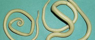
Drits Irina Alexandrovna. Parasitologist Helminth infections can lead to numerous health problems, shortening life by 15-25 years. Many parasites are extremely difficult to detect. They can be anywhere - in the blood, intestines, lungs, heart, brain. Symptoms of helminthic infestation can be confused with ARVI, gastrointestinal diseases and others. The main mistake in such cases is delay! If you suspect the presence of parasites, you need to contact a specialist. If we talk about medications and self-treatment, then this antiparasitic complex .
The life cycle of Leishmania is determined by the fact that it successively has two hosts - an insect (sandfly) and a vertebrate (including humans).
An insect becomes infected with a parasite by sucking blood from the host animal, from where they enter the digestive organs through the bloodstream, and there the flagellated forms transform into motile flagellated ones. The parasites multiply at a rapid pace, and after about 7 days they affect the upper part of the digestive system, and the protozoa completely cover it.
Life cycle
When a mammal is bitten, the insect's saliva, in which Leishmania has accumulated, penetrates the skin of the new host, and immune blood cells - neutrophils - immediately rush to the site of the bite, capturing the parasites.
They remain inside the neutrophils until they die naturally, after which the parasites emerge from them and enter the blood of a new host, turning into a flagellaless form and actively multiplying.
Why is leishmaniasis difficult to treat?
Leishmaniasis is a destructive, disfiguring disease. If infection occurs in a specific area on the leg, other parts of the body can become infected. Leishmania spreads through the bloodstream throughout the body, affecting one by one the organs and tissues of the body.
Due to the fact that the parasite invades cells, the disease is difficult to treat. The purpose of the drugs is to suppress the proliferation of Leishmania, thereby increasing the immune system's chances of victory. Even if you manage to get rid of the infection, the parasite can remain dormant for many years and then suddenly strike. Relapses in leishmaniasis are common, and drugs that poison leishmania are not always effective. The causative agents of this disease are very difficult to kill.
The human immune system fights parasites, but they are able to penetrate white blood cells, multiply and again attack the body’s defenses. If treatment is not started in time, the number of leukocytes will drop to the minimum acceptable level. This threatens serious violations, because the body becomes defenseless against the negative effects of any infection. For a weakened body, this will be a real blow. If the medications do not give the desired result within 3 or more weeks, the doctor increases their dosage.
Prevention of leishmaniasis is associated with the fight against mosquitoes, the use of sprays and other means against the attacks of these insects, as well as with the treatment of animals infected with the infection.
Epidemiology
What is cutaneous leishmaniasis? As indicated in the specialized medical literature, this is an infectious disease caused by the flagellated parasites Leishmania (Leischmania), and manifests itself as ulcerative skin lesions, which ends in the formation of rough scars. The causative agent of visceral and cutaneous leishmaniasis is Leishmania, but only of different species (L. donovani, L. infantum, L. tropica, L. major, etc.). The agglutination reaction helps to identify the type of pathogen. Mosquitoes are the specific carriers of leishmaniasis. The bite of this insect leads to infection of humans and insects.
In infectious practice, it is customary to distinguish between two forms of the disease, which have fundamental differences in epidemiological and clinical terms:
- Zoonotic type. It is also called rural. Rodents and dogs become a source of infection. L. tropica major provokes the development of a zoonotic type of disease. Seasonality is observed. The incidence rate increases in summer and decreases in winter.
- Anthroponotic type. The second name is urban. The carriers of the infection will be sick people with open lesions on the skin. L. tropica minor is responsible for the occurrence of the anthroponotic type of disease. Seasonality is not noted. Can be recorded all year round.
In both cases, mosquitoes perform the vector function. Despite the fact that the treatment of various forms of the disease is almost identical, the prevention of each type of cutaneous leishmaniasis has its own characteristics.
Visceral leishmaniasis, which affects internal organs, often ends in death without adequate treatment.
Clinical signs of visceral leishmaniasis
After entry of the causative agent of leishmaniasis, a long incubation period is observed, which can last up to 1 year, usually 3-5 months. During this time, the first sign of visceral leishmaniasis can be observed - the appearance of a pink papule at the site of the mosquito bite. After the spread of parasites throughout the human body, characteristic signs of visceral leishmaniasis develop:
- The disease begins with a feeling of weakness, fatigue, accompanied by recurrent fever. A significant increase in temperature during visceral leishmaniasis lasts in different cases from several days to several months, after which it is replaced by long periods of remission.
- In the first stages of development of visceral leishmaniasis, a rapid enlargement of the liver, spleen and lymph nodes is observed, which is associated with the multiplication of the pathogen in the cells. Palpation is painless; as the disease progresses, the growth of the affected organs slows down.
- Damage to bone marrow cells in visceral leishmaniasis leads to impaired hematopoiesis, which is clinically manifested by anemia. The skin and visible mucous membranes turn pale and acquire a bluish, sometimes porcelain or earthy tint.
- In visceral leishmaniasis, an enlarged spleen or liver can displace internal organs. The heart is confused, there is constant tachycardia, as well as a decrease in blood pressure.
- Leishmaniasis of the visceral type can be complicated by the development of secondary microflora in the lungs, in which case pneumonia develops.
- In the final stages of visceral leishmaniasis, an enlarged liver and the contours of the spleen are visible through the skin. Decreased muscle tone and dehydration are also characteristic.
- A blood test for visceral leishmaniasis shows a decrease in the number of red blood cells, leukocytes, platelets, and eosinophils. The erythrocyte sedimentation rate is increased.
After recovery, stable immunity to the visceral type of leishmaniasis is formed. Timely diagnosis and referral to a specialist will help to suppress the development of the disease in a short time and prevent complications from occurring.
Leishmania in humans
Leishmaniasis in humans can have many forms, and the methods of infection are always the same - a mosquito bite. The most common are the following:
- Visceral is one of the most complex forms of the disease, leading to death without urgent treatment. Parasites penetrate through the blood into any organs, including the liver, spleen, lymph nodes, bone marrow and begin to multiply rapidly, damaging them.
- Skin is one of the most common forms, in which the bite site immediately begins to hurt. The treatment is very long, lasting several months; scars remain on the affected areas.
- Diffuse cutaneous is also a widespread form, which in appearance is very similar to leprosy and is difficult to treat.
- Mucous - begins with the appearance of ulcers on the skin, which causes tissue damage, especially the nose and mouth.
Manifestation of cutaneous leishmaniasis
There are other, less common, but no less dangerous types of leishmaniasis for humans:
- Sequential leishmanioma;
- Tuberculoid and diffuse leishmaniasis.