Are adhesions after surgery a problem for those who have undergone abdominal or pelvic surgery? This problem still remains relevant in surgery, since there are a huge number of methods for preventing the appearance of new adhesions and treating existing ones. However, despite all efforts, often after extensive surgical interventions the process of adhesions continues to develop. This is largely determined by the characteristics of the human body and the nature of the intervention. However, even after the appearance of postoperative adhesions, the intestines can be treated, reducing the symptoms of the disease.
What causes adhesions?
Adhesive disease is a condition that occurs when a large number of individual adhesions are formed or a significantly pronounced adhesive process is formed, which leads to disruption of the functioning of internal organs.
In most cases, intestinal adhesions occur after surgery. Most often they appear after major operations performed by laparotomy (through a large incision in the abdominal wall).
Doctors who operated at the dawn of surgery noticed that when repeated operations were necessary, adhesions between individual organs were found in the abdominal cavity. Even then, it was clear to surgeons that numerous complaints presented by patients after surgical interventions on the abdominal organs were associated with adhesions. Since then, the complex history of studying this problem began.
Even more interesting:
Why is Unidox prescribed to women?
Balls hurt after arousal
The adhesive process (intestinal adhesion) is currently one of the most studied pathological processes in the human body. The main reactions of the internal environment that play a decisive role in the occurrence of adhesions include:
- inflammatory tissue reaction;
- coagulation of blood and the proteins it contains;
- anti-coagulation.
During surgery, trauma to the peritoneum is inevitable. In the event that only one of its leaves was damaged, and the one with which it is in contact remained intact, adhesions will not form. But even if such an injury causes a fusion between organs, it will be superficial, easily stratified and will not lead to dysfunction of the organs.
If 2 adjacent leaves were injured, then a whole cascade of pathological reactions is triggered. Due to a violation of the integrity of the blood capillaries, the release of individual blood proteins occurs. Globulins (namely coagulation factors) play a major role in the adhesion of organs. When these proteins come into contact with exposed intestinal tissue, a cascade of clotting reactions is triggered. The outcome of this cascade is the precipitation of fibrinogen in the form of fibrin. This substance is the universal “glue” of our body, which leads to the formation of early intestinal adhesions after surgery.
In the process of blood clotting, the anticoagulation system plays a significant role, which is activated somewhat later than the coagulation system. In most cases, blood that gets onto the peritoneum of the intestinal loops first coagulates and then returns to the liquid phase precisely thanks to the fibrinolysis system (dissolution of precipitated fibrin). But sometimes, upon contact with the peritoneum, this process can be disrupted, and fibrin does not dissolve. In this case, polar cods may appear.
https://youtu.be/6cX_AnqWItY
The reason for the appearance of adhesions
After surgery, you should not lose your vigilance and assume that all problems are behind you, because such treatment can have serious side effects. The fact is that the peritoneum, which covers the pelvic (small) organs and the abdominal cavity, creates conditions for displacement of the uterine loops or fallopian tubes. When the intestines function smoothly, there are no problems with the egg reaching the fallopian tubes, and the increase in the size of the uterus does not interfere with the positive functioning of the intestinal and urinary system. But such harmony does not always happen. It is disrupted after surgery.
Among these complications, the most dangerous is peritonitis. This is a pathology in the peritoneum that poses a health threat. The disease depends on the level of inflammation, but the body has a mechanism that inhibits the development of this disease. These are spikes. During the inflammatory process, organic tissues become swollen, the peritoneum becomes covered with a sticky coating, which consists of the substance fibrin.
Fibrin is a protein. This is the basis of blood mass. In contact with the film in the catalyst of the inflammatory process, fibrin connects the disconnected areas to each other. A unified system is created that blocks the path to the development of the disease process. Transparent films are formed. These are adhesions that form in places of adhesion after inflammation is completed.
The main function of these transparent films is to protect internal organs from suppuration and inflammation in the peritoneal area. However, such adhesions do not always form during inflammation. If surgical treatment was carried out correctly, then the likelihood of complications is sharply reduced. Adhesions are a protective reaction of the body. They form when the disease becomes chronic and its treatment is delayed over time.
Although adhesions are useful in inflammation as a protective measure, their appearance after recovery is negative because they:
- interfere with the good functioning of internal organs;
- disrupt the mobility of the intestinal organs, threatening to create obstruction in the intestinal area;
- disrupt reproductive function, destroy the functioning of the ovaries, fallopian tubes, and the movement of bodies in the body.
When they stagnate, they negatively affect human health, but in most cases they do not pose any threat to life. But their appearance creates discomfort and causes serious problems.
Symptoms of adhesions
The formation of intestinal adhesions takes quite a long time, sometimes it can take several months after surgery. This often happens almost unnoticed. To find out about the existence of adhesions, it is necessary to undergo a detailed medical examination. Patients turn to a specialist only when these formations have formed and become entrenched.
In order not to start the process and react in time, you need to know the symptoms of this process:
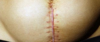
- Pain occurs sporadically, appearing in the area of surgical intervention, especially where there is a postoperative scar. They are sharply worsened by exercise or heavy lifting.
- Violation of the proper functioning of the gastrointestinal tract. Outwardly, this is expressed in an enlarged abdomen and difficulty in bowel movements. There is a feeling of discomfort in the navel area.
- Slowing down the process of passage of substances through the intestines. This is also called defecation disorder. Manifests itself in constipation.
- Nausea and vomiting after eating.
- Loss of body weight.
Each of these individual points can be confused with the manifestation of other pathological processes. But taken together, they indicate adhesions with extreme accuracy. In some rare cases, such processes can even pose a threat to human life, requiring immediate intervention by specialists and a surgeon. Especially notable among them are:
- Acute intestinal obstruction. This happens when adhesions compress the intestinal tube, which severely limits the passage of substances into the intestines. Accompanied by a gag reflex, acute pain, and accumulation of gases. Blood pressure decreases, tachycardia is likely to occur. In this situation, medical assistance becomes mandatory.
- Intestinal necrosis. It is characterized by the fact that adhesions sharply clamp the arteries, interfering with blood supply and depriving the intestinal walls of inflow, and this can lead to their death. In this case, immediate surgical intervention is required.
Diagnosis of adhesions
The patient or the specialist observing him may suspect the presence of an adhesive process in the body. In this case, it is necessary to undergo a comprehensive medical examination. It consists of the following stages:
- The doctor must conduct a primary (digital) medical examination, interview the patient for pain defects, and ask about past surgical interventions and injuries. After this, the doctor gives a referral for laboratory and technological tests.
- Ultrasound. The patient undergoes an ultrasound examination. The method has proven itself well in detecting the presence of adhesions.
- Radiography. Before the session, the patient drinks a glass of barium salt so that his stomach is empty. After which, appropriate photographs are taken, where disturbances in the functioning of the intestines will be visible and objects causing complications will be clearly visible.
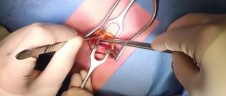
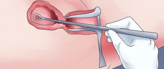
- Laparoscopy. When the procedure begins, a small hole is made in the patient's abdominal cavity, then a device with a camera is lowered there, with the help of which adhesions and their location are recorded. After this, the surgeon can simply cut them out.
- CT scan. A method that allows you to study in detail the adhesive process and its factors. This is the most effective method that helps you see the process practically from the inside.
After passing the examination, the doctor determines how to treat adhesions after surgery. There are several ways to deal with a possible relapse.
How to remove adhesions after surgery?
Treatment of adhesions after surgery is carried out by two methods: conservative and surgical. Once adhesions are diagnosed, treatment begins. In some cases, repeated surgery can be avoided.
If adhesions are not actively annoying, then it is quite possible to do without serious treatment, limiting yourself to preventive measures and visiting a specialist. The doctor will indicate how specifically to treat adhesions after surgery.
How to treat adhesions after surgery? If minor pain occurs or interruptions in the normal functioning of the body, the doctor prescribes special medications - aloe, various antispasmodics and enzymes. For problems with constipation, laxatives are taken. Proper nutrition after surgery is the key to avoiding serious problems in the future.
Symptoms after surgery
In most cases, the resulting adhesions are small in size and do not actually affect the functioning of the internal organs. However, when deformation of the structure occurs, symptoms of adhesions occur. The clinic depends on both the size and localization of the pathological process. The most common symptoms of adhesions include:
- abdominal pain;
- feeling of discomfort in the abdomen;
- constipation;
- general weakness;
- mental disorders.
Pain in the abdomen is the main manifestation of adhesive disease. The cause of the pain is a serious disruption of the functioning of the intestines. The nature of the pain may also differ from patient to patient. For some it is permanent, for others it is cramping. A feature of pain receptors in the intestinal wall is their increased sensitivity to stretching. Therefore, physiological bowel movements (peristalsis) can lead to significant tension in the intestine and provoke pain.
This is also the cause of pain after eating certain foods, which contribute to increased gas formation or increased peristaltic movements of the intestine. Separately, it is worth mentioning the pain, which intensifies with physical activity.
More often it occurs when the adhesion is located between the loops of the intestine and the anterior abdominal wall. Due to the contraction of the abdominal muscles, tension occurs in the intestinal tissue and its mesentery. With excessive physical exertion, this can lead to the formation of obstruction. The appearance of discomfort is due to approximately the same reasons as pain.
Diagnosis of adhesions is based on the collection of numerous complaints. Some patients may experience no pain or discomfort at all. But constant constipation and the presence of major abdominal surgery in the past should suggest an adhesive process. Abnormal bowel movements occur due to chronic damage to the intestinal wall and decreased motor activity. The consequence of such changes is a slowdown in the movement of chyme along the intestinal tube. Subsequently, the process of final formation of feces is delayed and the frequency of feces is reduced.
Diagnostic methods
If symptoms appear, the patient should consult a specialist. First, an examination and history taking of the patient is performed. Diagnosis of the disease must be comprehensive.
Analyzes
First of all, the patient needs to get tested.
- A general blood test is prescribed for any disease. The analysis can show the presence of inflammatory processes, as well as the general condition of the body. With adhesive disease, there is leukocytosis, which indicates an inflammatory process in the body. There is also anemia.
- Biochemical blood test - can tell about the functioning of internal organs, especially the liver and kidneys. There are abnormalities: elevated urea levels, low hemoglobin levels and C-reactive protein (in acute inflammation).
If intestinal obstruction is suspected, a stool test may be required. Additional tests may include a blood test for hormones and a semen analysis.
Instrumental diagnostic methods
The main diagnostic methods are instrumental studies. They are more informative than analyses. If adhesions are suspected after surgery, the following studies are prescribed:
- Ultrasound - the study shows the presence of adhesions;
- CT is the most informative method;
- radiography with a contrast agent - before the procedure you need to drink a special solution, which, when photographed, shows disturbances in the intestines and other complications;
- laparoscopy – a small incision is made in the abdomen and then a tube with a camera and light is placed. This allows diagnostics to be carried out from the inside.
After conducting instrumental diagnostics, the doctor may prescribe treatment or refer you for additional examination.
Differential diagnosis
Adhesive disease can be easily confused with diseases of the internal organs, since many clinical symptoms are similar. You need to know how to distinguish adhesions from another disease:
- pinched hernia - characterized by protrusion, pain and tension in the affected area;
- acute pancreatitis and cholecystitis - uncontrollable nausea and vomiting, increased body temperature, girdling pain;
- peptic ulcer of the gastrointestinal tract - paroxysmal pain in the abdominal area;
- acute appendicitis - pain in the right iliac region. High body temperature and increased leukocytes in the blood;
- torsion of an ovarian cyst - paroxysmal pain in the lower abdomen.
This diagnosis is carried out if the presence of adhesions is suspected. Diagnosis must be comprehensive and after clarification of the diagnosis, treatment is prescribed.
General manifestations of the disease
Intestinal adhesions manifest themselves as symptoms - both local and general. These include constant weakness, a number of mental disorders and decreased immunity. There are several reasons for these manifestations:
- Constant pain and discomfort in the abdomen lead to exhaustion of the nervous system and form the so-called “core” of psychological changes in consciousness.
- Disruption of normal intestinal motility leads to a decrease in the flow of nutrients into the bloodstream.
- Prolonged presence of feces in the large intestine promotes increased proliferation of microorganisms in its lumen.
The occurrence of pain both during movements, physical activity, and at rest contributes to the formation of protective behavior. It manifests itself in the fact that the patient tries to avoid a certain movement, posture or behavior. Accordingly, the normal spectrum of activity is limited. This may affect the sphere of professional activity, which ultimately leads to some withdrawal from social contacts.
Diet for adhesive bowel disease
If the presence of postoperative adhesions is suspected, the doctor prescribes the patient a series of laboratory and instrumental examinations:
- A clinical blood test will show the presence or absence of an inflammatory process in the body.
- Ultrasound examination (ultrasound) – visualizes the presence of adhesions.
- X-ray of the intestines.
- Diagnostic laparoscopy.
The research results allow the doctor to determine the presence of adhesions, examine their shape, thickness, determine how the internal organs work, and prescribe the necessary treatment.
- Acute stage. The first manifestations of the adhesive process occur in this form. The patient suffers from pain that does not go away after taking medications, intestinal obstruction develops, and peristalsis worsens. With prolonged constipation, severe vomiting is possible. The temperature rises, tachycardia appears. The longer this stage goes on, the more severe the symptoms become. Gradually, dehydration begins, blood pressure rises, and the exchange of microelements deteriorates.
- Intermittent form of the disease. In this condition, the patient experiences severe to moderate pain. Constipation or diarrhea appears - it all depends on the position of the adhesions.
- Chronic form. The adhesive process occurs with sluggish symptoms, sometimes nagging pain appears in the lower abdomen. The patient loses weight for no reason and suffers from digestive problems. In women, the chronic form of adhesions leads to infertility.
To identify the process, medical diagnosis and instrumental examinations are required.
In a normal state, the organs of the abdominal cavity and small pelvis are in motion, the intestines move when digesting foods. If scarring occurs, it interferes with natural processes, including the movement of an egg through the tubes into the uterus. This causes infertility. In severe cases, intestinal adhesions contribute to the development of obstruction.
Intestinal adhesions manifest themselves as symptoms - both local and general. These include constant weakness, a number of mental disorders and decreased immunity. There are several reasons for these manifestations:
- Constant pain and discomfort in the abdomen lead to exhaustion of the nervous system and form the so-called “core” of psychological changes in consciousness.
- Disruption of normal intestinal motility leads to a decrease in the flow of nutrients into the bloodstream.
- Prolonged presence of feces in the large intestine promotes increased proliferation of microorganisms in its lumen.
The occurrence of pain both during movements, physical activity, and at rest contributes to the formation of protective behavior. It manifests itself in the fact that the patient tries to avoid a certain movement, posture or behavior. Accordingly, the normal spectrum of activity is limited. This may affect the sphere of professional activity, which ultimately leads to some withdrawal from social contacts.
In addition, the belief is formed in the mind that this condition was caused by the actions of medical personnel, so in the future you should avoid seeking medical help. All this together leads to a delay in proper care and worsening of the condition.
Abdominal adhesions, disrupting intestinal motility and reducing the absorption of nutrients, are mainly associated with a violation of a person’s nutritional status. Chronic deficiency of proteins, fats and carbohydrates occurs. The consequence is weight loss and decreased immune status. However, this is not typical for all individuals who have developed adhesions as a result of surgery.
- General blood analysis. Necessary to check whether you have inflammation in the body. Also evaluate the activity of the fibrinolytic system of the blood.
- Ultrasound of the abdominal and pelvic cavity. A visual examination method helps to say with a 100% guarantee whether there is an adhesive process in the pelvis after hysterectomy.
- X-ray examination of the intestines using contrast (coloring) substances. An auxiliary method that allows one to judge the patency of the intestine and the degree of narrowing of its lumen.
- Laparoscopic diagnostics are also used, during which individual adhesive formations are dissected and removed, and the issue of repeated surgical intervention is also decided.
- pain;
- constipation;
- vomit;
- temperature increase.
Intestines
Adhesions in the intestines appear after surgical operations. They can be asymptomatic, but sometimes they manifest themselves very clearly. The most dangerous sign is intestinal obstruction. Due to severe prolonged pain, lack of stool, and bloating, the fear of death appears. Symptoms of the adhesive process are:
- painful vomiting;
- heaviness in the stomach;
- flatulence;
- constipation
Symptoms of the appearance of adhesions in the pelvis differ little from the manifestations of gynecological diseases. It is even more difficult to determine them because the cause often lies in inflammatory processes and hormonal diseases. Adhesions cover the adnexal organs - the ovaries, fallopian tubes and the uterus itself. This condition leads to ectopic pregnancy and infertility. Symptoms of adhesions:
- bleeding;
- aching pain in the lower abdomen;
- decrease in pressure.
In case of adhesive disease of the abdominal cavity, diagnosis begins with an external examination of the patient. The doctor conducts a survey about the symptoms and the nature of the pain. General tests are prescribed. In case of intestinal adhesions, a digital examination of the rectum is performed. More accurate diagnosis is carried out using ultrasound examination and radiography. What is prescribed:
- In gynecology, diagnosis is carried out using ultrasound and magnetic resonance imaging (MRI).
- Laparoscopic examination provides an accurate diagnosis. It is prescribed in severe cases.
- For adhesions in the fallopian tubes, a special contrast is injected and an x-ray is taken. Check how widespread the obstruction is.
- Another diagnostic method involves injecting saline solution through the cervical canal and examining it using ultrasound.
One method of treating intestinal adhesions is to follow a diet. It is necessary to exclude foods that irritate the stomach and intestines: spicy, sour, with coarse fiber. Food should not cause gas contamination. Use is not allowed:
- carbonated drinks;
- spices;
- confectionery products;
- hot and cold food;
- smoking;
- sweets;
- sausages;
- alcohol;
- fried.
It is advisable to eat in small portions, chewing food thoroughly. Meals should be fractional - at least 5 times. Steamed food is healthier. Should be eaten:
- vegetarian, milk soups;
- lean meat;
- fish;
- lactic acid products;
- porridge with water;
- black, white bread;
- ripe fruits, berries;
- greens, vegetables;
- tea with milk, juices.
Why are adhesions dangerous?
In addition to nutritional disorders, vitamin deficiencies and mental disorders that develop over years, the course of the adhesive process may be complicated by severe and often life-threatening conditions:
- acute intestinal obstruction.
- intestinal necrosis.
Acute intestinal obstruction develops when adhesions deform the intestine so much that its patency virtually completely disappears. In this case, acute cramping pain in the abdomen occurs. Quite clear localization of pain at the site of obstruction is possible. This pain is easy to distinguish from the usual course of the disease, which is associated with its severity and suddenness, and not with any movement or position of the body.
Vomiting follows very quickly. At first, the vomit has signs of previously eaten food, but after a while bile impurities appear. And if left untreated, the vomit becomes fecal (since the intestinal contents can no longer move in a physiological direction). Occasionally, blood appears in the stool. Common manifestations include the following:
- in the first place is pronounced general weakness;
- body temperature rises;
- the patient’s facial features become sharper;
- the skin takes on a gray tint;
- eyes are sunken;
- in the absence of emergency surgical care, death occurs within a few days.
An equally serious complication is necrosis of a section of the intestine. The pathogenesis of this condition involves tissue pinching the adhesions of blood vessels and disruption of blood flow in the intestinal area with the development of ischemia (oxygen starvation), and subsequently tissue death.
The main manifestation is increased abdominal pain and severe bloating. Vomiting may occur. The temperature rises significantly and chills appear. Due to disruption of the intestinal barrier functions, microorganisms gain access to the systemic bloodstream. As a result, sepsis develops, which requires emergency medical interventions. Otherwise, death will occur within a few hours or days.
How to remove adhesions, treatment methods
Treatment of adhesions after surgery is a serious, lengthy and controversial issue. The occurrence of complications is an absolute indication for surgical treatment. At the moment, numerous techniques are used for this purpose: starting from the intersection of individual elements of adhesive tissue (in the absence of necrosis in the intestinal wall) and ending with excision of a section of the intestine that has undergone necrotic changes.
If the issue of surgical treatment of intestinal adhesive disease has been decided, then full and comprehensive preparation of the patient for surgical intervention is necessary, aimed at correcting the disturbed parts of metabolism and compensating for all concomitant diseases. The surgeon's goal is to remove as much of the connective tissue that forms the adhesions as possible. However, this procedure is only temporary, because even after removing adhesions, areas of tissue remain that can later “stick together” again, and the symptoms of adhesive disease return.
There are many controversial opinions on how to treat adhesions formed after surgery conservatively (without surgery). However, all experts agree that a radical cure is possible only by removing the adhesions themselves. The attending physician can suggest a number of techniques that, as a rule, will alleviate the patient’s condition, but will not get rid of the cause. These include:
- dietary nutrition;
- periodic forced bowel cleansing;
- symptomatic drug treatment.
The peculiarity of nutrition is to eat food throughout the day in small portions, but often. It is necessary to avoid foods that increase the formation of gases (legumes, foods containing significant amounts of fiber).
Forced bowel cleansing means performing cleansing enemas. This procedure should be carried out as needed, but not more than 3 times a week. Drugs that can reduce the manifestations of the disease include antispasmodics (No-spa and its analogs), painkillers (Ketanov, Fanigan).
Mechanism of development of pathology and clinical picture
At first, the connective fiber is weak and thin; in the first stages of the disease, it is easy to disconnect. Over time, the film thickens, possibly even ossification. In later stages, fibrous tissue can only be excised surgically.
The clinical picture is established based on two criteria: the strength of the lesion (number of growths) and the location. The patient is often tormented by symptoms reminiscent of the progression of intestinal diseases or damage to the gastrointestinal tract. Often a negative condition is preceded by an asymptomatic period when the disease is at an initial stage.
Prevention of adhesions after surgery
Most patients are interested in how to avoid adhesions and prevent the development of pathology. Recommendations in this regard concern both the doctor and the patient. It is up to the patient to seek medical help in a timely manner in order to prevent the development of complications that significantly aggravate the course of surgical pathology. In some cases, timely prescribed conservative treatment can have a sufficient effect and surgical intervention is not required.
If, nevertheless, it is not possible to refuse the operation, then the prevention of the development of adhesions largely depends on the surgeon. However, it is worth noting that even the most modern methods of surgical treatment and the best techniques do not provide an absolute guarantee. The likelihood of adhesion formation is reduced if minimally invasive interventions are performed, and all actions are carried out with the utmost care. Even if a section of the intestine has to be removed, all measures must be taken to prevent the development of adhesions. Thus, the prevention of adhesions depends on both the doctor and the patient.
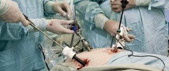
What happens to our body during operations? First, the tissues are cut, then connected, and they are forced to grow together again. It is believed that laparoscopic surgery, which is performed through several small incisions (“punctures”), is much less traumatic, since the surface of the surgical field is significantly smaller than with a conventional strip “open” operation.
During laparoscopy, damage occurs on the thin membrane covering the inner surface of the abdominal wall where instruments, incisions, or clips are inserted. After removing the instrument, this area of the damaged membrane (called serosa) heals on its own.
Indications
Infertility is caused by various factors, and some of them can be eliminated with the help of laparoscopy.
Indications for surgical intervention are:
- Endometriosis. Pathological growth of the endometrium outside the uterus, for example, in the abdominal cavity, on the ovaries, in the tubes. The properties of endometrioid tissue are preserved even outside the uterus, that is, it menstruates every month. This leads to the release of blood into the abdominal cavity, the development of an inflammatory and adhesive process. During laparoscopy, the endometriotic lesion is excised, and the adhesions that have already appeared are removed.
- Myoma. If small myomatous nodes are “husked” through hysteroscopy from the inside of the uterus, then large or subserous formations are removed through the abdominal cavity. Laparoscopy is indicated if the fibroid nodes do not exceed 5-6 centimeters.
- Obstruction of the fallopian tubes. It occurs due to the formation of adhesions in the abdominal cavity or in the lumen of the oviduct against the background of inflammatory processes. External adhesions change the course of the tube, create kinks, glue together the fimbriae of the ampulla, which prevents the passage of a fertilized egg into the uterus and is diagnosed as infertility. Therefore, even if conception occurs, there is a high probability of an ectopic pregnancy. During laparoscopy, the adhesions are dissected, restoring the anatomical patency of the oviduct.
- Hydrosalpinx. This is a pathology in which fluid accumulates in the lumen of the fallopian tube. Usually, in this case, restoring the patency of the oviduct does not make sense, since the risk of ectopic pregnancy and relapse of the disease is high. Damaged tubes are removed, and patients are recommended to undergo IVF.
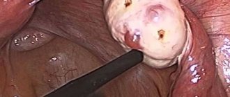
- Adhesive process. Occurs after surgical interventions in the abdominal area, inflammatory processes in the reproductive organs, and after peritonitis. The adhesions are cut, the blood vessels passing through them are ligated.
- Polycystic ovary syndrome. Endocrine pathology, in which the ovulation process is disrupted, which causes infertility. Many cystic formations form in the ovaries, causing low-grade inflammation. This leads to thickening and sclerosis of the ovarian membrane, which prevents ovulation. The essence of laparoscopy is to apply incisions to the membrane and dissect cystic formations. The method of choice is wedge resection of the ovary.
- Ovarian cyst. Cystic formations compress the ovarian tissue, thereby disrupting its function. In addition, during pregnancy the risk of cyst rupture increases, which is dangerous for the expectant mother and fetus.
Sometimes laparoscopy is performed to prepare for IVF. In such a situation, it is recommended to remove the non-functioning fallopian tubes. This is done to reduce the risk of developing an ectopic pregnancy after embryo transfer.
Diagnostic laparoscopy is prescribed if the cause of a woman’s infertility is not established, or the diagnosis needs to be confirmed. The fact is that small foci of endometriosis and adhesions are difficult to detect using other research methods.
As for tubal infertility, it is possible to confirm the diagnosis and check the patency of the fallopian tubes during laparoscopy. To do this, a colored sterile liquid is injected into the uterus through the vagina. If the pipes are passable, you can see on the monitor how the solution flows out of the ampullary section.
There are methods for restoring the patency of the fallopian tubes using the laparoscopic method. But research shows. That after surgery there is a high probability of recurrence of adhesions and ectopic pregnancy.
Therefore, for tubal factor infertility, the most effective treatment method is in vitro fertilization.
How adhesions and scars form
However, our fabrics have one natural, irrevocable property - they strive to protect our body. And sometimes the development of so-called protective factors after damage occurs intensively - with a reserve.
In practice, it looks like this: in places where the serous membrane is damaged, collagen and elastic fibers and connective tissue cells are intensively produced. If at this time any internal organ (for example, a loop of intestine) touches the area of the damaged serosa, it is involuntarily involved in this process. A cord of connective tissue is formed, which leads from the wall of the internal organs to the inner surface of the abdominal wall. This is called adhesions.
Why are adhesions bad?
Nature made sure that in our harmonious body the organs were equipped and arranged clearly and correctly, like in Tetris. They occupy the entire internal space and touch each other with suitable sides, like a carefully fitted puzzle. If you consider all the organs separately from the body, you will be amazed at how much space they take up and how they fit inside us! It is precisely because postoperative scars and adhesions disrupt this initial harmony that they affect our body.
What is the negative impact of adhesions? They:
- interfere with the mobility of the organ, which affects its function. Moreover, both external mobility, which depends on the movements of the diaphragm, suffers, as well as internal mobility, which is active and does not depend on the movement of the diaphragm;
- disrupt blood circulation in the affected organ;
- disrupt the innervation of the organ;
- contribute to the occurrence of pain and spasms in the organ.
Sometimes the adhesion is so powerful that it can disrupt the anatomically correct position of the organ. All of the above reasons lead to other disorders in the body. And yet, which at first glance are not related to the affected area. Adhesions and scars that arise after abdominal surgery can cause pain in various parts of the spine, joints, lead to changes in posture and disruption of the body’s position in space, etc.
How are adhesions treated?
According to the timing of formation of adhesions, they are distinguished:
- 7-14 days after surgery – the phase of young adhesions, when the adhesions are still very loose and easily torn;
- 14-30 days after surgery is the phase of mature adhesions, when the adhesions thicken and become strong.
Starting from the 30th day after the operation and further, for several years, the process of restructuring and formation of scars and adhesions occurs. The process is individual, much depends on the properties of the organism itself, its anatomical structure, and the functioning of internal organs.
The doctor may suspect the presence of adhesions in the abdominal cavity based on clinical data, medical history, and the results of studies such as ultrasound, CT, and colonoscopy. Adhesions in the abdominal cavity and pelvic cavity can be treated with medication or surgery. During surgery, the adhesions are separated, but this method should be used only in extreme cases, if the cords are so thick and rough that they severely disrupt the function of the organ, and more loyal and gentle treatment does not help.
Diagnostics
Diagnosis begins with collecting anamnesis. The patient describes in detail the nature of the pain. The doctor must definitely clarify whether abdominal surgery has been previously performed, and what inflammatory processes are (or were previously present). To confirm the diagnosis, you will need to undergo ultrasound diagnostics, x-rays or magnetic resonance imaging.
The most accurate and at the same time therapeutic method is laparoscopy. This is a minimally invasive intervention during which the surgeon sees all the smallest details inside the abdominal cavity. If an adhesive process is detected, the specialist can immediately excise the overgrown fibrous tissue.
Laboratory tests are required: blood and urine sampling for general clinical and biochemical tests.
How osteopathy affects adhesions
An osteopathic doctor is able to feel with his hands where the adhesions are located and where they lead, where they are attached and what they are pinching. He is also able to loosen their tension in a few sessions, and can restore, balance and balance damaged organs, and therefore restore their function to the fullest possible extent.
An osteopathic doctor is also able to interrupt the chains of damage and pain in parts of the body that seem to be unrelated to the operated area. After all, our body is an integral system where everything is interconnected. The osteopath acts on the adhesions directly, without violating the integrity of the body tissues, and therefore without an additional factor stimulating the formation of connective tissue. By restoring and harmonizing the function of the suffering organ, the body releases energy to initiate complete recovery in possible individual conditions for the entire organism.
Any surgical intervention, no matter how minimally gentle it may be, leaves behind a lot of negative changes, injuries and stress, which the body is forced to fight alone. What the body will do to heal itself, what it will sacrifice, how it will limit itself is always individual. But within the framework of self-preservation, this is always expressed in loss of function to one degree or another, and therefore subsequent suffering of the entire organism with loss of compensation and the expenditure of much greater effort on normal functioning throughout life.
Therefore, if you have had surgical interventions on the abdominal organs in your life, consult an osteopathic doctor. It does not matter whether the operation was conventional or performed using a gentle laparoscopic method. Any discomfort has a reason, which means there is an opportunity to solve it.
An osteopathic doctor can use pulse diagnostics to determine the significance of adhesions or scars on the body. This means that if, when pressing on a postoperative scar, the properties of your pulse change, then this zone is important and significant for the whole organism, and this adhesion or scar must be worked with.
Adhesions and scars have the following significance and prevalence of influence:
- local (the effect is limited to the area where the scar or adhesions are located);
- regional (the effect extends to the entire thoracic or abdominal region where the commissure is located);
- global (affects the entire body, even to the point of disturbing its position in space).
How long does osteopathic treatment last?
If the patient has undergone surgery, then the osteopathic doctor will tactically act as follows. 10 days after the operation, when the stitches are removed, the doctor will work with the scar itself layer by layer, work with the tissues directly around the scar itself and restore that independent mobility of the organ, which does not depend on the movement of the diaphragm. This period of work ranges from 10 days to 3 months after the operation.
If the duration after surgery is 3 or more months, then the doctor will pay attention to all surrounding organs and tissues in the operation area, influence the mobility of all internal organs in general and directly to the localization of the adhesions themselves.
The information was prepared by the leading specialist of the osteopathy and family medicine clinic Osteo Poly Clinic Gulyants Maria Alexandrovna, osteopathic doctor, chiropractor, endoscopist surgeon.
Prevention of adhesions after surgical interventions on the ovaries and fallopian tubes
05.07.2013 I.B. Manukhin, A.A. Kolesov, L.K. Bekmurzaeva, E.A. Petrovich
Moscow State Medical and Dental University
A comprehensive examination was carried out in 108 patients after surgical interventions on the appendages and fallopian tubes with varying amounts of rehabilitation measures. It was revealed that the inclusion in the complex of measures for the prevention of postoperative adhesions of drugs that, along with an immunomodulatory effect, have prolonged hyaluronidase activity, makes it possible to compensate for the negative impact of surgical trauma on the immune system. Clinical confirmation of the effectiveness of enzyme immunotherapy was a decrease in both the frequency and severity of adhesions, which made it possible to reduce the number of reconstructive operations for infertility in this category of patients. Key words: adhesions, prevention, Longidaza
The formation of adhesions in the postoperative period is one of the pressing problems from both a medical and social point of view. Postoperative intraperitoneal adhesion occupies a leading position among the causes of tubo-peritoneal infertility. A high frequency of adhesive complications is observed in 55–97% of patients after abdominal operations and is due to an increase in the volume and severity of surgical intervention, increased resistance of microflora to antibiotics, and changes in the immunological reactivity of the body [1, 8].
The main trigger for adhesions is the inflammatory reaction of the peritoneum with subsequent destructive changes due to various intraoperative effects (mechanical, thermal, chemical, etc.) on the peritoneum and abdominal organs. The immune system, including both cellular and humoral components, plays a decisive role in the formation of the response of the peritoneum and tissues to injury [2, 5].
The development of adhesions after abdominal interventions is primarily associated with the risk of developing intestinal obstruction, as well as various symptom complexes, including pain, dysfunction of organs covered by the peritoneum, and others. When performing repeated surgical interventions, especially laparoscopically, adhesions in the abdominal cavity and pelvis formed after surgery are a potential risk factor due to possible damage to internal organs [9, 15].
Postoperative intraperitoneal adhesion of the small pelvis is one of the most common causes of tuboperitoneal infertility, chronic pelvic pain syndrome, and ectopic pregnancy [3, 11].
Adhesion formation is especially common after appendectomy for destructive forms of appendicitis, ovarian resection, and salpingectomy due to tubal pregnancy [7, 13].
Along with gross destructive changes, even in the absence of adhesions after abdominal interventions, the development of functional disorders of the abdominal organs, manifested in an imbalance of ciliary, secretory and muscular activity, is possible. Thus, in the fallopian tubes, the advancement of sperm, the capture of the egg and its transport after fertilization to the uterus are disrupted, which may be the reason for the insufficient effectiveness of surgical methods for correcting tubal occlusion [4, 6], as evidenced by the relatively low pregnancy rate (21–28%) after laparoscopic reconstructive plastic surgery [3, 14].
Considering the role of inflammation in the implementation of the adhesive process, to prevent the formation of adhesions in the postoperative period, along with antibacterial treatment, various methods of physiotherapy, desensitizing agents, enzyme preparations, bio- and immunostimulants of various origins are widely used.
At the same time, the literature contains ambiguous data on the effectiveness of their use and influence on the immune system, while the state of the latter largely determines the nature of the post-traumatic reaction in the area of the postoperative wound [12].
In connection with the above, it is of particular interest to study the possibility of using immunomodulators in order to increase the effectiveness of the prevention of adhesions in gynecological patients after laparoscopic operations.
Purpose of the study
– study of the effectiveness of using Longidase (an immunomodulator with prolonged hyaluronidase activity) for the prevention of adhesions after surgical interventions on the ovaries and fallopian tubes.
Patients and methods
We examined 108 patients who had undergone surgical interventions on the uterine appendages. Depending on the surgical approach, two groups of patients were divided: 1st group - 49 patients who underwent laparotomy, 2nd group - 59 patients who underwent laparoscopy. Taking into account the scope of postoperative rehabilitation measures, a comparison subgroup and a main subgroup were identified in each group of patients.
The comparison subgroup consisted of patients who underwent laparoscopic interventions of various volumes for infertility. All patients had a history of previous operations on the fallopian tubes and ovaries, after which preventive measures were carried out in the traditional scope.
Patients of the main subgroup in the postoperative period, along with the traditional scope of preventive measures, were treated with Longidase. Dynamic laparoscopy was performed in patients of the main subgroup in the absence of pregnancy 6 months after the end of treatment.
Longidaza (Petrovax Pharm). Registration number: LS-000764. Trade name: Longidaza. Chemical name: conjugate of hyaluronidase (Lidase) with the active derivative of poly-1,4-ethylenepiperazine N-oxide (analogue of Polyoxidonium). Longidase is a conjugate drug containing lidase and polyoxidonium. The drug was administered intramuscularly from the first day after surgery at a dose of 3000 IU, every 5 days, for a course of 10 injections.
All patients were examined in accordance with the standardized WHO protocol. The examination program, along with generally accepted clinical and laboratory tests, included determination of immune status indicators, laparoscopy, and ultrasound.
All patients underwent a clinical blood test, biochemical blood test and immunological study before the start of treatment and over time. Hematological parameters were assessed based on a clinical blood test using visual microscopy with Romanovsky-Giemsa staining of a smear. Biochemical blood analysis was carried out using standard biochemical diagnostic kits.
The immunological study included determination of the number of T-lymphocytes, T-helpers, T-cytotoxic lymphocytes, NK cells, B-lymphocytes using DAKO reagents using the labeled streptavidin-biotin (LSAB) method using monoclonal antibodies CD 3+, CD 4+, CD 8+, CD 16+, CD 20+.
The functional activity of peripheral blood neutrophils was assessed by the adhesive activity of neutrophils to plastic, the production of superoxide anion in the nitroblue tetrazolium reduction test using the spectrophotometric method (spontaneous and stimulated NBT test). Determination of serum immunoglobulins was carried out by the method of radial immunodiffusion according to Mancini.
When assessing the severity of the adhesive process, the classification of J. Hulka et al was used. (1998) and the American Fertility Society (AFS) [10]:
- Grade 1: minimal adhesions, tubes are patent, most of the ovary is visible
- Grade 2: more than 50% of the ovarian surface is free, ampullary occlusion with preservation of folds
- 3rd degree: less than 50% of the surface of the ovary is free, ampullary occlusion with destruction of the folds
- Grade 4: the surface of the ovary is not visible, bilateral hydrosalpinx
Research results and discussion
Based on clinical characteristics data, we can conclude that patients operated on for pathology of the ovaries and fallopian tubes have a whole complex of factors predisposing to the development of peritoneal adhesion. Thus, among those examined, the majority of patients (74.1%) underwent emergency surgery, which is associated with an increased risk of postoperative complications, in particular the development of purulent-septic complications. A significant proportion consists of surgical interventions for ovarian tumors (35.1%) and ectopic pregnancy (21.3%), which are classified as operations most often accompanied by the development of adhesions.
It should be noted that quite often in 19.6% of cases, surgical interventions were performed against the background of hemoperitoneum, which is one of the etiological factors in the formation of postoperative adhesions. The rather high frequency of previously suffered inflammatory diseases of the genitals in the examined patients is noteworthy (in 40.7% of cases - salpingo-oophoritis, in 23.5% - vulvovaginitis, in 18.6% - endometritis).
The presence of inflammatory diseases of the genitals should be considered not only as a risk factor for the development of adhesions, but also as a background for the occurrence of immunodeficiency states in the examined patients.
The combination of these factors, in our opinion, was the reason for the significant incidence of infertility in the examined patients (51.8%).
Reproductive dysfunction is closely related to the development of postoperative adhesions, as evidenced by the frequency of secondary infertility in the examined patients after surgical interventions (41.7%).
The comparability of subgroups according to the main anamnestic characteristics made it possible to conduct a comparative assessment of the effect of Longidase therapy on the formation of postoperative adhesions during laparotomy operations on the ovaries and fallopian tubes.
The leading criterion for the effectiveness of the therapy was the results of laparoscopic examination. It is noteworthy that in 30.0% of cases after laparotomy, patients in the comparison subgroup had severe forms of pelvic adhesion, indicating destructive changes in the fallopian tubes. In 2 (10.0%) patients, the formation of hydrosalpinxes was noted (Table 1). During laparoscopy in 19 patients of the main subgroup, performed in the absence of pregnancy 6 months after the end of Longidase therapy, adhesions in the pelvis were detected in 10 (52.6%) patients.
When carrying out basic measures to prevent adhesions after endosurgical treatment during repeat laparoscopy, adhesions in the pelvis were detected in 7 (35.0%) patients, when using enzyme-linked immunosorbent therapy - in 6 (27.3%) patients.
It should be noted that in patients of the main subgroup, the adhesive process was not accompanied by the development of destructive changes in the fallopian tubes and did not exceed grade 2, in
Table 1. Characteristics of the adhesive process in patients after operations on the fallopian tubes and ovaries
| The severity of the adhesive process | Laparotomy | Laparoscopy | ||
| comparison subgroup | main subgroup | comparison subgroup | main subgroup | |
| No adhesions | 1 (5,0%) | 9 (47,4%) | 13 (65,0%) | 16 (72,7%) |
| 1st degree | 7 (35,0%) | 7 (36,8%) | 4 (20,0%) | 4 (18,2%) |
| 2nd degree | 6 (30,0%) | 2 (10,5%) | 2 (10,0%) | 2 (9,1%) |
| 3rd degree | 4 (20,0%) | 1 (5,2%) | 1 (5,0%) | 0 (0%) |
| 4th degree | 2 (10,0%) | 0 (0%) | 0 (0%) | 0 (0%) |
Table 2. Chromosalpingoscopy data in patients after operations on the fallopian tubes and ovaries
| Condition of the fallopian tubes | Laparotomy | Laparoscopy | ||
| comparison subgroup | main subgroup | comparison subgroup | main subgroup | |
| Both fallopian tubes are patent | 8 (40,0%) | 14 (73,7%) | 16 (80,0%) | 16 (72,7%) |
| One fallopian tube is patent | 3 (15,0%) | 4 (21,1%) | 3 (15,0%) | 4 (18,2%) |
| Both (or only) pipes are impassable | 9 (45,0%) | 1 (5,2%) | 1 (5,0%) | 2 (9,1%) |
In the comparison subgroup, one patient (14.3%) had a severe adhesive process. In the main subgroup, dynamic laparoscopy was performed in 22 (54.1%) patients in the absence of pregnancy within 6 months after the Longidase course (Table 2).
In patients who underwent laparotomy, impaired patency of the fallopian tubes during chromosalpingoscopy was noted in 12 (60.0%) when using the traditional scope of rehabilitation measures, while in 45.0% of cases there was bilateral occlusion or occlusion of a single tube. In the main subgroup, tubal obstruction occurred in 5 (26.3%) patients, and bilateral occlusion occurred in only one woman (5.2%).
Impaired patency of the fallopian tubes after laparoscopic operations was detected in 3 (15.0%) patients in the comparison subgroup and in 2 (9.1%) patients in the main subgroup.
The inclusion of Longidase in the complex of measures to prevent postoperative adhesions after laparotomy made it possible to reliably ( p
<0.01) reduce the frequency (28.6%) of reconstructive interventions for infertility in patients of the main subgroup compared to the comparison subgroup (95.0%).
The results obtained, indicating the clinical effectiveness of Longidase, were confirmed when analyzing the reproductive function of the examined patients. During the observation period (12–18 months) in patients who underwent an expanded complex of rehabilitation measures, pregnancy occurred after laparotomy in 65.6% of cases (19 patients), after laparoscopy – in 77.0% (30 patients). While in the comparison subgroups, 6 (30.0%) and 12 (60%) patients became pregnant, respectively.
Taking into account the above data on the role of the functional state of the immune system in the implementation of pathological intraperitoneal adhesion, we carried out a comparative assessment of the indicators of humoral and cellular immunity in the postoperative period in patients undergoing basic therapy (control group) and when including a course of therapy with an immunomodulator with enzymatic activity (main group ).
In the early postoperative period, a deficiency of CD3, CD4 T-lymphocytes in patients of both groups indicated the presence of a chronic inflammatory process, which is both a consequence of surgical trauma and a manifestation of immunodeficiency in patients with adhesions.
Changes in the functional state of B-lymphocytes (increased CD20 concentration) were observed in 50% of patients in the main group and in 52% in the comparison group. In 78% of the examined patients there was a decrease in the phagocytic activity of neutrophils. The indicators of the humoral component of the immune system did not differ significantly from the normative values ( p
< 0,05).
In a dynamic study of indicators of the cellular component of immunity in the main group, a decrease in the concentration of CD3 and CD4 T-lymphocytes occurred in 57.7% of patients, while in the comparison group the deficient state remained in 93% of patients. Thus, normalization of these indicators was detected in more than half of the patients in the main group.
It should be noted that the group average values of CD3 lymphocytes in patients who received Longidase therapy did not reach standard values, but had statistically significant differences ( p
<0.05) from the indicators in the comparison group (53.5 ± 1.1% and 46.3 ± 1.3%, respectively).
The inclusion of immunomodulators in the postoperative management program made it possible to restore the phagocytic activity of neutrophils in the majority of patients in the main group (only in 3 patients this indicator was below normal), with no changes in this indicator in the comparison group.
The data presented above allow us to conclude that surgical aggression, especially against the background of a chronic inflammatory process, is closely related to the destabilization of the immune system, characterized by a deficiency of CD3, CD4 T-lymphocytes, a decrease in the phagocytic activity of neutrophils, changes in the functional state of B-lymphocytes (increased concentration of CD20) .
The inclusion of drugs that combine immunomodulatory and hyaluronidase activity in the postoperative management program for gynecological patients ensures the adequacy of the immune system response to surgical trauma, as evidenced by significant differences ( p
<0.05) in terms of CD3, phagocytic activity of neutrophils,
It is important to emphasize that the positive dynamics of immunological parameters correlated with clinical data and, first of all, with the severity of the adhesive process.
Thus, correction of the immune status in the postoperative period in patients who have undergone interventions on the ovaries and fallopian tubes should be considered as an obligatory component of complex therapy to prevent the development of adhesions.
Literature
- Beburishvili A.G., Vorobiev A.A., Mikhin I.V. and others. Adhesive disease of the abdominal cavity. Endoscopic Surgery 2003; 1:51–62.
- Gasparov A.S., Volkov N.I., Korneeva I.E. Tubal-peritoneal infertility in women. Problems of Reproduction 1999; 5(2): 43–4.
- Kulakov V.I., Ovsyannikova T.V. The importance of laparoscopy in the infertility clinic: the structure and frequency of pathology, the effectiveness of treatment. Problems of Reproduction 1996; 2:35–8.
- Selezneva N.D. Surgical treatment of tubal infertility and sterilization. In the book: “Operative gynecology”, ed. IN AND. Kulakova. M.: Medicine, 1998; 336–42.
- Serov V.V. Inflammation. Guide for doctors. Ed. V.V. Serova, V.S. Pa-ukova. M.: Medicine, 1995; 219.
- Sukhikh G.T., Vanko L.V., Kulakov V.I. Immunity and genital herpes. NGMA, 1997.
- Yunda I.F., Ivanyuta L.I., Imshenetskaya L.P. Infertility in marriage. Kyiv: Healthy, 1990.
- Brill AI, Nezhat F., Nezhat CH, et al. The incidence of adhesions after prior laparotomy: A laparoscopic appraisal. Obstet Gynecol 1995; 6:269–72.
- Chang F., Chou H., Lee C., et al. Extraumbilical insertion of the operative laparo-scope in patients with extensive intraabdominal adhesions. J Am Assoc Gynecol Laparoscop 1994; 2(3): 335–7.
- Hulka JF, Reich H. Text book of laparoscopy Philadelphia: WB Sounders. 1998.
- Healy DL, Trounson AO, Andersen AN Female infertility: couse and treatment. Lancet 1994; 343:1539–44.
- Larsson B. Prevention of postoperative formation and reformation of pelvic adhesions. In: Treutner KN, Schumpelick V. eds. Peritoneal adhesions. Berlin: Springer 1997; 331–4.
- Mueller BA, Lus-Jimenez V, Daling JR, et al. Risk factors for tubal infertility: influence of history of prior pelvic inflammatory disease. Sex Trans Dis 1992; 19:28–34.
- Murphy AA Reconstructive surgery of oviduct. In: Rock JA, Murphy AA, Jones HW, eds. Female reconstructive surgery. Baltimore: Williams and Wilkins 1992; 146–69.
- Pelosi M., Pelosi A. A simplified method of open laparoscopic entry and abdominal wall adhesiolysis. J Am Assoc Gynecol Laparoscop 1995; 3(1): 91–5.
More information about the drug Return to list
Mechanisms and causes of pathology formation
During the adhesive process in the abdominal cavity, the membranes of the internal organs, consisting of serous matter, stick together. As a result, the fusion of connective tissue begins. The process is caused by various congenital anomalies, as well as acquired diseases.
Adhesions are formed in response to inflammatory processes with mechanical damage - this is how the body protects itself from injury. But pathology is not always caused by internal inflammation; it often develops against the background of surgical interventions, characterized by a long recovery process.
If we take a closer look at the pathologies that provoke adhesions, we can identify several of the most common provocateurs of adhesions:
- infectious diseases of the peritoneum;
- hemorrhages into the cavity;
- endometriosis, salpingoophoritis and other gynecological diseases;
- chemical intoxication;
- tuberculous peritonitis;
- removal of appendicitis, diverticulitis;
- numerous abortions and caesarean sections;
- long-term use of intrauterine devices;
- sexually transmitted diseases and pathologies in the pelvic organs.
When the inflammatory process begins, damaged tissues are surrounded by protein dissolved in the blood plasma. Fibrin, a substance that binds harmful substances, is released. For some time, fibrin fibers surround the surface of the peritoneum and begin to stick together in areas where the leaves of the cavity are connected to each other (the fibrin layer protects healthy organs from contact with diseased ones).
With minor pathologies, serous-fibrin adhesions resolve on their own. With deep damage, stable connections are formed, which may include traces of connective tissue, nerve fibers and blood vessels. This condition can only be eliminated surgically.
The adhesive process in 98% of cases is associated with a traumatic factor after surgery, which makes it a serious problem in abdominal surgery. Long-term growth of adhesions can involve the large and small intestines in the process.
What are adhesions
Adhesions are overgrown connective tissue that forms inside and between organs. An adhesive process occurs as a result of previous inflammation in organs or after incisions during surgical procedures. In the second case, adhesions occur more often.
The danger of connective tissue overgrowth is that it takes up free space in other organs, which can cause a number of problems. The most common example is infertility in women. The overgrown fiber blocks the path of sperm in the fallopian tubes, making conception technically impossible.
In more severe situations, formations block the intestinal lumen, which creates partial or complete intestinal obstruction.
If there are too many tissue connections, the risk of internal organs sticking together increases, which is also very dangerous. If you feel unwell, you should not delay visiting your doctor.
Symptoms of adhesions
Adhesive disease practically does not manifest itself at the initial stage. In many ways, the clinical picture depends on how many foci have formed during the process of tissue scarring. There are several forms of pathology:
- Acute stage. The first manifestations of the adhesive process occur in this form. The patient suffers from pain that does not go away after taking medications, intestinal obstruction develops, and peristalsis worsens. With prolonged constipation, severe vomiting is possible. The temperature rises, tachycardia appears. The longer this stage goes on, the more severe the symptoms become. Gradually, dehydration begins, blood pressure rises, and the exchange of microelements deteriorates.
- Intermittent form of the disease. In this condition, the patient experiences severe to moderate pain. Constipation or diarrhea appears - it all depends on the position of the adhesions.
- Chronic form. The adhesive process occurs with sluggish symptoms, sometimes nagging pain appears in the lower abdomen. The patient loses weight for no reason and suffers from digestive problems. In women, the chronic form of adhesions leads to infertility.
To identify the process, medical diagnosis and instrumental examinations are required.
Symptoms of adhesions in the pelvis
- Periodic nagging pain in the lower abdomen,
- Sometimes lower back pain
- Cycle disorders
- Pain during sexual intercourse
- Painful menstruation,
- Infertility.
Adhesions on the ovaries are characterized by different symptoms, which can be single or combined. A letter from one patient will help you understand, displaying characteristic signs of tight appendages:
“... During the examination, the gynecologist felt adhesions on my left ovary, which was a complete surprise for me. I wanted to know why they appear, why they are dangerous and what means can be used to remove them? The doctor explained that the reasons could be hypothermia, for example, cold appendages, abortions, infections. And how they affect the ability to have children and pregnancy. Neglected adhesions can be removed by surgery, but in my case, not everything is so bad and you can be treated with conservative methods; he will tell you what exactly after taking tests and an ultrasound. Now it became clear why I was bothered by periodic pain in the ovaries, sometimes on the right, sometimes on the left and in the lower abdomen, especially during intimacy and after physical exertion in the gym. Once you get a little cold, the next day all the signs are there—immediately cystitis and abdominal distension. I’m going to urgently take care of my health, I don’t want it to affect my fertility, or anything else too...”
Stages and degrees
Symptoms of adhesions in the area of the appendages (ovaries and tubes) depend, first of all, on the severity of the pathological formation. In gynecology, the following degrees of adhesions of the appendages are distinguished:
Stage I. Single thin adhesions are localized near the ovary, fallopian tube, uterus and adjacent organs, but practically do not displace the affected structures and prevent the movement of the egg. During vaginal examination, these ovarian adhesions give less pronounced sensitivity in the right and/or left half of the abdomen, in the area of projection of the appendages. Having this degree of ovarian adhesions, treatment is better and more effective in combination with the methods listed below. Stage II. The ovary is connected by dense adhesions to the fallopian tube or other organs, while more than 50% of its surface remains free. Adhesions interfere with the capture of the egg by the fimbriae. On palpation during a gynecological examination, pain in the area of the appendages along the lateral arches is noted, their mobility is limited. Stage III. More than half of the ovary is covered with numerous dense adhesions. The fallopian tubes are impassable due to deformation and blockage of the lumen. Displacement during examination is almost impossible due to fixations and a sharp painful reaction.
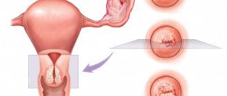
PHOTO OF ADHESIONS OF PELVIC ORGANS IN WOMEN
What could be the consequences of untreated adhesions of the ovaries to the uterus and neighboring organs? Most often these are infertility, pain in the lower abdomen and during sexual intercourse, displacement and bending of the uterus, obstruction of the fallopian tubes, ectopic pregnancy, and various menstrual irregularities. Therefore, given the variety of causes that cause pain in the lower abdomen and other manifestations similar to the symptoms of adhesions, you need to consult a good gynecologist! A formal approach to the problem will not give an effective and lasting result.
Detection methods
To effectively treat postoperative adhesions in the abdominal cavity, it is first necessary to accurately determine their position. To do this, the following examinations are prescribed:
- X-ray of the abdominal cavity - almost all places of fusion of connective tissue will be visible in the image;
- gastroscopy and colonoscopy - necessary to detect hidden processes;
- Ultrasound of the abdominal cavity;
- X-ray with a contrast agent to determine intestinal obstruction and the presence of small adhesions along the organ;
- Laparoscopy – at the same time acts as a method for correcting the adhesive process.
CT and MRI with contrast agents are also prescribed to determine all sources of the pathological process.
The essence of the method
The laparoscopy method is a minimally invasive intervention using a special device - a laparoscope. The bell is inserted into the abdominal cavity through a small incision on the front wall of the abdomen. It has a mini video camera at the end, the image from which is sent to the monitor. To carry out manipulations, the doctor uses micro-instruments, which are also inserted through small incisions. All actions are carried out under visual control.
Laparoscopy for infertility is of two types:
- Diagnostic. It is used to identify pathologies that prevent conception.
- Therapeutic. Aimed at restoring reproductive function and eliminating the pathological focus.
Both methods can be used simultaneously, that is, after diagnosis, therapeutic measures are carried out.
Laparoscopy is used in gynecology not only for infertility, but also for the treatment of other diseases.
Treatment methods
If adhesions can be detected immediately after the onset of inflammation, their resolution can be promoted without surgical intervention. If the cause of tissue scarring is eliminated, fresh marks will quickly disappear without having time to turn into tissue requiring surgical excision.
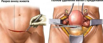
The operation is prescribed when the adhesive process is advanced:
- use a laparotomic or laparoscopic method of operation (large incision in the first case and manipulation through punctures in the second);
- adhesions are excised with an electric knife, pressurized water or a laser.
At the same time, all tumors on the walls of the organs are removed, but this does not guarantee protection from relapses. The more surgical interventions, the higher the risk of developing pathology.
Enzyme treatment
Enzyme therapy is prescribed in the first stages of the pathological process; it will not help with dense connective adhesions. During enzymatic treatment, the patient is prescribed injections of lidase, lipase, ribonuclease and other substances that resolve soft tissue adhesions. Anti-inflammatory ointments are also prescribed.
Massotherapy
If adhesions are detected as compacted areas accessible for deep massage, sessions with a specialist are prescribed. Massage relieves tension, improves tissue condition and blood circulation, and promotes the resorption of adhesions. You can do massage at home, but only some time after the operation.
Prevention
To avoid the development of adhesions, the patient must follow the recommendations that the doctor gave after the operation. A couple of days after the operation, the patient needs to restore motor activity. Even minor movements act like a massage on the internal organs, which prevents them from sticking together. A combination of physical activity and special massage will help avoid the formation of adhesions after surgery. It’s easier to carry out prevention so that you can then wonder how to remove adhesions after surgery.