The most common vision abnormality is myopia or nearsightedness. According to statistics, every third person on the planet suffers from this change, children and adults alike. There are so many people with myopia that it has long been perceived as a variant of the norm, which makes no sense to pay attention to. In fact, even minor pathology affects the quality of life and everyday comfort. Therefore, it is important to know how to improve vision with myopia and help yourself or others with this vision anomaly.
general characteristics
How to treat myopia
Decreased visual acuity can be caused by various refractive errors of the eye. Myopia is one of the most common. Myopia or myopia can be congenital or acquired.
Prevention of myopia
Myopia (myopia) is a disease in which a person has difficulty distinguishing objects located at a far distance. With myopia, the image does not fall on a specific area of the retina, but is located in the plane in front of it. Therefore, it is perceived by us as fuzzy. This happens due to the discrepancy between the strength of the optical system of the eye and its length.
Usually with myopia, the size of the eyeball is increased (axial myopia), although it can also occur as a result of excessive refractive power (refractive myopia). The greater the discrepancy, the greater the myopia.
Myopia (eye myopia) is a vision defect in which refraction, that is, the refractive function of the eye, is impaired. With this disorder, rays of light that come from distant objects are brought into focus not on the retina, as in normal vision, but in front of it.
In this case, only a fuzzy projection reaches the shell that perceives light. Because of this, a person sees a blurry image. With myopia, near vision remains normal, since the rays that come from nearby objects have a divergent direction. And when a person looks into the distance, his eyes catch parallel rays that are incorrectly projected onto the retina.
What is myopia?
Myopia is a pathology in which a person is unable to clearly see objects located at a distance. The patient sees objects nearby well. Most often, the reason for the development of the anomaly lies in a change in the shape of the eyeball. It lengthens and as a result the image is projected not on the retina, but in front of it.
To get a good look at an object, the myope has to bring it as close to his face as possible. The disease develops in one or both eyes at once. The anomaly was first described by Aristotle in the fourth century BC, he noted that some people squint to look at distant objects, and called this phenomenon “myops.” The word translated from ancient Greek means “squint.”
Symptoms and signs of myomia
The main symptoms of myopia are:
- Deterioration of distance vision.
- Blurred outlines of objects.
- The surrounding world literally merges.
- At the same time, good near vision will be maintained.
With myopia, a person has poor distance vision, the image of objects is blurred, but near vision remains good. Patients with a high degree of myopia see well almost right up to the nose, when the further point of clear vision lies not at infinity, but a few centimeters from the eye. You've probably seen people who, having taken off their glasses, literally “rest their nose” on the object they are looking at.
Signs of myopia, when it is combined with various types of astigmatism, are not only the blurring of the image, but also:
- double vision;
- distortion of objects;
- straight lines may appear curved.
If eye myopia begins to develop in adulthood, it can occur without symptoms for a long time, and often a person learns about visual impairment during a preventive examination. In childhood, the first signs of myopia, as a rule, appear with the beginning of educational activities.
The child is less able to distinguish objects in the distance, has difficulty seeing the lines on the board, and complains of a headache. Also, with myopia, weakening of twilight vision is observed. With this condition, a person notices difficulties with orientation in the dark.
According to many ophthalmologists, its long-term presence contributes to the growth of the anteroposterior size of the eyeball and true myopization of the eye.
The most dangerous complication of myopia is retinal detachment and blindness. In order to prevent irreversible damage, it is important to recognize the first prerequisites in time and take appropriate preventive measures.
Glasses and lenses for myopia
To correct myopia, ophthalmologists select optics with concave diverging glasses with a minus sign. They help to “reconfigure” the focus so that the light flows directly onto the retina.
The number written in the prescription next to the minus sign means the optical power of the eyepieces, which is necessary so that the patient can easily see the tenth line on the Golovin, Sivtsev table. For each eye an individual value is indicated.
There is no single answer to the question of whether glasses are needed for myopia. The final decision is made by the myopian, how comfortable he is in the optical product, and whether he can do without correction. However, it is also worth listening to the doctor’s recommendations.
Do not select glasses and lenses yourself, without first consulting an ophthalmologist. It uses eyepieces of different powers to determine visual acuity.
| The duration of use of corrective optics depends on how much the disease progresses. If you manage to stop its development, you can wear optics when absolutely necessary, otherwise you will have to wear them all the time. |
Types and degrees of myopia
Myomia has a different degree of development and expression in each person. Let's look at this in a little more detail, focusing on the nature and degree of development of the disease.
By the nature of development
According to the nature of development, myomia is:
- Progressive. This myopia develops at a rate of 1 or more diopters (D) per year. If the disorders become permanent and there is a significant increase in the degree of myopia, there is a high risk of developing myopic eye disease in a person, which can lead to visual disability.
- Transitional. It is temporary and lasts no more than a month. The appearance of myopia can be influenced by swelling of the lens, due to which its refractive power will be increased. May be caused by diabetes mellitus, the initial stage of cataract development, pregnancy, or taking certain medications;
- Stationary. This myopia does not progress and can be easily corrected by optical means (glasses or lenses).
By degree of development
The degree of development expresses how deep the visual impairment has become. We can highlight:
- weak myopia - in this case, the refractive error does not exceed 3 diopters. Such myopia can be called not a disease, but a feature of vision. As a rule, mild myopia does not require correction and can be completely eliminated by strengthening the eye muscles by performing visual exercises;
- average myopia - the disorder is within 3-6 diopters. Signs of myopia are revealed during examination by an ophthalmologist. These may be changes in the fundus, macula, narrowing of retinal vessels;
- high myopia - refractive error exceeds 6 diopters. A high degree of myopia allows a person to clearly see only those objects that are in close proximity. At this degree, the eyes are under constant tension, which causes visual discomfort and headaches.
Is myopia a minus or a plus?
Optical correction products purchased to eliminate myopia must have a “minus” rating. Glasses are made from curved eyepieces; lenses must be selected individually based on the structural features of the eye.
| As a person gets older, the anomaly most often progresses and can go from a weak to a strong degree in a few years. Therefore, it is extremely important to see your eye doctor regularly, as your optics may need to be replaced at any time. |
The use of corrective products in children significantly improves or even completely restores visual acuity. Elderly people buy glasses or lenses for temporary wear so as not to feel discomfort in everyday life.
False myopia
Myopia can be true, when the eye itself increases in size, and false, called a spasm of accommodation. The spasm does not require wearing glasses or contact lenses and can be treated with medication or exercise.
False myopia (spasm of accommodation) is caused by overstrain of the muscle that controls the lens of the eye. When a person looks into the distance, this muscle is relaxed and the lens is flat, allowing light to focus on the retina.
When we look at objects or work close up, the ciliary muscle is in constant tension and forces the lens to take a convex shape. This is necessary for good near vision.
Spasm of accommodation
A spasm of accommodation caused by prolonged work at close range does not allow the ciliary muscle to relax when looking into the distance. The optics of the eye remain tense for near. Constant tension of the ciliary muscle when working at close range leads to its spasm, and false myopia occurs.
In order for a person with an accommodation spasm to see well into the distance, he needs to have a minus lens placed on him, as with myopia. This is where the term came from - false myopia, which is not related to the size of the eye or the strength of its optics, but is caused precisely by a spasm of the ciliary muscle.
Treatment of myopia
Glasses and contact lenses
Myopia can usually be corrected with glasses or contact lenses. They cancel out the increased curvature of the cornea or the increased length of the eye so that distant objects no longer appear blurry.
Laser treatment
Laser surgery involves reshaping the cornea so that the eye can focus correctly. It is usually suitable for treating mild myopia.
Most people pay for laser treatment privately.
Current evidence suggests that laser surgery for myopia correction is safe and effective for use in patients who have been appropriately selected.
However, as with any surgery, there are possible complications that you need to be aware of (see "complications" section below). The two available procedures are described below.
Laser in situ keratomileusis (LASIK)
A small flap is made in the cornea and a tiny piece of tissue is removed using a laser. This is usually an outpatient procedure that takes about 15 minutes per eye. Vision may be foggy or blurry for several days after surgery.
Photorefractive keratectomy (PRK)
A laser is used to remove tissue from the cornea and change its shape. Treatment may be painful for 48 hours after. It can only be used to treat less severe myopia.
Lens implant surgery
In lens implant surgery, contact lenses are implanted into the eye to correct severe myopia (high myopia). It is sometimes called intraocular lens insertion or corneal implant placement.
There are two main ways to perform lens implantation surgery:
- Placement of a phakic implant —a contact lens is permanently inserted into the eye without removing the natural lens (“phakic” means that the eye contains a natural lens)
- Replacing a natural lens with an artificial one , sometimes called refractive lens replacement.
Inserting a phakic lens implant
This procedure is usually performed for people aged 25-45 years for whom laser surgery is not suitable, or for people who have difficulty wearing glasses, for example due to a disability or occupational requirement.
This is the preferred method for younger patients (under 45 years of age) because it preserves natural vision when reading without assistance.
Causes of myopia
Myopia is very common. According to statistics, more than 1 billion people on the planet suffer from myopia. The most common causes of myopia are:
- Irregular shape of the eyeball - when the length of the anteroposterior axis of the eye is greater than normal, and light rays, when focused, simply do not reach the retina. With an elongated shape of the eyeball, the posterior wall of the eye is stretched, and this state of the visual system can provoke changes in the fundus of the eye.
- Too strong refraction of light rays by the optical system of the eye (lens, cornea). In this case, the size of the eye corresponds to the norm, but due to strong refraction by the optical apparatus, light rays converge into focus in front of the retina, and not on it.
In addition to the causes of myopia, ophthalmologists also identify factors that can influence the development of this disease.
- Hereditary factor. According to experts, it is not poor vision that is inherited, but a physiological predisposition to it. Those at risk are those who have both parents suffering from this disease. The presence of myopia in only one of the parents reduces the likelihood of myopia by 30%.
- Weakening of the scleral tissue leads to an increase in the size of the eyeball under the influence of high intraocular pressure (18–24 mmHg) and, as a result, contributes to the development of myopia.
- Primary weakness of accommodation, leading to compensatory stretching of the eyeball.
Weakening of the body as a result of poor nutrition, overwork, and a number of diseases. Such as:
- violation of the musculoskeletal system: flat feet, scoliosis, etc.;
- allergic and infectious diseases: measles, scarlet fever, diphtheria;
- tuberculosis, infectious hepatitis, etc.;
- birth injuries;
- brain injuries;
- diseases of the nasopharynx and oral cavity: tonsillitis, sinusitis, adenoids; rickets;
- general decrease in immunity.
Unfavorable visual working conditions:
- Excessive strain on the eyes, eye strain;
- Reading in a moving vehicle, in the dark, while lying down;
- Sitting for many hours at the computer, TV;
- Insufficient lighting;
- Incorrect sitting position while reading and writing.
Causes
There are several factors that can lead to the development of myopia:
- Genetic predisposition. One of the main reasons why myopia occurs in children. If mom or dad suffered from pathology, then the baby automatically falls into the risk group,
- Enormous loads on the visual apparatus of students also negatively affect eye health. For this reason, the disease is most often activated in patients aged seven to twenty years. This period is when the workload is at its maximum, as children are forced to spend a lot of time doing homework. Vision may decrease to a certain level and no longer fall or progress rapidly,
- Incorrectly selected products for correcting refractive errors can further aggravate the situation. It is important that glasses or lenses are selected by an experienced ophthalmologist, based on the individual structural features of your eye. In addition, do not forget about the rules for wearing and storing optics and regularly check with a doctor,
- If, due to duty, a person is forced to spend a lot of time working with small parts (for example, watchmakers or jewelers), then sooner or later he will face a problem with visual acuity,
- The habit of reading while lying down or in a moving vehicle is something for which parents not in vain scold their children. Since such habits increase the risk of developing myopia,
- The cause of increased myopia may be a lack of vitamins and an unbalanced diet.
| Risk factors also include congenital anomalies (for example, weak extraocular muscles) and a number of ophthalmological diseases (astigmatism). |
Return to contents
Diagnosis of myopia
The diagnosis of myopia is made by an ophthalmologist after checking visual acuity using special tables. The fundus is also examined with a special mirror. Before this procedure, the patient is usually given a medication that dilates the pupils by dropping them into the eyes. These are traditional procedures.
A complete examination includes:
- accurate determination of the patient’s visual acuity and refraction,
- measurement of intraocular pressure,
- conducting an eye examination under a microscope (biomicroscopy),
- pachymetry (measurement of corneal thickness),
- echobiometry (determination of eye length),
- ultrasound examination of the eye,
- computer keratotopography and a thorough examination of the retina (fundus of the eye) with a wide pupil,
- determining the level of tear production,
- detailed examination of the patient's visual field.
The main vision problems in children develop before the age of 7 years. After 7 years, the child’s visual system begins to experience increased stress associated with studying at school. It is during this period that diseases that were not noticed or prevented before begin to develop.
After 12 years of age, vision problems usually cannot be treated. It will be possible to eliminate myopia and astigmatism in a teenager only after the age of 18, when the young person can undergo laser vision correction.
But a correct and timely forecast of vision development in some cases makes it possible not to put glasses on a child at all. In order to correct his vision, it will be enough for him to undergo a therapeutic course of treatment. For an accurate diagnosis of vision, you should contact the children's departments of ophthalmology clinics, and only those that have special diagnostic equipment.
After a thorough and complete examination, the doctor conducts a conversation with the patient and, based on all the data, prescribes treatment.
Myopia in children
Myopia is a “childhood” disease. It usually occurs between the ages of 7 and 10, with the second peak occurring between 12 and 15, the time of puberty. In adults, manifestation occurs less frequently. Sometimes myopia occurs already in infancy.
Until a certain age, radical vision correction is undesirable. Therefore, it is important, on the one hand, to detect the disease in time, and on the other, to take action. These include:
- Selecting glasses with the necessary diopters that will help reduce the load on the visual organs.
- Physiotherapy - laser massage, electrical stimulation, and other procedures that relieve spasms and help the normal development of the visual system.
- Drops for myopia - prescribed to children with caution. Proven means are recommended: Ujala - from 6-7 years, Visioned - from 3 years. Drops are not recommended for infants.
- Correction of the load - it is advisable to exempt the child from physical education, ask to sit on the first desk, and not allow him to use the computer and smartphone for a long time.
Childhood is the best age to prevent the development of myopia. Even if minor changes persist, they will not cause serious discomfort and will not affect the person’s quality of future life.
How to treat myopia
There is no universal cure for myopia, since some people inherited the disease, while others developed it at a certain stage of life. Measures aimed at getting rid of hereditary myopathy do not give the desired results in all cases.
As for the methods of treating myopia in people of different ages, they may differ slightly.
It is better to start treating myopia in children in the early stages. Inaction can lead to progression of the disease. As a rule, these are procedures that relieve excess tension from the eyes and improve blood circulation in the tissues of the eye. If everything is done in a timely manner, a positive result will be visible after 4-6 months.
Treatment of myopia in children with surgery is contraindicated. If all the treatment methods undertaken do not lead to a stable result, then you will have to wait until the child reaches 18 years of age to have the operation performed. But until this moment comes, you should not stop correcting your vision using approved methods.
To determine the treatment method for myopathy in adults, you must first identify the cause of myopia. This may be damage to the nerve that is responsible for oculomotor function, alcohol abuse, nicotine intoxication, various diseases of other internal organs, insufficient or improper lighting of the workplace, etc.
If the cause is found, then, of course, it must be eliminated and, at the same time, treated for myopia.
Many people find that it is easier to purchase glasses or contacts, and the problem with myopathy is solved. It should be remembered that these devices only help a person see what was previously blurred before the eyes, but they do not relieve myopia.
Treatment for this disease usually takes a long time. All of its methods are aimed at slowing or stopping the development of myopathy and preventing the development of various kinds of complications.
The most effective methods are:
- Special gymnastics for the eyes.
- Treatment with medications.
- Physiotherapeutic procedures.
- Surgery.
Special gymnastics for the eyes
There are many different programs aimed at improving accommodation (the ability to adjust to different distances) of the eyes. These are exercises for focusing the gaze and training its range, strengthening the eye muscles, and relaxing the visual organs after heavy loads.
The main requirement for these exercises is their regular repetition with the same frequency and duration. For example, every 4 hours or daily in the evenings for 15 minutes.
These days, there are even sets of eye exercises, presented in the form of computer programs and applications for Android and IOS.
Gymnastics for the eyes
Eye gymnastics can guarantee complete relief from myopia only at the initial stage of this disease. In other cases, it should be used as an auxiliary method for drug and (or) physiotherapeutic treatment.
Simple exercises for myopia
- You need to look down and fixate, then raise your eyes up. Stop for a few seconds.
- Raise your eyes up, look at the bridge of your nose, move your gaze to the tip of your nose.
- Draw a diamond shape with your eyes. At the same time, stop at each point for 2 seconds so that your gaze focuses.
- Look out the window and find an object in the distance that you like. Turn your gaze around the room to an item you like.
Do the exercise for 1 minute.
A set of exercises to improve vision
- You need to sit comfortably with your eyes closed. Open them and blink quickly for 10 seconds. Relax with your eyes closed. Do at least 3 times.
- Leave your eyes closed for 5-7 seconds. Open for a few seconds, as wide as possible. You can't blink.
- Using three fingers, starting from the little finger, lightly press on the upper part of the eyes, which should be closed.
- The skin near the eyebrows is gently lifted with your fingers. And at this moment you need to try to close your eyes.
- The fingers are placed at 3 points of the eye: inside (near the bridge of the nose), in the middle (above the upper eyelid) and in the corner. You need to gently stretch the skin with your fingers for a few seconds.
- The following exercise is performed on a chair. The head is straight and does not move. You need to move your eyes from the object on the ceiling to the objects on the floor.
- Run the index finger of your right hand at eye level from right to left. Repeat the same movement with your left hand. The head remains motionless.
Chinese gymnastics for myopia
The ancient method of such gymnastics is based on only 4 exercises that are done at certain points:
- The Qing Ming point is located at the intersection of the eyebrows. Massage the point with your fingers 35 times.
- The Syb-ai point is located in the middle of the cheeks. She is stimulated 35 times in a circular motion. Use index fingers for massage.
- The Tai Yang point is located between the edge of the eyebrow and the eye. Gently press the point 36 times with your fingers.
- The next exercise is a smooth transition with massage from the upper eyelid to the lower.
A set of effective exercises to restore vision
- Blink your eyes quickly for 60 seconds. Rest and repeat the exercise.
- Take a comfortable position, close your eyes tightly and count to 10. Eyes wide open while counting to 10. You can repeat this exercise 4-6 times.
- Stand up, extend your arm forward. Rest your gaze on your fingertips until they begin to double. Rest your eyes for 10 seconds. Do the exercise 6 times.
- Imagine drawing circles with your eyes clockwise and counterclockwise.
- Enjoy the energy of the sun. On a clear day, go to the window and expose your face to the sun. Stand in this position for 10 minutes.
- You need to sit straight in a chair. Look at the ceiling for 30 seconds. Then lower your head and look at your knees for 30 seconds.
- You need to attach a circle the size of a match head to the glass. Move your gaze from the circle to the object you like outside the window. It should be in line with the circle. This exercise is done 2 times a day for a month. You need to start with 3 minutes. By the end of the month the time reaches 8 minutes.
Treatment with medications
There are several drug treatments for myopia.
- The action of some agents is aimed at strengthening the sclera (ascorbic acid, calcium gluconate).
- Others - to accelerate metabolism in the eyeball and retina (taufon solution, aloe, injection of ATP solution).
- Third - to relieve spasm of accommodation, which is caused by excessive stress on the eyes (1% mesatone solution).
- Fourth - to improve blood circulation in the eyeball (trentap, nicotinic acid).
The best results with drug treatment can be achieved if it is carried out in courses every six months.
Physiotherapy
Physiotherapeutic methods for treating myopathy include: phonophoresis, electrophoresis, electrical or laser stimulation of the ciliary muscle, as well as glasses that massage the eyes.
The duration of various types of influences in different cases ranges from 7 to 15 minutes. The course of treatment is about 10 procedures.
For progressive myopia, physiotherapy includes instillation of sermion, 4% taufon, vitreous or aloe and exposure to PeMP LF-10 mT, continuous mode, duration of exposure 7-8 minutes; There are 10 procedures per course of treatment. Tissue preparations should not be prescribed during puberty.
At the same time, endonasal electrophoresis of no-shpa, calcium or 0.5% proserin solution can be performed. Current strength is 0.5-1.5 mA, duration of procedures is 15-20 minutes; for a course of treatment 10-15 procedures.
For chorioretinal changes, physical therapy in the form of endonasal electrophoresis of riboflavin mononucleotide, trental is recommended. The effects of SMV with a power of 2 W are carried out by placing the emitter on closed eyelids, the duration of the procedure is 10-15 minutes; for a course of treatment up to 20 procedures.
In case of hemorrhages in the fundus of the eye, endonasal electrophoresis of fibrinolysin or chymotrypsin is performed in the early stages for resorption (method No. 10). In the later stages (2-3 weeks after hemorrhage), especially with the development of scarring, endonasal electrophoresis of lecozyme and lidase is most effective.
For resorption purposes, to the physiotherapy described above, you can also add electrophoresis of iodine, steroids, and biogenic stimulants (aloe and vitreous).
Surgery
Surgical intervention is used in cases of severe myopia (from 6 diopters). The result of using surgical methods is a guaranteed prevention of disease progression and correction of refraction.
Operations used to treat myopia are divided into:
- Refractive, aimed at correcting the optical ability of the eye.
- Sclero-strengthening – when a substance is injected into the back of the eyeball that stops or slows down the process of eye expansion.
Most often, surgeons perform operations on lens extraction, keratotomy, keratophakia, and keratomileusis. Some clinics have begun to introduce more modern methods of surgical treatment, as a result of which the myopathy indicator is reduced by 12 diopters.
Classification of myopia
Most often, the cause of the anomaly is dysfunction of the eyeball and other elements of the eye involved in the refraction of light fluxes. Depending on which structure is damaged, myopia is divided into the following types:
- Axial. Diagnosed by elongation of the anteroposterior part of the eyeball. At the same time, the refractive systems work properly,
- Lenticular. It appears when the refractive power of the lens increases. This phenomenon is often observed in diseases such as diabetes. Some medications can also provoke deviations,
- Damage to the cornea. The reason for the anomaly lies in the increase in the degree of curvature of the element and the excessive expression of its refractive power.
True myopia
This includes the pathological condition of the patient, in which damage to the eyeball, lens, and cornea is observed. The anomaly may be congenital or acquired. If you ignore the pathology and do not start treatment in a timely manner, the risk of complications increases.
False myopia (spasm of the ciliary muscles)
Accommodation is responsible for the ability to clearly view objects located at different distances from a person. False myopia can occur as a result of myopia spasm, which causes excessive eye strain. Usually manifests itself in children, in rare cases the disease affects an adult.
When viewing an object located nearby, the ciliary muscles contract and the refractive power of the lens increases. If accommodation is strained for several hours, the metabolic process and nervous regulation are disrupted, which leads to spasm.
When trying to see an object at a distance, the spasmed muscles cannot relax, the refractive power of the lens does not decrease, so the object takes on a blurry outline. This phenomenon is called a spasm of accommodation. The following factors can cause an abnormal condition:
- Spending long periods of time at the computer without breaks,
- Working on a PC in poor lighting,
- Non-compliance with sleep and rest schedules,
- Unbalanced diet
- Lack of sleep,
- Reading or watching television for a long time.
| Since muscle spasm is a temporary phenomenon that goes away without complications after eliminating the abnormal condition, such a disease is called false myopia. |
At the same time, no destructive processes are observed in the organ of vision. However, if the spasm is not eliminated or a similar phenomenon occurs regularly, the disease can develop into true myopia.
Hereditary myopia
Numerous studies have confirmed that the disease can be transmitted along a genetic line. The human body has twenty-three chromosomes located in cell nuclei. In addition, a huge number of different genes are isolated; it is the activation of one or another gene that affects the functioning of the entire organism or its individual structures.
At conception, male and female cells unite, resulting in the formation of an embryo that inherits twenty-three chromosomes from the mother and the same number from the father. If the resulting “elements” have defects, the risk of developing pathology increases.
Weak and moderate myopia is inherited in an autosomal dominant manner, i.e. If the baby gets at least one “spoiled” gene, he will develop one or another disease. The risk of increased myopia depends on which parent is sick. If the mother and father suffered from refractive problems, then the probability of a new myope being born is from 75 to 100%. If only one parent has the disease, the risk will be from 50 to 100%.
High myopia is inherited in an autosomal recessive manner. Thus, if a mother or father is sick, their baby will be healthy, but if he inherits one defective gene, he will become an asymptomatic carrier of the anomaly. If both parents suffer from the disease, then with a 100% probability their child will have vision problems.
Acquired myopia
The development of this pathology is caused by unfavorable environmental factors that affect the eyes:
- Poor visual hygiene. When reading or working at a PC, tension in the ciliary muscle occurs; if this condition persists for a long time, muscle hypertrophy is observed. The process of changes in the accommodation system can last for several years, but if it occurs, then disturbances occur in the relaxation mechanism. As a result, the lens capsule is constantly relaxed, and the element itself does not thicken to the required extent for proper focusing,
- Working in unfavorable conditions,
- Lack of vitamins. Lack of nutrients, especially riboflavin, which is responsible for many functions in the visual apparatus,
- Primary weakness of the ciliary muscles. This anomaly hides a condition in which insufficient refractive power of the lens is diagnosed. Light rays passing through the eyeball do not fall on the retina, but behind it,
- Damage to the organ of vision.
Return to contents
Night myopia
This condition can hardly be called an anomaly, since it often occurs in healthy people. The development of night myopia is due to the fact that in low light conditions the pupil dilates and accommodation contracts, causing an increase in the refractive power of the lens. All these processes lead to the fact that the objects in question, located at a distance, are focused in front of the retina.
It is believed that such a reaction of the body allows one to increase visual acuity in the dark, since when the pupil dilates, more photons reach the membrane, and slight myopia forces one to look at objects at a closer distance.
In the daytime and in good lighting, the unpleasant symptoms disappear completely.
Prevention of myopia
In fact, there is nothing difficult in observing preventive measures for such vision pathology. It is necessary to monitor your eye health from an early age. For example, school-age children need to avoid visual fatigue, especially while reading, watching TV or playing on the computer.
Prevention of myopia
Every forty minutes you need to give your eyes a little rest to prevent the development of myopia. In addition, you should not read while lying down or sitting in transport. This is due to the fact that when reading in such conditions, the distance to the eyes constantly changes, that is, the lens bends. As a result, the visual organs become very tired, and myopia progresses.
General measures to prevent such pathology include proper lighting, good ventilation, adherence to sleep and rest patterns, and frequent walks in the fresh air. When exposed to direct sunlight, you must wear protective glasses. Do not forget also to comply with generally accepted hygiene standards: do not use other people's towels, dirty handkerchiefs, and do not rub your eyes with unwashed hands.
Proper nutrition as the main method of preventing myopia
Undoubtedly, a balanced diet has a positive effect on the condition of the entire body, including the visual organs. Therefore, in order to prevent any eye diseases, your diet should contain foods high in vitamins A, B1 and B12 and also microelements - potassium, calcium, silicon.
Try to eat vegetables and fruits, avoid processed foods. Eat as much fish and greens as possible. Pay special attention to fresh lettuce leaves, which contain minerals that are beneficial for vision.
Exercises to prevent myopia
Therapeutic exercises for the eyes are the simplest and most effective method of combating various pathologies of the visual organs. Moreover, the exercises can be performed by people with 100% vision, and those who already wear corrective glasses.
Such prevention is also indicated for children. By devoting some time to special eye exercises, you will improve your vision and prevent the development of many diseases of the visual organs.
Such exercises include rotational movements performed clockwise and back, looking at objects located near and far, squeezing and frequently unclenching the eyelids.
Prevention of myopia in the ophthalmology center
If you experience minor complications in your vision, you should seek help from a treatment center. In this place you can consult a doctor, carry out effective prevention and treatment of the disease. For example, a separate room is set up for reading, where a special font is read at a maximum distance from the eyes.
They spend a few minutes doing therapeutic exercises and carry out procedures using massagers and light radiation. If you complete a whole course of such training, the prevention of myopia will be successful.
Glasses and lenses for myopia
Wearing special glasses will allow you to avoid overstraining your visual organs, so your vision will improve significantly. Lenses to prevent myopia are worn during the day and removed at night. Glasses for vision correction are prescribed by a doctor.
Now you know what methods will help prevent the development of myopia and cure the disease at an early stage, without surgical intervention.
Kinds
There are types of the disease that help diagnose it and determine the treatment strategy for myopia. They are determined depending on the time of development and other features. The following forms are distinguished:
- Congenital - detected from the first months of life, completely determined genetically.
- Combinatorial - this form of myopia in children also occurs quite often; it is not very pronounced, but is associated with the structural features of the eye, and therefore can only be corrected surgically. Most often, ophthalmologists consider surgery inappropriate.
- Spasmodic - also false, it affects schoolchildren, students, and office workers. Spasm of accommodation associated with visual fatigue, however, progressing to more severe types.
- Transient - associated with other diseases, usually chronic, such as diabetes. This type of myopia can be cured if you focus your efforts on the provoking factor.
- Night - associated with emmetropic refraction of the eye, manifests itself only in low light conditions.
- Complicated - a rare but dangerous form associated with serious pathological changes in the eye. The only type of myopia that can lead to complete loss of vision without proper medical intervention.
Diagnosing and determining the type of myopia not only increases the chances of curing myopia at an early stage, but also preventing the development of severe forms of the disease. Despite the fact that 90% of people suffering from this eye feature live with it all their lives, getting used to it over time and not paying attention, a primary diagnosis must be made by an ophthalmologist in order to exclude complicated and destructive types of the disease.
Treatment with folk remedies
- Take three parts each of black currant berries, carrots and rose hips, add one part of stinging nettle leaves. Pour two tablespoons of the vegetable mixture into four hundred ml of hot water and bring to a boil, reduce the heat and turn off after fifteen minutes.
- Take three parts of stinging nettle and one part of rowan leaves along with the fruits. Brew one tablespoon of the mixture with two glasses of hot water, bring to a boil, reduce heat to low and simmer for fifteen minutes. After two hours, strain. Take half a glass warm four times a day before meals. You can add a little honey or sugar as a flavoring agent.
- Make juice from fresh calamus root. Take one teaspoon three times a day for three months.
Also, do not forget that before using traditional methods of treating myopia in practice, you must consult an ophthalmologist.
Complications of myopia
As the disease progresses, the risk of unpleasant consequences increases. Complications are most often encountered by myopes with severe disease:
- Detachment of the retina. The most dangerous consequence of myopia. The first symptom is the formation of a foggy curtain before the eyes. A person sees practically nothing due to the veil that has arisen. The anomaly does not cause pain, appears suddenly,
- Pathological processes in the visual apparatus. For example, clouding of the lens,
- Formation of Fuchs spots. They are formed by hemorrhage into the macula. Such a symptom signals a rupture in the vascular system; newly formed branches that have an irregular structure are usually damaged. The cells responsible for light sensitivity are destroyed,
- Dystrophic processes in the retina, for example, lattice dystrophy.
To avoid complications, it is important to seek medical help when the first signs of the disease appear.
Forecast
The leading role in the development of myopia is played by hygiene in education in children and hygiene in adults. Maintaining visual hygiene in combination with a proper diet and taking vitamins will significantly reduce the risks.
Preventive examinations are also important in maintaining healthy vision. They should not be ignored or neglected by the ophthalmologist’s recommendations.
The prognosis for the immediate treatment of myopia of all degrees is comforting. Patients' vision is restored and preserved. In the treatment of progressive myopia there is a risk of difficulties and complications. Therefore, ophthalmologists strongly recommend compliance with conservation, prevention and treatment measures.
Surgical treatment of myopia
It may be required if the disease progresses rapidly - more than 1 diopter per year - and there is no effect from other treatment methods. Surgical treatment is aimed at strengthening the posterior wall of the eyeball. This prevents its stretching and the occurrence of related complications - retinal detachment, thinning of the choroid.
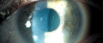
In severe cases of myopia, lens replacement surgery may be prescribed. It is performed when myopia is no more than −20 diopters. Also, the lens is replaced when it becomes cloudy, that is, with cataracts. The surgeon removes the natural lens and installs an artificial (intraocular) lens in its place. This operation is performed on only one eye. The second can be operated on no earlier than six months later, when the eye tissues have finally healed and the person has adapted to the IOL.
Degenerative myopia
In most cases, nearsightedness is only a minor inconvenience and poses little or no risk to eye health. But sometimes myopia can be so progressive and severe that it is considered a degenerative condition.
Degenerative myopia (also called malignant or pathological myopia) is a relatively rare condition that is considered hereditary and usually begins in early childhood.
In malignant myopia, elongation of the eyeball can occur rapidly, leading to rapid and severe progression of myopia and vision loss. People with the condition have a significantly increased risk of retinal detachment and other degenerative changes in the back of the eye, including bleeding in the eyes from abnormal growth of blood vessels (neovascularization).
Degenerative myopia can also increase the risk of cataracts.
Types of myopia (myopia)
The most common types of myopia include the following:
- Congenital (pathology from birth associated with an abnormal development of the eyeball; quite rare) / Acquired (myopia developed during life under the influence of the factors listed above);
- Stationary (vision remains stable, no deterioration occurs) / Progressive (vision deteriorates over time; very often myopia progresses in childhood and adolescence due to the child’s growth);
- Weak (up to 3.0 D (diopters) inclusive) / Medium (from 3.25 to 6.0 D) / High (more than 6 D, and can reach very significant values up to 30 D);
- Night blindness (occurs when there is insufficient lighting, popularly called “night blindness”);
- Professional (occurs due to frequent, prolonged visual strain when examining objects at close range);
- School myopia (occurs in students due to prolonged visual strain at close range, is a subtype of professional myopia);
- False (occurs with an increase in the tone of the ciliary muscle - spasm of accommodation - and disappears with its normalization);
- Complicated (with myopia, the eyeball can lengthen, which leads to stretching of the inner membranes of the eye, disruption of the nutrition of its tissues and a negative effect on the retina. The progression of myopia can lead to dystrophic changes in the fundus, retinal tears and detachment. Therefore, people suffering from myopia should It is recommended to undergo a fundus examination at least once a year by an ophthalmologist to prevent retinal detachment, which can lead to blindness);
In addition, there is a distinction between progressive myopia - a condition in which the degree of myopia increases by one or more diopters per year - is considered progressive myopia.
Top
Prevention
To prevent vision defects, it is necessary, first of all, to comply with the requirements of visual hygiene: avoid eye strain when working at a computer, reading in transport or lying down, and ensure that the lighting is correct.
Of no small importance in overall eye health is a healthy diet, with the presence of natural vitamins necessary for metabolic processes. And preventive examinations by an ophthalmologist can help detect any abnormalities at the earliest stages.
Differences between myopia and farsightedness
Many people are interested in the question of myopia and farsightedness - what is it and what is the difference between these diseases. Myopia is characterized by the fact that a person sees poorly in the distance, objects and contours are unclear. With farsightedness, everything happens exactly the opposite: distant pictures are clearly visible, but near objects are not clearly visible.
Myopia usually develops at school age. The main cause of myopia is a genetic predisposition, while farsightedness is a disease that is caused by age-related changes; over the years the lens becomes less mobile. This causes vision problems to arise.
Stages of myopia
The stages of myopia development represent several degrees of vision deviations from the norm. They are determined by the distance the focus is removed from the retina. There are such degrees of deviation:
1st. Low myopia is a violation of visual focus by 3 diopters. The length of the eye increases by 1.5 mm. No significant changes in the image are detected. It may only appear that the pictures are blurry when looking into the distance.
2nd. The focus distance range in this case is between 3 and 6 diopters. At the same time, the eyeball increases by 3 mm. The vessels in it stretch and become thin. The image becomes blurry from a distance of 30 cm.
3rd. The distance between the retina and the focus becomes more than 6 diopters. Through the greatly thinned retina and blood vessels, the sclera of the eye can be seen. In this case, the patient sees his fingers no closer than at arm's length.
Stretching of blood vessels during myopia can lead to complete loss of vision. Sometimes myopia occurs as follows:
Other ways to restore vision for myopia
For mild myopia, hardware treatment methods can be prescribed. With their help, the development of the disease is stopped. Special eye exercises help restore vision after visual stress. A lot of them have been developed. They are also used as prophylaxis.
Before choosing a specific set of exercises for yourself or your child, you should consult with an ophthalmologist. Vitamins and minerals help improve visual function. Eye drops relieve symptoms of fatigue after prolonged visual work. Also, these drugs can be used to eliminate spasm of accommodation.
Is it possible to cure myopia?
Myopia is treated with surgical and laser methods. Laser operations are performed for any degree of refractive error. They are the most effective because they help get rid of myopia forever or for many years. Several laser techniques have been developed today: PRK, LASEK, LASIK. There are many modifications of these methods: SuperLASIK, FemtoLASIK, FemtoSuperLASIK, EpiLASIK. Each of them has its own characteristics. A particular procedure is prescribed based on the medical indications and financial capabilities of the patient.
Before laser surgery, the patient undergoes a thorough examination, during which contraindications to the procedure are excluded. After this, the preparation stage begins. It does not last very long - 1-2 weeks. During this period, the patient should not wear glasses, contact lenses, including orthokeratological and colored ones. During this time, the cornea will take its natural shape. Drinking alcohol is prohibited 2 days before surgery.
Laser correction is performed on an outpatient basis. Its duration is 20-30 minutes, depending on the chosen technique. During the procedure, the surgeon exposes the inner layers of the cornea by excision of its upper epithelial layer and evaporates the eye tissue with a laser beam, giving it the desired shape. Vision is restored quickly after laser correction. So, after LASIK a person sees well within a few hours. Complete healing of the cornea will occur in 2-4 weeks, during which it is necessary to strictly follow all the doctor’s instructions: instill eye drops, do not lift heavy objects, do not load your eyes with reading, do not visit baths and swimming pools.
Eye examination methods for myopia
If you suspect myopia, you can undergo a duochrome test. It is a two-color table - a poster divided into two halves: green and red. There are also letters in the table. They must be read first with one eye and then with the other eye. The test is loaded directly on the computer. You must position yourself at a distance of one meter from the monitor. While reading optotypes, write them down so that you can check that they are reproduced correctly later.
The test results are deciphered very simply: if you can clearly see the letters on a red background, but did not correctly name all the optotypes located on the green half, most likely you are developing myopia. This test is not objective, and therefore cannot provide maximum accuracy, but if the results are unfavorable, it should be a reason to visit the clinic.
If myopia is suspected, the doctor checks visual acuity using the standard Sivtsev table. To diagnose the disease in preschool children who may not yet know the alphabet, the Orlova table is used. In it, images of animals, geometric shapes and other symbols understandable to a child act as optotypes.
Once myopia has been identified, it is necessary to find out its degree. This can be determined using the same table that was used to test visual acuity. The ophthalmologist gives the patient special glasses with replaceable lenses. He reads optotypes with these glasses. The lenses are changed until the subject can correctly name all the letters of the line shown by the doctor. The optical power of the lenses determines the degree of refractive error. For example, if lenses with an index of −4 diopters are required to accurately reproduce all the letters in the table, the patient is diagnosed with second degree myopia.
These research methods are subjective. Today, other methods of studying the organs of vision are used, which help to identify not only the indicator of visual acuity, the degree, but also the causes of myopia, the rate of progression, the form of pathology, and so on. Let's look at these methods in more detail.
Treatment
To determine the degree of myopia, the most traditional test is used - visometry. The patient, at a distance of 5 m from a table with letters of various sizes, names them according to the doctor’s instructions, and the doctor, using lenses with different D values, determines the degree of myopia.
Carrying out visometry
The following methods are used as additional methods to obtain more complete results:
- Ophthalmoscopy;
- Biomicroscopy;
- Refraction test;
- Ultrasound examination.
Drug therapy
Conservative methods are effective in treating myopia only in combination and may include the use of:
- Medications. These are mainly means for relieving spasms of accommodation and restorative drugs;
- Vitamins: B, C, A, PP, as well as microelements: zinc, manganese, magnesium;
- Physiotherapy;
- Gymnastic complexes for the eye muscles.
Complex treatment is recommended for all patients with varying degrees of myopia 1-2 times a year in order to prevent the progression of myopia.
Correction methods:
- Spectacle correction is one of the most common methods in the treatment of the disease, which has undoubted advantages: accessibility and ease of use. For young children, glasses are always prescribed as the safest remedy;
- A variation of the method is contact lenses, which can be worn by the older generation of children who understand the responsibility when using them. Contact lenses come in a wide range of quality and design, but they are also not suitable for everyone, as they can cause persistent discomfort. And this is fraught with the development of damage to the eye structures;
- Orthokeratology is a technique of using special lenses when the patient wears them only at night. During sleep, the lenses act on the cornea, flattening it, and during the day its shape is maintained, ensuring the quality of visual perception.
Orthokeratology lenses
When eliminating vision defects in childhood, techniques using special equipment can be used - the so-called hardware treatment.
Surgically
For progressive severe myopia that cannot be treated with other methods, the doctor may recommend one of the types of surgical intervention. For children, such operations are allowed only from the age of 18, since the formation of the visual system has not been completed before.
To correct myopia, the following types of surgery are performed:
- Laser correction – is carried out using a laser beam, which corrects the degree of curvature of the corneal layer to the required level;
- Implantation of phakic lenses - to correct the curvature of the cornea, a specially selected lens with the necessary properties is placed into the space of the anterior (or posterior) chamber of the eye;
- Lensectomy (refractive lens replacement) - during the operation, the lens is replaced with an intraocular lens. This method is used most often for elderly patients, when the function of accommodation is significantly reduced or lost altogether;
- Scleroplasty – used for progressive myopia (more than 1 D per year). A special plate is inserted inside the eye to prevent retinal tears as the size of the eye rapidly increases.
Carrying out laser correction
Folk remedies
The use of folk remedies can be used as an additional therapy in the treatment of myopia:
- Take 2 tbsp. l. finely chopped eyebright herb and pour 0.5 liters of boiling water over them, then infuse for 20 minutes. The strained infusion is consumed 50 ml shortly before meals three times a day;
- Spruce or pine needles are finely chopped, and 5 tbsp. l. raw materials are poured with 600 ml of boiling water, then simmered in a water bath for 6 hours. Add 1 tbsp to the resulting strained infusion. l. honey and take 1 tbsp. l. three times a day after meals. The course of treatment is 3 months. ;
- At 10 tsp. warm boiled water diluted with 0.5 tsp. honey Place 2 drops in each eye three times a day. You can store the solution for no more than 2 days in the refrigerator, warm it up slightly before use;
- Plantain infusion (2 tsp of dry raw material per 250 ml of water) is prepared over low heat for 30 minutes. The strained solution is taken 100 ml three times a day before meals.
The use of folk remedies can be effective with long-term use, but before using them, consultation with a doctor is required, since they may have a negative effect in the presence of certain diseases.
Possible complications
The progression of myopia, caused by age-related changes and increased loads during this period, most often stops upon reaching 20 years (for women) and 22 years (for men). The visual defect itself is not dangerous, but with the development of high degrees of myopia, the risk of developing severe complications increases:
- Retinal disinsertion. An increase in the size of the eyeball entails its stretching and the threat of detachment and rupture, which can lead to loss of vision partially or completely;
- Glaucoma. With dystrophic changes in the ocular structures (most often age-related), an increase in IOP is possible, which in most cases leads to damage to the optic nerve and a decrease in visual acuity;
- Retinal dystrophy. It is the result of degenerative processes in the tissues of the eye, changes in the functioning of the circulatory system and disturbances in the nutrition of nerve fibers;
- Cataract. Violation of the passage of light rays through the lens is most often of an age-related nature, but eye strain when trying to look at distant objects plays an important role in disrupting metabolic processes.
The threat of complications can arise at any age, even after surgical correction. Therefore, people even with a mild degree of myopia are recommended to undergo preventive examinations by an ophthalmologist.
Non-surgical treatment
Treatment of myopia without surgery in adults can slow the progression of the disease and suppress its symptoms.
Conservative treatment includes:
- use of medicines;
- physiotherapeutic procedures.
Gymnastics for the eyes
The essence of gymnastics for myopia is correct and clear focusing on objects that are located at a distance from the person.
Examples of exercises:
- Focus on the sticker. Glue a small sticker with a diameter of no more than 5 mm to the window glass. You need to move 50 cm away from the window and look at an object located in the distance (a house, a tree). Then look at the sticker. Repeat the exercise about 20 times.
- You need to slowly “draw” the number 8 in the air with your gaze with maximum amplitude. Repeat 15-20 times.
It is recommended to complete the complex by massaging closed eyes for two minutes.
Eye drops
Treatment of myopia in adults with eye drops allows you to relieve spasm of accommodation, improve the condition of the retina and cell nutrition, as well as eliminate the unpleasant manifestations of the disease (discomfort and pain in the eyes, redness of the mucous membrane).
When consulting patients on how to stop myopia in adults, doctors recommend eye drops from the group of mydriatics - agents that relax the muscles of the visual organs.
How to treat myopia in adults? Drops are used for this purpose:
- Mydriacyl;
- Midroom;
- Cyclomed;
- Irifrin.
Drops containing lutein are also prescribed. This substance affects accommodation, relaxing the lens.
Physiotherapy
For accommodation spasms, the following physiotherapeutic procedures are prescribed:
- electrophoresis with the introduction of nicotinic acid;
- magnetic therapy;
- photochromotherapy;
- laser puncture.
Myopia in an adult can be cured using a comprehensive conservative method. But, if the listed methods do not produce an effect, and the disease progresses, a decision is made to undergo surgery or laser correction. .
What other tests are prescribed for myopia?
There are a lot of modern methods for identifying the causes of myopia and its degree. Sometimes it is necessary to conduct all types of examinations. As mentioned above, with myopia various changes in the fundus and other structures of the eye can occur. In order to exclude the presence of these changes, ophthalmoscopy is performed to examine the fundus.
When examining him, the doctor uses a special device with a mirror, which is placed on the head. He sits opposite the patient, places a magnifying glass in front of his eye and directs the rays of light reflected from the mirror into the patient’s pupil area. This is how the condition of the optic nerve head and the inner wall of the eye fundus are studied. In order for all its structures to be better visible, drops that dilate the pupil - mydriatics - are first instilled into the eyes.
Without such drugs, the pupil will narrow under the influence of a beam of light directed into it. Ophthalmoscopy is not always prescribed. It has contraindications, including glaucoma. With this disease, mydriatics cannot be instilled.
To study the state of refractive structures, that is, the cornea and lens, skiascopy is used. It helps to determine the degree of myopia as accurately as possible. Skiascopy is performed as follows. The ophthalmologist sits one meter away from the patient and directs a beam of light reflected from the mirror into his eye. On the side of it there is a special lamp. The light beam passes through the cornea and lens, is refracted and ends up on the retina.
With myopia, a shadow will fall on it, which moves in the opposite direction of the movement of the light source. Next, the doctor places a skiascopic ruler with lenses of different optical powers between the mirror and the patient’s eye. The lens reading in diopters, at which the shadow disappears from the retina, will reveal the degree of myopia. In addition to these methods, computer methods, for example, keratotopography, can also be prescribed. It makes it possible to measure the curvature of the cornea and its refractive power.
How to stop progressive myopia?
Progressive myopia in adults is corrected by laser therapy. The rays are absolutely safe for the body and the organs of vision in particular. They affect the cornea, changing its shape. As a result, the shell becomes flatter and its optical power decreases. Thanks to this, the image falls directly on the retina, and the patient clearly sees distant objects.
Surgery
If myopia progresses in an adult and has reached a high degree, surgery is performed.
The surgery is called myopic keratomileusis. The essence of the manipulation is to excise the upper layers of the cornea, remove the optical disc and return the excised layers of the cornea to their place. This allows you to preserve the functions and structure of the stratum corneum.
Taking vitamins
For false myopia, the following vitamins are useful:
- A;
- IN 1;
- AT 2;
- AT 6;
- AT 12;
- WITH.
Of the microelements, calcium and zinc are especially important. Ready-made vitamins for adults, which are prescribed for myopia - Blueberry Forte, Vitalux Plus, Vitrum Vision.
Farsightedness
Farsightedness or hypermetropia is a vision pathology characterized by the fact that the focus of the image is behind the retina. In this case, the length of the eye decreases, so the person sees close objects poorly, but at the same time sees well into the distance. With farsightedness, the refractive power is quite weak, therefore, in order for the focus to fall on the retina, the muscles that change the curvature of the lens are overstrained.
The degrees of farsightedness are described in the material.
With hypermetropia, deterioration in distance vision may also be observed (especially with a high degree of hypermetropia).
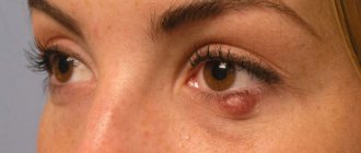
In addition, with excessive eye strain, headaches and burning sensations can occur, and various inflammatory diseases can often develop, for example, blepharitis, conjunctivitis in adults, chalazion of the upper eyelid, and so on. Children may experience amblyopia or strabismus.
How does a person see with and without glasses?
To treat farsightedness, methods such as:
- Glasses, contact lenses.
- Photorefractive keratectomy.
- Laser thermokeratoplasty.
- Lens replacement.
- Lens implantation.