First stage of the luteal phase
During ovulation, the granulosa membrane ruptures; to put it in medical terms, the wall of the Graafian follicle bursts. After rupture occurs, fluid from the follicle flows from the ovarian tubercles containing the egg into the peritoneum. This stage is called follicle decline. When there is a violation of the integrity of the vascularized epithelium, a process of destruction of the integrity of small vessels occurs in the membrane. They begin to bleed in the place where the follicle was ruptured due to the fact that the pressure inside it decreases. In this area, blood clotting occurs, and after some time, metamorphosis occurs with this blood clot.
The remaining part of the granulosa membrane, which is the follicular cells of the epithelium, connects with the theca interna, becomes profiled and hypertrophies. After which the process of germination into a clot begins, and the formation of large epithelial blastomeres begins along the edge of the burst membrane. They begin to grow larger, their bodies become rounder and their numbers increase.
Causes
Insufficiency of the gravidal corpus luteum occurs for various reasons. Scientists say that such disorders are most often caused by changes in the structure of the X chromosomes.
It is clear that in these cases we are talking about genetic disorders. Low levels of the hormone cannot but affect the functioning of the ovaries and pituitary gland. Disorders that cause a lack of hormones may be associated with pathologies such as cystic changes in ovarian tissue, oncology and polycystic ovary syndrome.
Pathologies associated with dysfunction of the pituitary gland can also lead to insufficiency of the corpus luteum. Problems with a gland such as the pituitary gland can arise against the background of genetic changes, injuries and in the case of growth of tumors in areas associated with the pituitary gland.
Moreover, changes in such disorders may not affect the entire gland, but only part of it. Some quite serious diseases, such as liver and kidney failure, can affect the synthesis of the hormone.
If all the problems associated with the functioning of the corpus luteum rest on diseases of the liver and kidneys, then it is completely impossible to change anything until the main problem is eliminated. In this case, liver and kidney failure.
Lutein is an important component of the corpus luteum
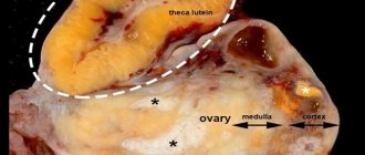
At this time, the cytoplasm accumulates spherosomes that have an orange-yellow tint (lutein) containing droplets of fat. Depending on where the cells came from, elements are distinguished that originate from the granulosa membrane (luteal follicular cells), as well as from the theca (thecal luteal blastomeres). This is how the process of formation of a fibroblast node occurs, in the center of which, in the place of the bursting gaaf follicle, a small part of a blood clot overgrown with connective tissue remains.
Pastila
Corpus luteum phase deficiency (CLP)
THIS DIAGNOSIS IS NOT MADE BY BASAL TEMPERATURE CHARTS!
If your doctor makes a diagnosis only according to BT charts, urgently change him to another doctor.
The most “popular” method of “treatment” today is, perhaps, maintaining the corpus luteum phase (the second phase, the luteal phase). Mainly - duphaston (we will pay special attention to it in our article, since it is this that receives the most complaints and the most problems arise when taking it). Unfortunately, most doctors prescribe it to “everyone and everyone” without any reason (relying more on the Russian word “maybe” than being guided by common sense or actual necessity).
The most common hormonal disorder leading to miscarriage is corpus luteum phase deficiency. In order for a fertilized egg to successfully implant and grow in the future, the duration of the corpus luteum phase should not be shorter than 10 days. If pregnancy occurs, the corpus luteum phase should continue until the placenta forms and takes over the function of feeding the fetus. This usually takes about 10 weeks from conception. If a miscarriage occurs before this period, this may indicate (attention - it does not indicate, but only may indicate!) a possible insufficiency of the corpus luteum phase.
A little theory...
As a result of hormonal processes in a woman’s ovaries, one follicle matures each cycle (very rarely - two, and even less often - more than two).
The first half of the cycle - from the 1st day of menstruation to ovulation - is called follicular (or estrogenic). Its duration can be very different.
For example, a woman, under the influence of stress or other external factors, ovulated only on the 30th day. As a result, her cycle lasted approximately 44 days (30+14). Thus, if a woman does not have her period before the 44th day, this does not mean that she is pregnant.
The second phase of the cycle, from ovulation to the last day before menstruation, is called the “corpus luteum phase” (or progesterone phase), and usually lasts from 12 to 16 days. The hormone produced by the corpus luteum is very important for the onset and successful course of pregnancy, since it prevents the release of other eggs during a given cycle and stimulates the growth of the endometrium (the inner lining of the uterus).
Therefore, we draw your attention to the fact that the key point in the problem of diagnosing and treating luteal phase insufficiency is mandatory monitoring of a woman’s ovulation in each individual cycle (regardless of whether there were previously disturbances or delays). Because By starting to take progesterone drugs before ovulation, a woman risks not only not achieving the desired effect (pregnancy), but rather the opposite - getting a contraceptive effect (progesterone in the body prevents the further release of eggs).
Diagnosis of NLF
Let us repeat once again that this diagnosis (like any other) should absolutely not be made solely on the basis of basal temperature charts. If your temperature chart shows that the corpus luteum phase is 10 days or less, you should pay special attention to this.
Does an increase in basal temperature always indicate that ovulation has occurred? No not always. Unfortunately, many women, despite an increase in basal temperature, do not ovulate, and, conversely, with a monophasic basal temperature schedule, ovulation can occur. Likewise, low (below 37°) basal temperature in the second phase does not mean insufficiency of the corpus luteum phase (progesterone) and a late rise in temperature does not mean a short length of the corpus luteum phase (BT can rise a few days after ovulation).
To confirm the diagnosis, a blood test (for progesterone) and an endometrial biopsy are done with ovulation tracking by ultrasound. If the progesterone level is really not enough, then the doctor prescribes hormonal medications.
Blood analysis. A blood test for progesterone is usually done about a week after ovulation, i.e. on day 7-8 of the corpus luteum phase. If after taking the test, menstruation occurs later than 10 days, and even more so - if more than 2 weeks later (if the test was taken without tracking ovulation using ultrasound or ovulation tests) - it is better to retake the test. To be sure that the results are correct, it is better to take this test several times in one cycle (with an interval of a couple of days) and several cycles in a row to eliminate laboratory error.
Endometrial biopsy. Endometrial biopsy - taking a piece of the endometrium for histological (under a microscope) examination. This study helps to identify a number of diseases that may be the cause of failure or infertility.
Ultrasound monitoring
To track the moment of ovulation and determine the duration of the corpus luteum phase (and, accordingly, to clarify the need or timing of taking medications), repeated ultrasound monitoring by a qualified specialist is necessary.
With an “ideal” 28-day cycle, the first ultrasound can be done on the 8-10th day of the cycle or immediately after the end of menstruation (with a longer cycle - correspondingly later). Next, an ultrasound scan is performed every two to three days (depending on the condition of the uterus and ovaries during examination, the doctor may prescribe the next examination earlier or later) until the day that ovulation has occurred or menstruation begins.
As a result of observation, the following information can be obtained about the development of follicles in the ovaries:
* follicles do not develop, the ovaries “sleep”, ovulation does not occur
* the follicle develops, then stops developing, not reaching the required size, then regresses (confirmed by ultrasound readings and tests for hormones, including progesterone), ovulation does not occur
* the dominant follicle develops, but does not grow to the required size and luteinizes (forming the corpus luteum), while the cycle is constant, progesterone is normal, but in fact, ovulation does not occur
* the dominant follicle develops, grows to the required size, but for some reason does not rupture (further regression of the follicle or the formation of follicular cysts occurs), ovulation does not occur
* the follicle develops, grows to the required size, ovulation occurs and a corpus luteum appears in place of the follicle
In the first four cases, it is better to read the article on our website about ovulation stimulation. In the latter case, the length of the corpus luteum phase is counted from the moment of ovulation to the first day of the next menstruation. The normal length of the corpus luteum phase is from 12 to 16 days.
Treatment
If the corpus luteum phase is shorter than 10 days or the level of progesterone in the blood is below normal (and ovulation definitely occurs), the doctor prescribes hormonal progesterone drugs (for example, progesterone injections or utrozhestan).
Progesterone drugs cannot be taken according to instructions from any specific day of the cycle (as well as according to a similar doctor’s prescription)! Such drugs can only be taken strictly after ovulation - otherwise, instead of treatment, the woman will receive a “contraceptive effect” (we already talked about this above - progesterone in the blood prevents ovulation).
Ovulation must be monitored in each individual cycle by ultrasound or ovulation tests, or (if there are no other possibilities) by a basal temperature chart (after a clear rise and other signs of ovulation, see Tony Weschler’s book “The Desired Child?..”).
The dosage of the drug is usually determined by the doctor depending on the circumstances and characteristics of the body. It is recommended to continue taking the drug until the presence or absence of pregnancy is determined (tests, blood test for hCG, ultrasound).
The most common drugs now are natural progesterone (in ampoules), utrozhestan (natural progesterone in capsules), duphaston (synthetic drug). The latter is most widespread in many government and commercial institutions, where the doctor’s qualifications leave much to be desired, and (due to the myth floating around the world about “no effect on ovulation,” which is not at all true) this drug is prescribed to all patients one at a time. the same scheme without any control and individual approach to each individual patient. We would like to draw the special attention of all our readers to this, since if there are any hormonal disorders in a woman’s body, such “treatment” can adversely affect the patient’s health.
“The Myth of Duphaston”...
It is believed that duphaston (unlike other progesterone drugs) does not affect ovulation. But THIS IS NOT SO! We received a large number of letters from women who, when taking this drug incorrectly (due to the incompetence of doctors) (before ovulation), instead of the effect of maintaining the corpus luteum phase, received anovulatory cycles. We would like to draw your attention to the fact that if such a “treatment” may not have any effect on a healthy body (after a cycle of taking the drug, the body will function the same as before the drug was prescribed without any disturbances), then if there are any ( already existing) hormonal disorders, it can greatly worsen the situation (keep up ovulation not only in the cycle with taking the drug, but also lead to further disorders). So, it is possible that taking this drug does not affect the onset of ovulation, but this is more likely to apply to very healthy women (those who become pregnant after just one half of the missed tablet). But the healthy, as they say, are usually not treated...
From blood clot to corpus luteum
This formation is called sanguinolentum. Lutein, represented by a yellowish substance, colors the membrane that begins to enlarge in a yellow-orange hue. That is why it is called the “yellow body”. The luteal phase, during which progesterone peaks and is secreted, is a favorable time for the formation of the corpus luteum. Along the edge, where the luteal cells are concentrated, thin vascular bundles and connective tissue begin to grow. During this process, a small organ is formed, the structure of which resembles an endocrine gland.
The luteal phase is represented by cellular formations with nodules and bars that fill the entire corpus luteum. The result of the appearance of these cells is the remains of a blood clot located at this site.
Treatment
Treatment of luteal phase deficiency occurs only conservatively. For therapy, it is first necessary to establish the cause of the disease or the factors that influenced its occurrence in the patient’s body.
For general strengthening, various vitamins are prescribed or the intake of certain foods is recommended. In order for treatment to occur as quickly as possible, you need to follow a diet and give up some physical activity.
Since the cause of the disease may be a hormonal imbalance, it is necessary to correct the hormonal levels with special pharmacological agents. These include antiestrogens, which reduce the concentration of estrogen in the female body.
One of the stages of treatment is the introduction into the body of human chorionic gonadotropin, which in another way, like progesterone, is called the pregnancy hormone. During natural processes, it begins to be produced by chorion tissue after implantation of the embryo (6-8 weeks). Follicle-stimulating hormone (FSH) may be added to therapy.
Degenerative processes in the corpus luteum
The corpus luteum, which forms and degenerates every 28th day, is called the “menstrual body.” When the luteal phase ends, progesterone, the norm of which is not exceeded, has a degenerative effect on the cells of the “yellow menstrual body”, in the place of which connective tissue is eventually formed.
After this process is completed, the corpus luteum is replaced by a whitish, slightly shiny scar consisting of dense connective tissue. It is called whitish or fibrous. If a woman becomes pregnant, then it not only does not leave the ovary, but also increases and performs its functions until the second trimester of pregnancy. Its endocrine activity suppresses the action of other membranes, in other words, it prevents blood clots from appearing. As a result, the norm of the luteal phase means that the reproductive system is functioning normally, which means that there are no obstacles to conceiving offspring.
For those wishing to get pregnant, the luteal phase, the cycle day of which falls in the middle of ovulation, is very important, because with the right calculations, the long-awaited conception can occur. But not everything is always so simple, because some women also have a disorder.
LLF or luteal phase deficiency is a fairly common disorder of ovarian function in the female half of humanity. This problem usually worries those who are unable to conceive, or those who have had repeated early miscarriages.
Luteal phase deficiency is caused by the fact that the corpus luteum does not produce progesterone in the required amount, which is necessary for fertilization and pregnancy.
Complications
If treatment is not started in a timely manner, this threatens, first of all, with the loss of the main function of the reproductive system to sexual reproduction. Luteal phase deficiency also threatens:
- isthmic-cervical insufficiency during pregnancy;
- spontaneous abortions;
- deviation in the placenta;
- failure of menstruation;
- cancer of the uterus;
- fibrocystic mastopathy;
- deviation in the functioning of the ovaries;
- fibroids.
Such complications can cause a significant blow to the patient’s psycho-emotional state and will only worsen the situation.
Since stress and depression are some of the causes of menstruation irregularities. Share:
About the cause of NLF
The main causes of deficiency may be several fairly common reasons, consisting in a violation of the central regulatory mechanisms of reproductive function, which has a detrimental effect on various levels of the system, from the hypothalamus and pituitary gland to the functioning of the ovaries.
In this case, the ovaries produce a small amount of hormones, and the doctor, upon examination, determines hyperprolactinemia and hyperandronemia - disorders in the thyroid gland and adrenal cortex.
Firstly, this condition causes hormonal imbalance.
Secondly, it has an inappropriate effect on the developing egg. Thirdly, degenerative changes occur in the endometrial receptor apparatus of the inner lining surface of the uterus.
This factor is influenced by infections and inflammation, adhesions after cesarean section, abortions and miscarriages, and abnormal development of the appendages and the uterus itself or delays observed during puberty may also be to blame.
In such cases, infertility will occur due to the fact that the egg fails to attach to the uterine wall, which will subsequently prevent it from receiving vital substances and other components for the development of the baby.
The second reason may be a lack of lipoproteins due to low blood density, as well as the amount of progesterone required in it, and a change in the biochemical composition of the peritoneal fluid.
Diagnostics
First of all, to make a diagnosis, the gynecologist needs to listen to the signs that worry the patient, and then study the medical history. In addition, the woman is examined, including measuring her weight and height.
If progesterone deficiency is suspected, the patient should be explained what luteal phase deficiency is and continue the examination. It includes: measurement of body temperature at complete rest, palpation, determination of diastolic pressure, examination of the external genitalia using a mirror. Then it is necessary to carry out differential diagnosis.
To determine the thickness of the inner mucous membrane of the uterus, ultrasound diagnostics of the pelvic organs is used. This procedure makes it possible to see a complete picture of the condition of all organs and make an accurate diagnosis.
After determining luteal phase deficiency, especially if a woman is pregnant, a follitropin test is used as a supplement. The function of this hormone is to ensure the maturation of eggs in the follicle. Determination of hormone levels in the blood.
About symptoms and diagnostic methods
With this problem, scanty discharge before menstruation, irregularities in the menstrual cycle, infertility, and frequent miscarriages in the first and second trimester of pregnancy appear.
In order to determine luteal insufficiency, you can use a couple of methods. Measure your basal temperature. If the hormone is observed in small quantities, then changes will be visible in the second phase. Do an ultrasound and examine the dynamics of follicular growth and the thickness of the endometrial layer, as well as examine the hormonal status and take a biopsy of the membrane lining the uterus. This method is also effective in that it will help identify other abnormalities in the menstrual cycle.
Sometimes doctors talk about hysterosalpintography to determine the level of patency of the fallopian tubes, as well as determine their tone.
Diagnosis of the disease
If symptoms of luteal phase deficiency (LPF) are detected, it is necessary to visit a gynecologist to determine the cause of this pathology. At the appointment, the doctor will conduct a general and gynecological examination, collect information for anamnesis, and find out what medications the woman is taking.
To determine the length of the second phase, the gynecologist will advise you to measure your basal temperature every morning - normally, progesterone increases the temperature. Instead of measuring it, you can use ovulation tests, which are sold in pharmacies. The diagnosis of second phase deficiency is confirmed if, after these methods, it turns out that it lasts less than 12 days.
The doctor will prescribe various tests:
- general and biochemical blood test;
- blood for sex hormones and thyroid hormones;
- coagulogram (clotting test).
If a tumor or inflammation is suspected, the patient will be referred for an MRI, ultrasound and biopsy. To obtain information about the condition of the endometrium, hysteroscopy (examination of the uterine cavity performed endoscopically) is used.
About the main methods of treating luteal phase deficiency
This complication is fraught with not only hormonal disruptions, but also problems with reproductive function, which usually cause miscarriages and long-term infertility.
Often, gynecologists prefer monotherapy with various drugs that restore progesterone, but this is practically to no avail. For it to be effective, you must first regulate the follicular phase, without which normal development and maturation of eggs is impossible. This means that in treatment it is necessary to use not only drugs to increase hormone levels, but also those that will reduce prolactin and androgens to normal.
Physiotherapeutic and sanatorium-resort treatment combined with acupuncture, vitamin therapy and the use of adaptogenic drugs are also prescribed. The emotional and psychological state of a woman is no less important. A month after the procedures, a new luteal phase will begin. Which day of the cycle will be the most successful for determining the correctness of treatment can be determined using a special test. If the deficiency has been overcome, you can begin planning your pregnancy.
Ascites and oncology
There are several main methods for treating ascites in patients with cancer:
- conservative therapy (aldosterone antagonists, diuretics) - aimed at normalizing water-salt metabolism and reducing the formation of fluid in the abdominal cavity;
- laparocentesis - puncture of the abdominal wall under ultrasound control; used not only for removing fluid, but also for installing drainage, which will serve for long-term removal of fluid;
- palliative surgical operations - peritoneovenous shunt, omentohepatophrenopexy, deperitonization of the abdominal walls and others.
Symptoms
Abdominal ascites, which develops as a complication of cancer, should be treated together with the underlying disease.
It is also important to begin eliminating excess fluid in the first two weeks of its formation, since delaying therapy leads to the development of a host of complications. Excess fluid can be removed by puncture and pumping it out - laparocentesis, by taking diuretics.
Following a special diet will help reduce intra-abdominal pressure and reduce the likelihood of further production of excessive exudate.
What is dangerous about hypofunction of the corpus luteum, or progesterone deficiency?
What are the effects of progesterone, which the corpus luteum is obliged to secrete during its heyday? Progesterone not only prepares the lining of the uterus, that is, the endometrium, for the implantation of a fertilized egg. Progesterone reduces the muscle activity of the layers of the uterus, preventing and suppressing uterine contractions so that a fertilized egg does not accidentally come off.
In addition, progesterone ensures that the maturation of a new follicle does not occur, and God forbid, ovulation does not occur for some unknown reason after fertilization has occurred. After all, now we need to deal with the fertilized egg! The corpus luteum does all this.
Therefore, it is clear why corpus luteum deficiency is dangerous. What does it mean? This is a lack of progesterone.
By the way, before moving on to diagnosis, it should be recalled that the normal size of the corpus luteum, on average, is from 15 to 29 mm. But even if the size of the corpus luteum, this hormonal gland of the 3rd phase of the menstrual cycle or 1st trimester of pregnancy, is normal, there may be a deficiency of progesterone for one reason or another.
What are the diagnostic criteria underlying the diagnosis of insufficient corpus luteum during pregnancy in terms of complaints and symptoms? This condition can manifest itself in different ways, for example:
- the inability of a fertilized egg to implant into the uterine wall, its rejection;
- improper formation of the placenta;
- lack of nutrients for the unborn fetus.
All this can lead to serious consequences, and even to late spontaneous abortion, for example, in the second trimester, miscarriage, infertility.
But even if pregnancy does not occur, progesterone deficiency leads to symptoms of corpus luteum deficiency such as bleeding, increased uterine tone, abdominal pain, and so on. How can you diagnose hypofunction? What should a woman know?


