Intestinal amebiasis - symptoms, diagnosis and treatment Amebiasis is a protozoal infectious disease characterized by the occurrence of ulcerative lesions in the colon area. Amebiasis, the symptoms of which include, in particular, the formation of abscesses in various organs, is prone to a protracted and chronic course.
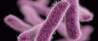
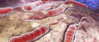
The causative agent of the disease is a dysenteric amoeba, the life of which consists of two stages: the vegetative stage (trophositis) and the resting stage (cyst). These stages are capable of transition from one to another, while depending on the living conditions in the body of their carrier.
Let us note that the disease is endemic; accordingly, it is characterized by concentration in a specific area and spreads in those areas characterized by a hot climate.
What it is?
Amebiasis is an intestinal infectious disease that has a long course and is characterized by the occurrence of ulcerative defects of the large intestine and damage to other organs.
Amoeba was discovered by St. Petersburg scientist F.A. Lesh in 1875 while examining the stool of a patient with bloody diarrhea. In Egypt, R. Koch (1883) isolated the pathogen from intestinal ulcers and purulent cavities in the liver. Amoebiasis, called “amebic dysentery,” was classified as an independent disease in 1891.
https://youtu.be/TpGRnNVaxPU
Who is at risk
Amebiasis is usually transmitted by the fecal-oral route. Can be transmitted indirectly through contact with dirty hands or objects, as well as through anal-oral contact.
The infection is spread through ingestion of the parasite, a semi-dormant and hardy structure found in feces.
Amoebas or trophozoites die quickly after leaving the body and may be present in feces. They are rarely the source of new infections.
Because amoebiasis is transmitted through contaminated food and water, it is often endemic in regions of the world with poor sanitation, including Mexico, Central America, western South America, southern Asia, and western and southern Africa.
Amoebic dysentery is often confused with traveler's diarrhea. In fact, most travelers' diarrhea is bacterial or viral in origin.
At risk:
- People who have traveled to tropical places with poor sanitation;
- Immigrants from tropical countries with poor sanitation;
- People who live in institutions with poor sanitation;
- Men who have intimate contact with men.
Find out more How to cure a child of parasites
How can you get infected?
A person can become infected with amoebiasis only from another person who has already had the disease and is a clinically healthy carrier of cysts. Amebiasis, like many other intestinal infections, can be called a “disease of dirty hands.”
If the carrier of the cysts does not adhere to the rules of personal hygiene, the cysts with his feces can end up in wastewater, in the soil, in the water of open reservoirs, and from there - on vegetables and fruits grown on private farms. If, after visiting the toilet, the cyst carrier does not wash his hands thoroughly, he can transfer the cysts to household objects or food; finally, it can infect another person simply by shaking hands. Without washing your hands before eating or eating unwashed vegetables and fruits, a healthy person brings cysts into his mouth, from where they spread further along the gastrointestinal tract.
This method of transmission of infection is called fecal-oral.
Prevention
Prevention of infection is possible in the only way - strict adherence to hygiene rules. Prevention of amoebic dysentery is especially important in endemic areas. Since infection occurs through the mouth, all foods and drinks must be properly prepared and thoroughly washed.
Drinking water can be disinfected in the following ways:
- boiling for 10-15 minutes. followed by storage in a closed container (the most reliable method);
- adding special cleansing tablets with an exposure of at least 15 minutes;
- filtering through special devices.
In disadvantaged regions, boiled water should be used for brushing teeth and washing, washing dishes, vegetables, fruits and protecting food from flies and cockroaches.
After hospitalization of the sick, the outbreak is disinfected. Carriers of amoebas and cysts are not allowed to work in children's institutions, water supply and public catering systems. Patients who have recovered from amoebic dysentery are observed for 1 year with quarterly laboratory examinations.
Amoebic dysentery is a protozoal endemic infection with ulcerative lesions of the large intestine, prone to chronicity of the process and severe metastatic damage to internal organs. If illness occurs, you should immediately consult a doctor.
Forms of amoebiasis
Pathomorphological changes and symptoms of the disease make it possible to distinguish invasive and non-invasive amebiasis. The first form is accompanied by pathological changes in the patient's body. Its characteristic features:
- symptoms of infection;
- endoscopic examination reveals characteristic changes in the intestinal mucosa;
- serological tests show the presence of specific antibodies;
- presence of parasites in feces.
The non-invasive form (passive) is defined as the “carriage” of amoebic cysts. Peculiarities:
- lack of a characteristic clinic;
- absence of antibodies and pathological changes in the intestines;
- absence of hematophagous trophozoites in feces.
In 90% of infected people, the non-invasive form is present. These individuals are asymptomatic carriers.
The clinical picture of invasive amebiasis has a wide range of symptoms, from mild manifestations of invasion to amoebic liver abscess.
Symptoms of amoebiasis
The duration of the incubation period is from 1–2 weeks to several months.
Intestinal amebiasis is manifested by certain symptoms: gradually increasing cramping pain in the abdomen (mainly in the left lower half of the abdomen) and frequent loose stools with a significant admixture of mucus and blood (raspberry jelly).
Fever is also characteristic, manifestations in the form of decreased performance, weakness, rapid heartbeat, and decreased blood pressure. Acute symptoms of intestinal amebiasis decrease within 4–6 weeks, but spontaneous recovery and cleansing of the body from the pathogen are rare.
In most cases, after remission, an exacerbation of the disease is recorded after a few weeks or months. In these cases, the total duration of the disease (chronic form of amebiasis) without adequate treatment is decades. This form is characterized by disorders of all types of metabolism (exhaustion, hypovitaminosis, hormonal imbalance, anemia, etc.)
Infection with amoebic dysentery
The causative agent of amebiasis is E. histolytica. Infection with amoebas occurs when cysts are ingested, most often with water or food. Cysts are resistant to exposure to the external environment and can remain outside the body for weeks or even months. Living conditions, water supply, sanitary conditions of populated areas and the general cultural level of the population can determine varying degrees of damage. Urban populations are generally less affected than rural populations. Recently, E. histolytica infection among schoolchildren has been increasing. The source of infection for amoebic dysentery is a sick person or carriers of amoebae. The incubation period for amoebic dysentery ranges from 1 week to 3 months.
Signs of the extraintestinal form
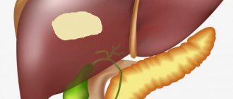
Of the extraintestinal manifestations of amebiasis, the most common is amoebic liver abscess. It is characterized by single or multiple abscesses without a pyogenic membrane, most often localized in the right lobe of the liver. The disease begins acutely - with chills, hectic fever, profuse sweating, pain in the right hypochondrium, aggravated by coughing and changes in body position. The condition of the patients is serious, the liver is sharply enlarged and painful, the skin is sallow in color, and sometimes jaundice develops.
Pulmonary amebiasis occurs in the form of pleuropneumonia or lung abscess with fever, chest pain, cough, and hemoptysis. With amoebic brain abscess (amebic meningoencephalitis), focal and cerebral neurological symptoms and severe intoxication are observed. Cutaneous amebiasis occurs secondarily in weakened patients, manifested by the formation of mildly painful erosions and ulcers with an unpleasant odor in the perianal area, on the buttocks, in the perineal area, on the abdomen, around fistulous openings and postoperative wounds.
Diagnostics
A quick diagnosis of amoebiasis is needed. The causative agent of the disease can be detected in feces or in tissues (for example, during sigmoidoscopy). It is necessary to conduct an analysis to detect antibodies to amoebas in the patient’s blood. But in order to refer the patient for a blood test, the doctor must still guess that the disease was caused by protozoa and not other microorganisms.
In the emergency room it is not always possible to make a correct diagnosis: the disease is too rare and its symptoms are very similar to other ailments. Suspicion of amoebiasis may appear immediately if the patient says that in the last two months he has been in Southeast Asia and his stool looks like raspberry jelly.
However, it is impossible to make a diagnosis and prescribe treatment based on one question. Next, diagnostics are carried out: feces, urine and blood are taken for analysis. You can find protozoa in the stool and guess about amoebic infection.
Complications of intestinal amoebiasis
If treatment for amoebiasis is not started on time, there is a high probability of developing complications of the disease. Perforation may develop in the cecum or rectosigmoid area, leading to peritoneal abscess or peritonitis. In some cases, amoebic appendicitis and ameboma, which is a tumor-like growth in the area of the rectum and cecum, may appear.
Sometimes an amoebic intestinal stricture develops, which has the appearance of granulation tissue. Strictures are usually single and located in the area of the sigmoid or cecum. They contain amoebic trophozoites and are often not accompanied by any symptoms. In some cases, strictures provoke the development of constipation or intestinal obstruction.
Treatment of amoebiasis
All drugs used in the treatment of intestinal amebiasis are divided into two groups - contact and systemic tissue amoebicides.
- The former are used in the presence of non-invasive amoebiasis and at the final stages of treatment to eliminate remaining amoebas. In addition, contact (luminal) amoebicides are in demand in cases where it is necessary to take preventive measures to prevent the spread of infection. Drugs in this group include: etofamide, paromomycin, clefamide, diloxanide.
- When invasive amebiasis is diagnosed, treatment involves taking systemic tissue amoebicides - metronidazole, secnidazole, tinidazole. For severe forms of amebiasis, antibacterial drugs that are active against intestinal microflora are recommended.
Advanced abscesses (more than 6 cm) require aspiration (percutaneous drainage). This procedure is necessary to prevent abscess rupture, as well as in cases where bowel chemotherapy does not lead to the expected results. Corticosteroids are contraindicated for patients diagnosed with amebiasis, as they are accompanied by numerous complications.
In general, with early diagnosis and adequate drug therapy, intestinal amebiasis is completely cured within a few months of starting treatment.
Life cycle of intestinal amoeba
Intestinal amoeba is capable of maintaining its vital functions outside its host. There are even cases of its reproduction outside of humans, but this happens rarely.
Basically, to maintain its vital functions and successfully reproduce, the amoeba needs the human intestine.
The life cycle of intestinal amoeba includes the following forms:
- Metacyst development.
- Translucent form.
- Pre-cystic state.
- Direct formation of cysts.
- Fabric form.
- Large vegetative phase.
The intestinal amoeba feeds on food debris, bacteria and fungi. It does not penetrate the intestinal walls and does not harm its carrier. Even if a large number of red blood cells appear in the organ, the amoeba does not swallow them. She moves very slowly, one might say, she is marking time in one place.