Types of myocardial infarction - what are they and what is the basis for the classification of the disease? A heart attack is the softening of tissues due to necrosis, i.e. their death due to lack of oxygen supply to them.
Characteristic signs of a typical form of myocardial infarction are intense burning retrosternal pain (medically defined as anginal), which radiates between the shoulder blades, into the arm, ribs or jaw and is not relieved by analgesic drugs.
A heart attack can occur in various internal organs, but the most common is myocardial infarction. What is this and why does it happen? Due to the functional importance of the heart and the high demand of the heart muscle for oxygen, myocardial infarction develops very quickly and is accompanied by irreversible consequences. Several types of classifications of myocardial infarction have been created, each of which has clinical significance.
The type of heart attack can only be determined after examination
Universal classification
According to the universal classification, the following types exist:
- Spontaneous. Occurs due to rupture, erosion or dissection of the plaque, which leads to thrombosis of the coronary vessel. There is a disruption of blood flow and subsequent necrosis.
- Due to ischemic imbalance. The cause of myocardial necrosis is a condition not related to coronary heart disease.
- Type 3 or fatal. This is a condition of sudden cardiac death.
- Obtained as a result of revascularization. Cause: thrombosis of a previously installed stent or coronary artery bypass surgery.
The universal classification determines the types of myocardial infarction depending on the causes and consequences.
Treatment
The sooner treatment for myocardial infarction begins, the better. With timely administration of drugs, the blood clot will quickly resolve and blood circulation will resume. Next, it is necessary to remove the possibility of new blood clots; for this purpose, medications are administered that slow down blood clotting. This is usually aspirin. Strict bed rest and the introduction of beta-blockers, which will relieve heart pain, are also necessary.
Further treatment of the heart will be as follows:
- Bed rest in the first days.
- If necessary, oxygen inhalations.
- Taking antiplatelet agents that prevent the formation of blood clots.
In severe cases of heart attack, surgical intervention is necessary - coronary artery bypass grafting and coronary angioplasty.
Treatment of a heart attack occurs in a hospital, since this disease is life-threatening. If symptoms of any of the listed stages of myocardial infarction are detected, you must call an ambulance. To determine the disease and clarify the characteristics of the pathology, an electrocardiogram is performed.
After recovery and discharge from the hospital, a long rehabilitation period is provided. People who have suffered a myocardial infarction should undergo sanatorium treatment, regularly visit a cardiologist for examination, and use diagnostic methods if their condition worsens. The goal of therapy in the treatment of myocardial infarction is to reduce pain, prevent the spread of the lesion, and eliminate the causes that caused the attack. The prognosis of the disease depends on the correctness of such therapeutic measures and the characteristics of the patient’s body.
Therapy goals:
- Restoration of blood circulation. If congestion is stopped in a timely manner, the prognosis for the patient’s life improves.
- Reducing the area of tissue susceptible to necrosis.
- Reduced pain.
- Prevention of the occurrence of other pathologies that manifest themselves against the background of a heart attack. With the help of medicinal and sometimes surgical methods, the likelihood of developing complications is reduced.
Many patients, aware of the increased risk of heart disease, are interested in what heart attacks are like. When studying the types and stages of this disease, it is necessary to promptly respond to the appearance of negative symptoms by seeking medical help. Signs of any form and phase of a heart attack are weakness, pain, shortness of breath, and increased sweating.
5 types according to the classification of the World Federation of Cardiology
This is the main type of classification used in cardiology. Developed by a group of scientists from the World Heart Federation. It is based on a complex division into causes, symptoms, and pathogenesis. A total of 5 types are identified:
- Spontaneous. Associated with a primary violation of the coronary circulation, damage to the supply vessel, significant narrowing of the lumen in the coronary arteries.
- Secondary. The cause is coronary spasm or thromboembolism. Arrhythmia and high blood pressure are also common causes of secondary heart attacks.
- Type 3 is sudden coronary death . Occurs with simultaneous disruption of the conduction system of the heart and acute ischemia.
- Type 4 has two varieties : 4a, which is associated with complications of percutaneous coronary intervention, and 4b, associated with stent thrombosis.
- Problems with coronary artery bypass surgery. They arise both directly during surgery and as late complications.
The last 2 types are of iatrogenic origin, since they are provoked by the action of doctors.
Secondary prevention
The disease cannot be called an unpleasant episode in a person’s life; for people who have suffered it, it becomes the line beyond which health problems begin. The most dangerous thing is that cardiac ischemia is undergoing accelerated progress.
The attending physician helps the patient achieve a high quality of life if he:
- follow a diet;
- take medications in a timely manner;
- independently monitor your health and report it to your doctor;
- lead a healthy lifestyle;
- take courses in cardiac rehabilitation.
The first time after a heart attack is fraught with serious complications, the development of which only specialists can prevent.
Stage of development and clinical picture of typical forms
Typical types of MI all go through similar stages of development. All stages of infarction progression can be seen by characteristic clinical signs. Each term has its own duration.
Damage period (initial)
This is a pre-infarction condition that can last from several hours to several days. It is at this stage that the blood supply to the myocardium decreases. Short-term pain appears behind the sternum and in the heart area.
As a result, angina attacks become more frequent, and the pain during these attacks intensifies. This condition is a serious reason to consult a doctor. The specialist will order an ECG and, based on its results, make a forecast of the development of the situation.
Acute
The acute stage of myocardial infarction can last from several days to two weeks. Some tissues die during this period, while others are restored. At this time, necrosis and softening of the tissue occurs. At the very beginning, the most acute stage develops, the characteristic clinical signs of which are: pain, cold sweat, a feeling of approaching death, shortness of breath. During the acute period, the pain goes away and becomes less intense. Clinical signs of the acute stage:
- dyspnea;
- headache;
- temperature 37-38;
- blood pressure decreases;
- heart rhythm is disturbed.
Fever occurs due to intoxication of the body with decay products. During this period, a scar begins to form. It is at the acute and most acute stage that most complications arise that can lead to the death of the patient.
Subacute
The stage lasts about two months. The risks of complications are significantly reduced. The pain returns if new areas of ischemia form. But in general, the patient’s well-being improves, and the scar forms and becomes denser. Low blood pressure may persist while the heart rate returns to normal. If the patient had hypertension before the heart attack, the blood pressure will gradually rise again.
In place of the cells that died, connective tissue appears. If the disease is not complicated by additional pathologies, then the subacute period will last 6-8 weeks. In other cases, recovery may take longer. This is an individual process.
Cicatricial (final)
In most cases it lasts for several years, but actually until the end of life. The focus of necrosis is completely overgrown with connective tissue, and the heart gets used to working in new conditions.
There are few clinical signs. There are some changes on the ECG, but the patient himself does not feel symptoms. In some cases, angina attacks may return. The most severe complication at this stage is considered to be chronic heart failure.
Classification of myocardial infarction and differences between types
Myocardial infarction is damage to the heart muscle due to disruption of its blood supply. In the part where oxygen starvation has developed, cells die, the first ones die within 20 minutes after the blood flow stops.
Heart attack is one of the top diseases that cause mortality among the population. Every year in Europe alone, 4.3 million people die for this reason.
Stages of development and clinical picture of typical forms
The classification of myocardial infarction implies four stages of disease development according to time and clinical picture - damage, acute, subacute, cicatricial.
Damage period (initial)
Symptoms occur within a few hours to 3 days . At this stage, transmural damage to fibers is observed as a result of circulatory disorders. The longer the latent phase, the more serious the disease.
An ECG allows you to recognize the disease. Potassium ions, leaving the dead cells, form damage currents. Then a pathological Q wave appears , which is recorded already on the second day.
If necrotic disorders have appeared in the heart, then the ST segment is much higher than the isoline , the convexity is directed upward, repeating the shape of the monophasic curve. At the same time, the fusion of this segment with a positive T-wave is recorded.
It is noteworthy that if there is no Q wave, then all cardiac muscle cells are still alive . This tooth may appear even on the 6th day.
Acute
The duration of the second stage is from 1 day to 3 weeks .
Gradually, potassium ions are washed out from the damaged area, weakening the strength of the currents. In this case, the damaged area decreases, since some part of the fibers dies, and the surviving part tries to recover and goes into ischemia (local decrease in blood circulation).
The ST segment descends to the isoline, and the negative T wave acquires an expressive contour. However, with infarction of the anterior wall of the left ventricle, ST elevation is likely to persist for some time period.
If a large transmural infarction has occurred, the ST segment rise lasts the longest, indicating a severe clinical picture and poor prognosis.
If in the first stage there was no Q wave, now it appears in the form of QS in the transmural type and QR in the non-transmural type .
Subacute
The stage lasts about 3 months, sometimes up to a year .
At this stage, deeply damaged fibers move into the necrosis zone, which stabilizes. Other fibers are partially restored and form an ischemic zone. During this period, the doctor determines the size of the lesion . In the future, the ischemic zone is reduced, and the fibers in it continue to recover.
Cicatricial (final)
Scarring of the fibers lasts the entire life of the patient. At the site of necrosis, the tissues of neighboring healthy areas are united . The process is accompanied by compensatory hypertrophy of fibers, the affected areas are reduced, and the transmural type sometimes turns into a non-transmural type.
In the final stage , the cardiogram does not always show a Q wave , so the ECG does not report the disease. There is no zone of damage, the ST segment coincides with the isoline (myocardial infarction occurs without its elevation). Due to the absence of an ischemic zone, the ECG shows a positive T wave, characterized by flattening or less height.
Division by anatomy of the lesion
Based on the anatomy of the lesion, the disease is distinguished:
- transmural;
- intramural;
- subendocardial;
- subepicardial.
Transmural
With a transmural infarction, ischemic damage occurs to the entire muscular layer of the organ . The disease has many symptoms that are common to other diseases. This makes treatment significantly more difficult.
In terms of symptoms, the disease resembles angina pectoris, with the difference that in the latter case ischemia is a temporary phenomenon, and with a heart attack it becomes irreversible .
Intramural
The lesion is concentrated in the thickness of the wall of the left ventricle and does not affect the endocardium or epicardium . The size of the lesion may vary.
Subendocardial
This is the name for a heart attack in the form of a narrow strip near the endocardium of the left ventricle. Then the affected area is surrounded by subendocardial damage , as a result of which the ST segment descends below the isoline.
During the normal course of the disease, excitation rapidly passes through the subendocardial sections of the myocardium. Therefore, a pathological Q wave does not have time to appear above the infarction zone. The main sign of the subendocardial form is that the ST segment horizontally shifts above the affected area below the electrical line by more than 0.2 mV.
Subepicardial
The lesion occurs near the epicardium. On the cardiogram, the subepicardial form is expressed in a reduced amplitude of the R wave, a pathological Q wave is visible in the leads above the infarction area, and the ST segment rises above the isoline . A negative T wave appears in the initial stage.
For more details about determining the disease on an ECG, watch the video:
Volume of affected area
There are large-focal, or Q-myocardial infarction, and small-focal, which is also called non-Q-infarction.
Large-focal
Causes large-focal infarction, thrombosis or prolonged spasm of the coronary artery. As a rule, it is transmural.
The following symptoms indicate the development of a Q-infarction:
- pain behind the sternum, radiating to the right upper part of the body, under the left shoulder blade, to the lower jaw, to other parts of the body - shoulder, arm on the right side, epigastric region;
- ineffectiveness of nitroglycerin;
- the duration of pain varies - short-term or more than a day, possibly several attacks;
- weakness;
- depression, fear;
- often - difficulty breathing;
- lower blood pressure in patients with hypertension;
- pale skin, cyanosis (blue color) of mucous membranes;
- profuse sweating;
- sometimes - bradycardia, in some cases turning into tachycardia;
- arrhythmia.
When examining the organ, signs of atherosclerotic cardiosclerosis and expansion of the heart in diameter are revealed. Above the apex and at Botkin's point, the 1st tone is weakened, sometimes split, the 2nd tone dominates, and systolic murmurs are heard. Both heart sounds become muffled . But if necrosis did not develop against the background of pathological changes in the organ, then the 1st tone prevails.
With a large-focal infarction, a pericardial friction rub is heard , the heart rhythm becomes galloping, which indicates a weakened contraction of the heart muscle.
a high level of leukocytes in the body , and a “scissors” effect is observed in the relationship between these two indicators. The large-focal form is accompanied by other biochemical abnormalities, the main one of which is hyperenzymemia, which occurs in the first hours and days.
In case of a large-focal form, hospitalization is indicated . In the acute period, the patient is prescribed bed rest and mental rest. Meals are fractional, limited in calories.
The goal of drug therapy is to prevent and eliminate complications - heart failure, cardiogenic shock, arrhythmias. To relieve pain, narcotic analgesics, antipsychotics and nitroglycerin (intravenously) are used. The patient is prescribed antispasmodics, thrombolytics, antiarrhythmic drugs, ß-blockers, calcium antagonists, magnesia, etc.
Finely focal
With this form, the patient develops small lesions of the heart muscle. The disease is characterized by a milder course compared to large-focal lesions.
The sonority of the tones remains the same , there is no galloping rhythm and pericardial friction noise. The temperature rises to 37.5 degrees, but not higher.
The level of leukocytes is about 10,000–12,000 , high ESR is not always detected, in most cases eosinophilia and band shift do not occur. Enzymes are activated briefly and insignificantly.
On the electrocardiogram, the RS-T segment is displaced, most often falling below the isoline . Pathological changes in the T wave are also observed: as a rule, it becomes negative, symmetrical and takes on a pointed shape.
A small focal infarction is also a reason for hospitalization of the patient . Treatment is carried out using the same means and methods as for the large-focal form.
The prognosis for this form is favorable, mortality is low - 2-4 cases per 100 patients . Aneurysm, cardiac rupture, heart failure, asystole, thromboembolism and other consequences of small-focal myocardial infarction occur rarely, but this focal form of the disease develops into a large-focal form of the disease in 30% of patients.
Localization
Depending on the location, myocardial infarction occurs in the following clinical variants:
- left and right ventricles - blood flow to the left ventricle often stops, and several walls may be affected at once.
- septal , when the interventricular septum suffers;
- apical - necrosis occurs at the apex of the heart;
- basal - damage to the high parts of the posterior wall.
Atypical types of disease
In addition to the above, there are other forms of this disease - atypical. They develop in the presence of chronic diseases and bad habits, due to atherosclerosis.
Atypical forms significantly complicate diagnosis.
There are gastralgic, asthmatic, asymptomatic and many other variations of heart attacks. We talked in more detail about atypical forms of myocardial infarction in another article.
Multiplicity
Based on this criterion, the following types of myocardial infarction are distinguished:
- primary - occurs for the first time;
- recurrent - the lesion is recorded within two months after the previous one, and in the same area;
- continued - the same as recurrent, but the affected area is different;
- repeated - diagnosed after two months or later, any area is affected.
Therefore, at the first symptoms that may indicate a heart attack, you should immediately seek medical help.
Division by anatomy of the lesion
Assessing how deeply the heart tissue is affected and where the focus is located, we can list what other types of heart attacks there are.
- Transmural. Penetrating type, characterized by damage to the entire thickness of the cardiac tissue. This is an acute variety, most often large-focal.
- Intramural. Necrosis occurs in the thickness of the myocardium. Pathological changes do not directly affect the epicardium or endocardium. The pathological Q wave on the ECG is most often absent in this situation.
- Subendocardial. With this type of MI, the layers of the heart muscle adjacent to the endocardium are affected.
- Subepicardial. This type of heart attack is different in that the layers of the heart muscle adjacent to the epicardium are affected.
Causes
Many believe that baldness in men is a kind of precursor to a heart attack, since baldness begins due to high levels of androgens. Also an indicator of the likelihood of an imminent heart attack is increased blood pressure and cholesterol levels due to changes in hormones.
Diseases such as atherosclerosis of the coronary arteries, which is characterized by loss of elasticity of the arteries and the appearance of plaques that are formed due to a mixture of fats and cholesterol, may also be the cause. Against the background of atherosclerosis, vasospasm occurs, it changes the activity of the arteries responsible for feeding the heart muscle, this occurs due to changes in sensitivity to external factors (for example, stress). When a plaque breaks off and is subsequently carried by the bloodstream into an artery, venous thrombosis can begin.
Atherosclerosis of the coronary arteries is one of the causes of heart attack
Volume of affected area
This is another type of classification. It is assessed by the size of the area of dying tissue. There are two types of heart attacks based on the volume of foci.
Large-focal
Large-focal myocardial infarction is often easier to recognize, since it has a more vivid clinical picture and more symptoms. This is a classic heart attack with a large focus of necrosis in the heart muscle, which goes through all 5 stages of development.
Finely focal
In this case, the volume of myocardial damage is significantly less. It differs from macrofocal in the absence of bright and numerous symptoms in the acute and subacute periods. The disease has an unclear periodicity and all clinical signs are not so pronounced. Not all patients develop tachycardia; anginal pain is also much less pronounced. In many cases, a small-focal infarction is a precursor to a more serious one - a large-focal one.
The small focal form accounts for about 20% of all clinical cases. And it develops into macrofocal in 30% of cases.
Clinical forms of heart attack
There are various options for acute myocardial thrombosis. The most common clinical picture of acute ischemia is painful. It accounts for 80% of cases of coronary ischemia.
Characteristic features:
- Severe unbearable pressing pain behind the sternum, radiating to the left shoulder, ear, jaw, teeth. The irradiation expands to the area between the shoulder blades, the hand and the tips of the ring and little fingers go numb.
- The pain syndrome lasts from 30 minutes to several hours, is not relieved by Nitroglycerin, and can only be relieved with narcotic analgesics.
- The skin is pale with acrocyanosis - blue discoloration of the tip of the nose and ears.
- Shortness of breath is pronounced, severe weakness occurs.
- Feeling of fear of death, excitement, anxiety.
An insidious type of heart attack is not so rare - the abdominal form. This means that acute coronary ischemia results in severe abdominal pain. The burning sensation causes patients to take Rennie, a soda solution, to no avail. Some patients experience nausea.
Vomiting without relief against the background of unabated pain in the epigastrium for 30 minutes should alert the patient and force him to call an ambulance. In such cases, it is strictly forbidden to take painkillers that will mask the true cause of the disease.
In such a situation, it is necessary to do a cardiogram. The abdominal form can occur after physical activity or overeating.
Emergency doctors encounter in their practice another deceptive disease - atypical pain syndrome during a heart attack. Sudden sharp pain occurs in the arm, leg, ear, or throat. Patients take painkillers that lubricate the true cause of the disease.
In 25% of cases, a painless form of heart attack occurs on the legs. It is discovered accidentally in the form of a heart muscle scar during a routine examination or examination for another disease.
Doctors are also aware of the arrhythmic variant of heart attack. This form is manifested only by a violation of the heart rate - paroxysmal tachycardia, atrial fibrillation or extrasystole.
The sudden appearance of a rare pulse may indicate the development of heart block. These signs cannot be ignored. You need to see a doctor. A cardiogram reveals existing acute thrombosis of the coronary vessels.
The asthmatic type of heart attack is manifested by suffocation, shortness of breath, and the discharge of foamy pink sputum from the mouth.
The reason for this is often an extensive focus of necrosis of the heart muscle, which leads to a sharp loss of contractility of the heart. According to the classification of myocardial infarction, this form is included in the group of “some ongoing complications” under code I23.
There are several classifications of cardiac infarction using different algorithms. All of them are designed to facilitate diagnosis and selection of treatment tactics. In Russia, ICD-10 criteria are used. Killip's principles are also used to determine the severity of the disease. During the diagnostic period, they are determined by clinical signs.
https://youtube.com/watch?v=qcC-9fuq-L0
Localization
The localization of the affected area is very wide and covers almost the entire organ. Based on this, there are the following types of MI:
- anterior - localized strictly on the anterior wall of the organ;
- anterior septal - in addition to the anterior wall, it also covers part of the septum;
- septal or septal - located in the thickness of the interventricular septum;
- in the region of the apex of the heart;
- anterior apical – located at the apex of the heart, involving the anterior wall;
- anterolateral – located on the front wall and goes to the left;
- anterobasal - the upper part of the anterior wall near the atrium;
- the lateral one is located in the left wall;
- rear - in the middle of the back wall;
- posterolateral;
- posterobasal.
There is also an infarction of the left and right ventricles, separately of the interventricular septum.
When diagnosing, it is not necessary to indicate the exact location of necrosis. It is enough to indicate the approximate area of the lesion.
What happens during a heart attack
From Latin, infarction is translated as “stuffed.” That is, this is a plaque (a place of particularly large accumulation of lipids), which, unexpectedly cracking, breaks and, as a result, a blood clot forms in this place in the vessels, which grows very quickly and soon clogs the entire distance in the artery. A plaque or blood clot can break off and reach the heart at any time.
The larger the artery, the more myocardial cells die as a result of hypoxia (impaired blood circulation). Hypoxia, in turn, causes the appearance of dead areas of heart tissue. The more of them, the more difficult the myocardial infarction.
Atypical types of disease
Atypical forms of the disease are most often found in elderly patients with various chronic diseases. Especially often against the background of heart failure or a history of heart attack. The following atypical forms of myocardial damage exist:
- Peripheral infarction with atypical location of pain. In such situations, pain may be in the throat, under the shoulder blade, or in the cervicothoracic spine. To diagnose, pay attention to other symptoms: dizziness, weakness, sweating, fear of death, problems with heartbeat.
- Abdominal form. It is a sign of a posterior infarction and is manifested by pain in the epigastrium, in the right hypochondrium or in the entire right half of the abdomen. Symptoms of the pathology are: cold sweat, drop in blood pressure, possible intestinal or stomach bleeding.
- Asthmatic form. The main symptom in such cases is a sudden and severe attack of suffocation. Foamy pink sputum and cold sweat may be produced. Develops with extensive transmural infarctions, as well as with mitral valve insufficiency.
- Collaptoid form. There is no pain, suddenly fainting occurs, darkening of the eyes, and a drop in blood pressure.
- Edema. Sudden onset of shortness of breath, weakness. Feeling of unstable heart function.
- Arrhythmic form. In this case, there is no pain and other signs of a heart attack may be mild. The main symptom is arrhythmia of various types and suddenness.
Diagnosis is carried out in a clinical setting and under the supervision of a specialist. Atypical forms are dangerous because most patients, not suspecting they are having a heart attack, do not seek qualified help.
Myocardial infarction on ECG: photos of films and principles of interpretation
Myocardial infarction (MI) is an acute form of coronary heart disease. It occurs as a result of a sudden disruption of the blood supply to the heart muscle, due to blockage (thrombosis) of one of the coronary arteries by an atherosclerotic plaque. This leads to necrosis (death) of a certain number of heart cells. Changes in this disease can be seen on the cardiogram. Types of infarction: small-focal – covers less than half the wall thickness; large-focal – more than ½; transmural - the lesion passes through all layers of the myocardium.
Signs of pathology on film
Electrocardiography today is the most important and accessible method for diagnosing myocardial infarction. The study is done using an electrocardiograph - a device that transforms signals received from a beating heart and turns them into a curved line on film. The recording is deciphered by the doctor, forming a preliminary conclusion.
General diagnostic ECG criteria for myocardial infarction include:
- Absence of the R wave in those leads where the infarction area is located.
- The appearance of a pathological Q wave. It is considered such when its height is more than one-fourth of the amplitude R, and its width is more than 0.03 seconds.
- ST segment elevation above the area of cardiac muscle damage.
- ST shift below the isoline in leads opposite to the pathological area (discordant changes). Drawing for points 3 and 4:
- Negative T wave over the infarct area.
Can an ECG fail to show a heart attack?
There are situations when the signs of MI on the ECG are not very convincing or are completely absent. Moreover, this happens not only in the first hours, but even throughout the day from the moment of illness. The reason for this phenomenon is the zones of the myocardium (the left ventricle behind and its high parts in front), which are not displayed on a routine 12-lead ECG. Therefore, a picture characteristic of MI is obtained only by taking an electrocardiogram in additional versions: according to Neb, according to Slapak, according to Kleten. Also used for diagnosis is a cardiovisor, a device that detects hidden pathological changes in the myocardium.
Determination of stage
Previously, there were four stages of MI:
In the latest classifications, the first stage is called acute coronary syndrome (ACS).
| Stage and its duration | ECG signs |
| OKS (2-4 hours) |
|
| Acute (1-2 weeks, maximum 3) |
|
| Subacute (from 2 weeks to 1.5-2 months) |
|
| Scarring |
|
The properties of the first three stages include reciprocal changes - when compared, in leads opposite to the pathological process, the opposite deviations are observed (elevation instead of depression, positive T instead of negative).
Photo with transcript
Rice. 1. Acute coronary syndrome.
Rice. 2. Acute coronary syndrome-2.
Fig 3. Acute myocardial infarction.
Rice. 4. Subacute myocardial infarction.
Rice. 5. Scarring stage.
How to determine the focus of a heart attack using an ECG
Each lead displays changes from a specific area of the heart muscle. For a better understanding of the localization of myocardial infarction according to the ECG, let’s consider the correspondence between the heart area and the lead:
- I – left ventricle (LV) from the front and side;
- II – confirms lead I or III;
- III – diaphragmatic surface, posterior;
- aVL – lateral wall of the left ventricle;
- aVF – the same as III;
- V1, V2 – interventricular septum;
- V3 – front wall;
- V4 – apex;
- V5, V6 – LV side;
- V7, V8, V9 – LV posterior.
Lead on the Sky:
- A – anterior wall of the LV;
- I – inferolateral wall;
- D – side and back;
- V3R, V4R – right ventricle (RV).
| Localization | Heart attack on ECG |
| Interventricular septum (IVS) anterior (septal) | For V1 – V3:
|
| Top of the heart | For V4 and A in the Sky:
|
| LV, anterior wall |
|
| Anterolateral |
|
| High sections of the anterolateral wall |
|
| Lateral wall of the left ventricle |
|
| Highly located left side wall MI |
|
| Lower wall of the LV – posterodiaphragmatic (abdominal type of MI) |
|
| Posterolateral (inferolateral) |
|
| pancreas |
|
| Atria |
|
Extensive infarction, in which damage covers large areas of the heart, is common. In this case, the manifestations on the ECG contain a combination of deviations from certain areas at the same time.
You should be careful when detecting a complete block of the left bundle branch, since it hides pathological forms of infarction.
Rice. 6. Anteroseptal MI.
Rice. 7. MI with transition to the apex of the heart.
Rice. 8. MI of the anterior wall of the LV.
Rice. 9. Anterolateral MI.
Rice. 10. Posterior diaphragmatic MI.
Rice. 11. RV infarction.
conclusions
The disease described is an extremely serious and life-threatening pathology. A person’s life depends on its timely diagnosis and treatment. Therefore, people should be very careful about their health and seek emergency help if symptoms of myocardial infarction appear. The examination of such a patient necessarily begins with an ECG. If latent forms are suspected, a cardiogram is taken in additional leads. If a patient who has previously suffered a heart attack has clinical signs of such a pathology, then a repeat episode of the disease is suspected. But often on the ECG, due to the scar from previously suffered atherothrombosis, new changes may not be seen. In such situations, additional research methods are used to determine markers of MI in the blood.
The following sources of information were used to prepare the material.
Sources: https://prodavlenie.online/infarkt/vidy.html https://cardiograf.com/diagnostika/electro/ehkg-pri-infarkte-miocarda.html
Multiplicity
After the first ischemic heart attack, in many cases the condition recurs. The statistics are as follows:
- 8-10% of patients who have had an MI during the first year experience it again;
- 30% of deaths from heart attack are patients of working age;
- the risk of death is reduced by 60% if the patient is rushed to hospital;
- Every third heart attack is asymptomatic.
To avoid the risk of recurrent heart problems, it is necessary to take preventive measures:
- quit bad habits;
- improve nutrition;
- monitor cholesterol;
- Provide the body with adequate but regular physical activity.
Myocardial infarction is one of the most common causes of death worldwide. This condition has several classifications depending on the location, depth of damage to the heart muscle, and localization. But in almost all forms, the disease goes through 5 characteristic stages, and recovery, in fact, lasts throughout life.
First aid for suspected heart attack
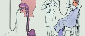
At home, a patient with symptoms of myocardial infarction can be provided with the following first aid:
- the patient must be seated with pillows under his back, his collar unbuttoned, his neckerchief, scarf, tie, etc. removed;
- open the window to provide access to fresh air;
- give the patient half a tablet of Aspirin (the tablet should not be swallowed, it must be chewed and left under the tongue). Patients who were routinely prescribed aspirin to prevent blood clots may be given one tablet;
- give the patient one tablet of Nitroglycerin, if necessary, after fifteen to twenty minutes the patient is given another tablet. This drug is contraindicated in patients with low blood pressure. Also, it is necessary to take into account that Nitroglycerin during a heart attack does not relieve pain symptoms, but is prescribed to reduce the severity of shortness of breath;
- in the absence of contraindications (severe forms of congestive heart failure, low blood pressure, decreased heart rate, etc.), you can give the patient 20 mg of propranolol;
- If necessary, the patient can be given a mild sedative.
Additionally, it is recommended to measure your pulse every five minutes and your blood pressure every ten minutes.
Attention! The emergency physician must be notified of all medications that were given to the patient, his blood pressure (if it was measured several times, all measurement results are reported), as well as all medications that are taken routinely.
Who is at risk of a heart attack?
Some conditions increase the likelihood of myocardial infarction. These include:
- hypertonic disease;
- diabetes;
- insufficient physical activity;
- increased body weight, obesity;
- smoking, alcohol abuse and other intoxications;
- cardiomyopathy (pathological increase in the volume and mass of the myocardium, which the coronary vessels cannot normally supply with blood);
- dietary errors (an abundance of foods rich in animal fats and cholesterol increase the risk of heart attack);
- male gender (this factor is important until about 60 years of age, then the likelihood of a heart attack in men and women is equal);
- old age (myocardial infarction occurs much more often in the elderly, although in recent decades it has also been observed in young people);
- increased psychological stress.
You can read about the causes of a heart attack in this article.
A healthy lifestyle is the best prevention of heart attack
Forecast
Myocardial infarction can be complicated by diseases of varying severity; therefore, MI most often gives a conditionally unfavorable prognosis. The worst consequences are expected in the case of extensive heart attacks, when large areas of the heart muscle are affected. So-called microinfarctions can be carried on the legs, but this does not mean that such patients are not in danger. Most likely, arrhythmias may appear over time. Therefore, it is extremely important to carry out timely treatment of MI, especially in the form of thrombolytic therapy, after which the prognosis of the disease significantly improves.
Video: What are the chances of survival after a major myocardial infarction?
Rules that must be strictly followed:
- take all medications prescribed by your doctor strictly according to the prescribed regimen;
- adhere to a diet depending on the severity and period of the disease;
- do not make sudden movements, do not worry, do not get irritated and do not tense up;
- sit down and get up in the first days only with the participation of another person;
- when the doctor allows the patient to sit up in bed, it is necessary to help him do this smoothly;
- monitor blood pressure and pulse - at least 3 times a day for the first 10 days;
- monitor bowel function and avoid constipation;
- control the amount of fluid you drink and excrete.
Symptoms
AMI can occur at rest or during exercise.
The myocardial infarction presentation may include any of the following:
- sharp pain in the chest, often described as “kicked by a horse’s hoof”;
- dull pain in the upper abdomen;
- back, neck, jaw, or arm pain;
- shortness of breath, suffocation;
- profuse cold sweat;
- nausea, vomiting;
- loss of consciousness.
Unlike men, for women chest pain is less likely to be the first sign of heart failure. These women report less common symptoms such as unusual tiredness, trouble sleeping, upset stomach and anxiety in the month before their heart attack.
In women with diabetes during acute myocardial infarction, the clinical picture may be blurry. They often suffer a myocardial infarction without any noticeable symptoms at all.
Rehabilitation
Rehabilitation after a heart attack will last a lifetime. It consists of the following stages:
Support of therapy, while you cannot refuse the prescribed medications, even if you do not feel pain. Prevention of heart disease.
Refusal of bad habits and unhealthy lifestyle, i.e. from smoking, drinking alcohol and drugs, introducing proper nutrition and light physical activity.
Return to work. However, you need to be prepared for the fact that it is not always possible to return to a full working state.
Consequences and complications
In any period from the onset of an acute infarction to its resolution in the form of the formation of a connective tissue scar, the following complications may be observed:
- cardiogenic shock;
- heart failure;
- ventricular fibrillation;
- wall rupture due to myomalacia;
- formation of a parietal thrombus and thromboembolism;
- pericarditis.
The lethal outcome of this disease is recorded in the early stages in approximately 35% of cases. Its cause is rhythm disturbance, cardiogenic shock and acute heart failure. Later death may occur as a result of the formation of an aneurysm with rupture, thromboembolic complications, or acute pericardial tamponade.
After the acute period, the heart muscle adapts to new working conditions, but there remains a high risk of repeated episodes.
Diagnostics
An ECG reveals changes in the heart characteristic of a heart attack, all risk factors and lifestyle are clarified. The presence of heart disease, heart attacks, and strokes in close relatives is clarified.
During the examination, attention is paid to the condition of the skin, the presence of wheezing in the lungs, and murmurs in the heart area. Blood pressure is measured, all irregularities and instabilities are noted. The presence of enzymes in the blood is sought, which get there when the cells that contain the myocardium die.
A general and biochemical blood test is taken, which will help determine cholesterol levels, the presence of increased leukocytosis, signs of anemia, and high blood sugar levels. A blood test will help determine your triglycerd (fat) levels.
An echoECG is performed, which will help examine the functioning of the heart, its size and structure, assess the condition of the valves and the degree of vascular damage. Find changes in the work of the contracting muscle.
Coronary angiography. Helps more accurately determine the location of narrowing of the arteries.
Chest X-ray is done to detect dilatation, atherosclerotic changes in the chest. Impaired lung function and some complications of the disease.
Kinds
This syndrome can occur with varying degrees of intensity, symptoms and localization.
Myocardial infarctions differ according to the size of the affected area:
- Finely focal.
- Large-focal.
Small-focal infarction, as the name suggests, has a small area of the affected area of the heart and a small number of foci of necrosis. They occur in a fairly mild form and have more favorable prognoses. There are no ruptures in the muscles or the appearance of a cardiac aneurysm. Very rarely, heart failure and ventricular fibrillation may occur after a heart attack.
Large-focal infarctions are characterized by correspondingly large affected areas. A huge number of myocardial cells die. Treatment and rehabilitation after this is very long, and subsequently complications may arise, including a second heart attack or death. The most dangerous is an infarction of the anterior wall of the myocardium.
During large focal heart attacks there is always a risk of death in the first 6-12 weeks.
Early signs of a heart attack
Most of the patients (approximately 60 to 80%) who were observed by me with this diagnosis indicate that the disease did not begin suddenly in them. It was preceded by precursors of a heart attack, or a prodromal period. The most favorable outcome was observed in those patients who sought help or were brought by the team in the first hours of the attack.
I would like to make a small disclaimer - early signs of myocardial infarction do not always occur, it all depends on the pain threshold and the state of the patient’s nervous system.
But in most cases I have seen the following symptoms:
- Pain along the anterior surface of the chest radiating to the left arm, part of the lower jaw, and scapula.
- The unpleasant sensation is not relieved by taking nitro-containing drugs and goes away only after the administration of narcotic analgesics.
- It has a constant, increasing or wave-like character with periods of subsidence and resumption.
- Lasts more than 20-30 minutes.
- The patient begins to sweat and tries to sit down or lie down as high as possible. This position makes the attack a little easier.
- Shortness of breath develops, the skin becomes pale, and the nasolabial triangle turns blue.
- When it comes to the first signs of myocardial infarction, rhythm disturbances cannot be ignored. According to my observations, they are observed in 90% of cases at the stage of ischemia development and before scar formation.
Any suspicion of a heart attack requires an immediate ECG, which will reveal all signs of ischemia, and at a later stage, necrosis and scar formation. Read more about what changes will be visible on film here.
Risk factors
For your information!
During myocardial infarction, the patient usually experiences severe pain lasting more than 15 minutes, which is not relieved by taking nitroglycerin. The pain occurs suddenly and quickly becomes less intense. At the slightest suspicion of a heart attack, immediate hospitalization is necessary. To establish a diagnosis, the following methods are used: ECG, echocardiography, coronary angiography. Treatment should only be carried out in a hospital.
- smoking
- fat metabolism disorders
- diabetes
- arterial hypertension
- stress and/or depression
- abdominal obesity
- sedentary lifestyle
- insufficient consumption of fruits and vegetables
- alcohol abuse (as well as complete abstinence from it)
How to prevent recurrent heart attacks?
Prevention of relapse consists of eliminating all possible risk factors:
- treatment of such disorders: diabetes mellitus;
- hypercholesterolemia;
- hypertension.
Doctors also prescribe the patient to exercise and avoid situations that cause stress.
It is recommended to adhere to treatment based on the following medications:
- beta blockers;
- aspirin;
- nitroglycerine.
Periodically, the patient must undergo examinations to diagnose the functioning of the heart and cardiovascular system (clinical examination, ultrasound, ECG, stress test).