Why do you need a puncture?
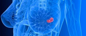
If the mammary gland has enlarged, hardened, or upon palpation the doctor has detected the presence of neoplasms, the mammologist may prescribe a puncture of the mammary gland cyst. Puncture is a puncture of the gland and analysis of the contents. To do this, the patient exposes her breast and a needle is inserted under the skin. This method has long been successfully used in modern medicine to diagnose dangerous diseases such as:
- malignant tumor;
- benign formations;
- cysts of different sizes and origins;
- lipomas (in everyday life these tumors are known as wen).
The insidiousness of these diseases lies in the fact that at an early stage they are practically asymptomatic, and without laboratory research methods, even a doctor cannot make a correct diagnosis. When the patient begins to feel pain, this indicates that the disease is already rapidly progressing, and most often it is necessary to resort to surgical intervention. Therefore, puncture is recommended for everyone who is at risk for developing cancer.
This method is one of the most effective, reliable, painless and easy to use. Also, one of the advantages of the puncture is that the tests come back quite quickly, already on the third to fifth day after the procedure.
Statistics claim that if the manipulation is carried out correctly, the tests show reliable results. Therefore, if for some reason the doctor prescribes a puncture of the mammary gland, you should not refuse.
https://youtu.be/XfEXIttykVc
Methods for diagnosing breast tumors
Mammography is performed both as a means of diagnosing the disease and as a preventive measure.
Various methods are used to identify cysts in the mammary gland. Their combination depends on how well it is possible to determine the type of pathology after standard procedures - mammography and ultrasound.
A mammogram is a special x-ray aimed at examining the breast. It can be used to assess the position of the cyst, shape and size. For prevention, after 35 years of age, mammography can be done once every 2-3 years, and after 45 years of age - every year.
Ultrasound helps measure the size of the cyst and identify mural formations inside it. Ultrasound is especially effective under the age of 30, when mammography cannot be used due to high breast density.
Additional diagnostic methods include:
- MRI. Allows you to accurately describe the structure of the cyst, and also indicates even the most minor changes in the surrounding tissues.
- Cytology. The material obtained during the puncture is taken for cytological analysis. You can find out about the presence of malignant processes.
- Pneumocystography. Helps to study the walls of the capsule. Air is introduced into the cavity, after which the boundaries of the neoplasm are examined.
- Dopplerography. It is carried out to identify blood impurities, which indicates the malignant nature of the formation.
Only after a biopsy is it possible to establish the exact nature of the cyst and ensure the absence of oncological processes. Diagnostic methods are almost always complemented by laboratory tests of blood and urine to identify inflammatory processes in the body.
How is the puncture performed?
Breast puncture is an important diagnostic method . There is no need to be afraid of this procedure. It is carried out as follows:
- The patient lies on the couch on her back and undresses to the waist.
- The nurse applies an anesthetic cream to the site of the intended puncture or administers an injection of a mild local anesthetic. General anesthesia is not required for the operation. If the patient is nervous, on the eve of the procedure the doctor may prescribe her to take sedatives in a dosage appropriate for her age.
- The surgeon inserts a needle into the mammary gland and takes out the contents, which are then placed in a test tube and sent for testing.
- The puncture site is covered with a sterile napkin. After this, the patient should spend some time at rest, and then can go home. The contents that are taken from the mammary gland are sent to a clinical laboratory.
Reasons for the development of neoplasms
Hormonal changes in the body during menopause can trigger the formation of cysts in the mammary gland
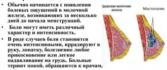
The formation of cysts is caused by hormonal imbalance, which can be caused by gynecological diseases. This is a common disease, occurring in almost every second woman. The following reasons can provoke a breast cyst:
- constant changes in the body caused by the menstrual cycle;
- sharp hormonal changes during pregnancy and lactation;
- period of menopause and puberty.
If a woman has diseases of the pituitary gland, which is responsible for the production of certain sex hormones, the risk of cysts also increases.
Gynecological diseases are a provoking factor for the formation of cysts in the mammary glands
Other accompanying factors that also affect hormones and breast condition can provoke the formation of a tumor:
- frequent abortions, childbirth after the age of 35, early onset of menstruation;
- refusal of breastfeeding, birth of a large fetus;
- gynecological diseases: endometrial pathologies, oophoritis, adenomyosis, tissue dysplasia and other disorders;
- sexual pathologies and changes, including the use of interrupted sexual intercourse as contraception;
- exposure to stress, frequent psycho-emotional overload;
- diseases of the liver and biliary tract, which interfere with the natural elimination of waste products from the breakdown of estrogen;
- diseases of the adrenal glands, thyroid gland, diabetes, hereditary predisposition.
Knowledge about the causes of cysts will help patients pay more attention to preventing its formation and take care of their health.
Preparation for the procedure
Many patients believe that getting a puncture is dangerous. This opinion is just a common misconception. This procedure is no more dangerous than a regular intramuscular injection. It is best to perform a puncture of the mammary gland under ultrasound guidance. The main thing is to properly prepare for the operation:
- Take general urine and blood tests. If tests reveal the presence of an inflammatory process in the body, the puncture is postponed until the source of inflammation is eliminated.
- Take blood clotting tests.
- Do not eat anything for at least several hours before surgery, as the administration of the medicine may cause vomiting.
- Do not wear a bra that compresses your breasts for at least a few days before the procedure, so as not to interfere with blood circulation. If blood circulation is impaired, puncture can lead to a large hematoma at the puncture site.
The manipulation can be dangerous if the patient does not properly care for the puncture site after the puncture. Therefore, before going for the procedure, it is necessary to obtain precise instructions from the doctor on how to speed up the healing of the wound after manipulation. Perhaps the doctor will recommend antibiotics to prevent inflammation, as well as antiseptics for external treatment of the wound.
Also, to prevent complications after puncture, the patient should avoid contact with patients with infectious diseases and not visit crowded places, because any medical intervention leads to a temporary weakening of the immune system, and the risk of “catching” an infection increases significantly.
Techniques and types of breast puncture
Breast puncture is performed on an outpatient basis and does not require specific preparation. Before puncturing, the patient will undergo an ultrasound, mammography, and cystography , which will clarify the location and size of the pathological focus. The next stage of the diagnostic search will be a puncture and removal of tissue for cytological examination.
It is not advisable to perform a puncture on the eve of or during menstruation, and the optimal interval is considered to be between 7 and 14 days of the menstrual cycle. A week before the scheduled puncture, blood thinners (aspirin, warfarin) and non-steroidal anti-inflammatory drugs should be discontinued. The day before the puncture, it is better not to drink alcohol.
To reduce discomfort, the puncture area is treated with local anesthetics, although in some cases the procedure performed without anesthesia does not cause pain due to the very thin needle.
You should come to the puncture in comfortable, non-restrictive clothing that is easy to take off and then put on. If a woman is very worried, and the procedure will be carried out after the administration of sedatives, then it is better to think in advance which of her relatives or friends can help her get home.
The most common option for puncture of the mammary gland is an ultrasound-guided puncture - a puncture biopsy. During the study, the patient can sit or lie on her back, the puncture area is treated with an antiseptic solution, then a syringe with a puncture needle is taken, which is inserted into the area of the pathological focus. To achieve high accuracy, the doctor uses an ultrasound machine, which shows not only the localization of the lesion, but also the movement of the needle towards it through the breast tissue. After collecting the changed tissue, the needle is quickly removed, and the puncture site is treated with an antiseptic and covered with a dry cloth.
The material taken through puncture is immediately sent for cytological examination, the result of which can be obtained in the coming days. A puncture biopsy allows for the most accurate diagnosis by microscopic analysis of the cells taken for examination.
After a puncture biopsy, some unpleasant consequences are possible - tissue swelling and hematoma at the puncture site. To minimize the likelihood of these effects, experts recommend applying an ice pack to the gland immediately after surgery. If pain is severe, painkillers may be prescribed.
If the pain is caused by the accumulation of exudate in the cystic cavity, which bursts and presses on the glandular parenchyma, then puncture of the breast cyst helps to significantly reduce pain and discomfort in the chest.
Fine-needle puncture biopsy is usually used for superficially located formations that are easily accessible to palpation. In diagnostically more complex cases, in addition to the one described, other methods of puncture of breast tissue are used. These include:
- Stereotactic puncture;
- thick needle;
- Incisional and excisional puncture;
- Trephine biopsy;
- Aspiration of a breast cyst.
Stereotactic (fine- and thick-needle) puncture in technique is very similar to that described above, but the patient must lie on her back, and the surgeon punctures several points in the mammary gland under ultrasound or mammography (X-ray) control. If a mammograph helps determine the exact localization of pathologically altered tissues, then before puncturing several pictures are taken in different projections so that the resulting image is three-dimensional. The method is informative for deeply located formations that are difficult to reach with conventional puncture.
A core needle biopsy involves the use of a large-diameter needle, which allows you to obtain a sufficient amount of tissue for microscopic analysis. This type of puncture is more informative, since there is enough tissue not only for cytological, but also histological examination. However, a core biopsy is more painful and necessarily requires local anesthesia.
An incisional biopsy is more extensive; in it, fragments of the affected parenchyma are excised under local anesthesia. This type of puncture is indicated in cases where it is impossible to exclude the presence of cancer or the result of a conventional puncture cannot be considered reliable. Tissues removed by incisional biopsy are subject to morphological examination.
Trephine biopsy is indicated for tumors that cannot be palpated. It is performed using a special biopsy gun under ultrasound guidance. Local anesthesia is required.
Puncture of a breast cyst is carried out by aspiration of its contents. Under ultrasound control, a needle is inserted into the cyst, then a syringe is attached to it, through which the contents of the cystic cavity are taken and removed. Complete extraction of exudate helps reduce pain, collapse and obliteration of the cyst cavity with recovery.
Excision puncture can be performed as not only a diagnostic, but also a therapeutic method for small breast formations. It is a minimally invasive intervention in which the entire tumor is removed if its size does not exceed 2.5 centimeters, otherwise the surgeon takes only the area of the tumor for histological examination. Excisional biopsy may be indicated for fibroadenoma, fibroma, oleogranuloma and other benign processes in the gland.
Video: trephine biopsy of the breast
The essence of the procedure
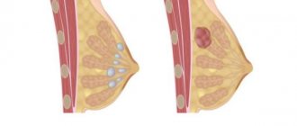
The cyst puncture procedure means a medical procedure that involves puncturing an abnormal formation, followed by drawing out the liquid contents from it.
This procedure is prescribed by a mammologist. It is necessary for:
- Determining the nature of the cells of the pathological formation, in other words, identifying their malignant form. Such a study is mandatory for use in cases where there is a change in the color of the skin of the breast or if various discharges occur from the nipples;
- Cyst treatment. This procedure can be used as a therapeutic treatment. However, it can only be used to eliminate single large formations.
Types of puncture
The main area of application of this procedure is puncture biopsy of the mammary gland, that is, carrying out diagnostic measures. Based on this, it is divided into several types of such a procedure, which make it possible to identify pathology parameters:
- Fine needle aspiration puncture. This type of procedure is performed on those anomalies that can be felt upon palpation. The essence of the procedure is to first outline the problem area, and then treat it with an anesthetic. Next, insert a thin corner located on the syringe into the gland, and then pump out a certain amount of liquid;
Worth knowing! This technique is minimally invasive and leaves no scars.
- Stereotactic fine needle puncture. This kind of technique involves taking images of pathological tissue from its different parts. When such a formation is small in size, the procedure is monitored by ultrasound;
- Thick needle puncture. A similar procedure is used to obtain a large area of tissue necessary for a more complete study and, accordingly, a more accurate diagnosis. To do this, use a thick needle with a cutting device;
- Stereotactic puncture. It is carried out in cases where the pathology cannot be palpated due to its deep location in the tissues;
- Incisional puncturing. This procedure involves excision of part of the tumor, which is performed under local anesthetics. It is prescribed when there is doubt that aspiration biopsy has given the correct results;
- Excisional puncturing. This type of procedure is considered a minor operation. During it, part or all of the pathology, whose size is less than 2.5 mm, is excised. And if atypical cells are detected, it is necessary to remove the lymph nodes.
Thick needle puncture
Stereotactic fine needle puncture
Important! Carrying out aspiration and incisional procedures does not always provide accurate information. Therefore, it is recommended to perform a puncture.
Indications
The main objectives of the puncture are:
- determining the type of tumor neoplasm;
- identifying the lesion;
- restoration of violations that may affect internal structures.
On this topic
- Breast
Can fibroadenoma hurt?
- Natalya Gennadievna Butsyk
- December 5, 2020
In addition, the procedure allows, in some cases, to detect the presence of microcalcifications. In addition, manipulation is carried out in the presence of specific discharge from the nipples or if there is deformation of the nipple area.
In the presence of large tumors, it is important to conduct a thorough diagnostic examination.
The puncture is indicated in the following situations:
- During an ultrasound examination, a lesion was identified without the manifestation of characteristic symptoms, which makes it possible to determine a benign tumor;
- mammography did not reveal cystic growths, while ultrasound showed their presence .
Microcalcifications
They are small areas of calcified tissue. Upon closer examination, dense structures that are not similar to others are clearly visible on mammography.
In this condition, only a fine-needle biopsy procedure is prescribed. Rarely can vacuum aspiration be used.
Violation of breast structures
It is necessary to understand that distorted ducts of the mammary glands are a clear sign of the development of a malignant neoplasm. Ultrasound examination rarely shows this condition in most cases. For this reason, specialists resort to puncture.
This procedure, if violations are detected in the structural structure of the organ, is carried out without fail under the control of an ultrasound machine.
If the study shows pathological cells, after puncture manipulation an additional surgical biopsy is performed.
Sizes of formations
Puncture is not indicated for all tumor sizes. If there is a suspicion of a cyst, then first of all they do an ultrasound and mammographic examination. If the tumor volume does not exceed 1-1.5 centimeters, a puncture procedure is allowed.
Aspiration
In cases where the development of a cyst is accompanied by unpleasant sensations and discomfort, the doctor prescribes a fine-needle type of diagnosis.
On this topic
- Breast
Do lymph nodes enlarge with mastopathy?
- Natalya Gennadievna Butsyk
- November 29, 2020
Indications include pathological signs such as:
- detection of wall thickening;
- presence of wall deposits;
- heterogeneity of structure.
An additional indication is the absence of enhancement of the acoustic shadow on ultrasound.
Interpretation of puncture results
Analysis of the punctate is usually done within 3-4 days, but if necessary, an urgent study can be carried out - within a few minutes.
Conclusion options may be as follows:
- results are within normal limits;
- a benign tumor was detected (adenoma, lipoma, fibroma, cystoadenopapilloma);
- a breast cyst was discovered;
- an inflammatory process is detected;
- a cancerous tumor has been identified - its detailed characteristics are given in accordance with the methods used;
- The diagnosis has not been clarified; additional research is required.
The results obtained are the basis for developing treatment tactics - conservative, surgical treatment, or combined treatment in an oncology clinic.
Preparation rules
There is no special preparation for puncture and breast ultrasound. It is recommended to conduct the examination on the 7-10th day of the menstrual cycle, that is, 2-3 days after the end of menstruation. During this period, tumors are enlarged to the maximum, which increases the accuracy of diagnosis.
No diet or medication is required. It is enough to carry out hygienic treatment of the breast. If a woman is constantly taking hormonal drugs or anticoagulants, treatment should be temporarily stopped. The possibility of a break is discussed with the attending physician.
There is no special preparation for the study. It is not recommended to take any medications before taking material unless prescribed by a doctor. It is forbidden to take painkillers based on non-steroidal anti-inflammatory substances, aspirin. If necessary, the doctor will perform pain relief with drugs that do not reduce blood clotting.
On the day of the procedure, you should not drink coffee or eat foods that increase blood pressure. A light breakfast is available. The analysis is carried out immediately after the end of menstrual bleeding, since this can be difficult in the second phase of the cycle.
What's happened
Puncture is one of the diagnostic examination methods.
Diagnostics is an important stage, which should be carried out regularly. The earlier the disease is detected, the easier it is to cure it without developing serious consequences.
Puncture of the mammary glands is a diagnostic measure, which consists of taking a piece of tissue (biopsy) for further cytological examination.
This diagnostic method is considered one of the most effective and most accurate. In addition, with its help it is possible to determine the nature of the origin of a benign formation and its nature.
Price
Puncture with ultrasound of the mammary gland is carried out strictly according to the doctor’s indications. It can be done at your place of residence free of charge if you have a medical insurance policy. You can also do the examination at a private center.
| City | Price, rubles |
| Moscow | 2500-3000 |
| Saint Petersburg | 2000-3000 |
| Ekaterinburg | 1700-2500 |
| Novosibirsk | 1900-3000 |
| average cost | 2025-2875 |
Ultrasound-guided puncture makes it possible to distinguish between benign and malignant tumors at an early stage. The technique is used to make women feel better with large breast cysts. The procedure is easily tolerated and is not accompanied by complications.
How they do it
The entire process must be monitored by an ultrasound machine. An exception may be the presence of neoplasms located close to the surface. The analysis does not require any additional preparation other than following the recommendations of a specialist.
On the day of the study appointment, the patient arrives and can leave the medical facility immediately after the procedure is completed.
Despite the fact that puncture is a painless method, a woman may experience minor discomfort during the insertion of the needle.
On this topic
- Breast
What breast fibroadenoma looks like on ultrasound
- Olga Vladimirovna Khazova
- November 29, 2020
To perform the manipulations, the patient must take a lying position. First, the specialist uses the camera head to determine the exact location of the cystic tumor. After this, the needle is inserted. Without stopping the ultrasound control, punctures continue, which allows the cyst to be gradually removed from the mammary gland.
Upon completion of the puncture procedure, the collected material is sent to the laboratory for further histological examination. It takes several days to receive results.
Such a diagnostic study does not pose a danger to the female body, and ultrasound control allows it to be performed correctly.
Progress
As already written above, such a procedure can be carried out as a therapeutic measure. In the case where the pathology can be felt with your own hands, then the puncture is also carried out by touch. If there are any problems, the procedure is carried out under ultrasound control.
During the puncture, a special needle is used to puncture the pathology and remove the contents through it. Next, he is sent for analysis to test for the presence of atypical cells or, in other words, cancer cells in the liquid.
When the procedure is carried out to treat a formation, then after the contents are evacuated from it, ozone is pumped into it, which leads to gluing of the walls. In some cases, ethyl alcohol can be injected there, although such a procedure is dangerous, due to the chance of developing tissue necrosis.
This procedure is performed on an outpatient basis. In the case of using the fine-needle method, the use of anesthetic is not required. And if a thick aspiration needle is used for this, local anesthesia is performed.
The last option is used when there are large pathologies. Moreover, before starting the procedure itself, the doctor must conduct a complete visual examination of the breast. His attention should be focused on identifying various skin defects, from wrinkling and thickening to redness. Read more about breast cyst removal.
Does it hurt?
The female breast is one of the most sensitive organs. That is why, in most cases, women refuse to undergo a puncture, because they are worried that they may experience unpleasant painful sensations.
However, most often the procedure is performed without the use of an anesthetic, since a thin needle is used during the procedure.
A puncture is not so much a painful as an unpleasant manipulation.
On this topic
- Breast
The role of breast puncture in fibroadenoma
- Olga Vladimirovna Khazova
- May 15, 2020
To a greater extent, the psychological aspect plays here. The patient imagines that they will make punctures in her chest and pump out the contents from it.
Subconsciously, she begins to convince herself that such events are quite painful. However, after the first puncture, fears usually disappear.
The puncture is done using anesthesia directly at the deep location of the cystic neoplasm.