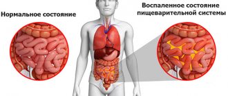
One of the most insidious acute life-threatening conditions is internal bleeding. They are a pathological condition accompanied by the release of blood from the vessels into the free abdominal cavity, retroperitoneal space or hollow organs (stomach and intestines). The whole complexity of the situation is that usually most diseases cause concern in a person in connection with the signaling pain syndrome. With any bleeding there are no such signs. This leaves this problem unnoticed for a long time. They begin to pay attention to it only when the condition of patients sharply worsens. But there are specific symptoms of internal bleeding, knowledge of which can help in the timely diagnosis of this complex problem.
Frequency of internal bleeding
Most often, bleeding occurs when the gastrointestinal tract is affected. Therefore, it is worth getting acquainted with emergency treatment methods for gastrointestinal bleeding. In total, there are about 20 diseases, the most significant and common among which are: acute erosive gastritis and stomach cancer, varicose veins of the esophagus, cirrhotic liver disease. In the case of stomach cancer, the danger is a disintegrating tumor. The only sign that allows the patient to suspect something is wrong is the darkening of the stool, which occurs due to the coagulated blood contained in it. If vomiting occurs, the vomited masses, again due to coagulated blood, have the color of coffee grounds.
Symptoms of internal bleeding depend on its location and the degree of blood loss. In some cases, it is necessary to distinguish bleeding from the esophagus or stomach from bleeding from a damaged lung. In case of lung pathology, foamy, unchanged scarlet blood is released.
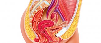
Diseases of the female reproductive system can also cause internal bleeding. The most common cause is tubal abortion. When a fallopian tube ruptures, blood accumulates in the abdominal cavity, creating a feeling of tension and some pressure in the pelvis, especially on the rectum. By the way, bleeding from the anus is also quite common. Subsequently, irritation of the peritoneum occurs with blood, which leads to the development of a state of shock, loss of consciousness, and fainting. The pulse in this case becomes frequent and thread-like. Upon examination, abdominal bloating, stool and gas retention are revealed. The patient becomes pale and covered in cold sweat.
First aid for bleeding: rules for applying a tourniquet
When bleeding, the rate of blood loss can be a concern, so in many cases you need to act quickly. First aid measures depend on the type of bleeding, its location, the nature of the injury and some other factors. In this article we will talk about ways to combat blood loss in different situations.
Guidelines and a poster for stopping bleeding on the stand are available after the article.
Types of bleeding
Most often, bleeding is grouped according to anatomical principles, taking into account the damaged blood vessel.
According to this classification, there are 3 main types of bleeding :
- Arterial. The blood stream is pulsating and scarlet in color. It is characterized by a high rate of blood loss and is the most dangerous.
- Venous. The blood is dark and may flow more slowly.
- Capillary. The blood is bright red, appears slowly and in a small volume. Sometimes it appears in the form of small drops on the surface of the skin.
Separate topic:
Types of external bleeding
There is also parenchymal bleeding that cannot be seen. It occurs when the integrity of the liver, pancreas, and kidneys is damaged. Parenchymal bleeding is similar in nature to capillary bleeding, but poses a great danger to life. With deep penetrating wounds or damage to the integrity of internal organs, bleeding can be mixed.
Internal and external bleeding are also distinguished according to the direction of blood release. In the first case, blood accumulates in the cavities of the body, in the second, it comes out through wounds.
We recommend additional material:
Providing first aid for wounds
Rules for applying a tourniquet
A tourniquet is applied only to stop arterial bleeding, and also if an arm or leg was amputated as a result of injury. In other cases, the use of a tourniquet is not advisable due to the high degree of injury to the skin and soft tissues. To temporarily stop bleeding, you can use an Esmarch tourniquet or a handy rubber material.
Rules for applying a tourniquet for bleeding
Basic rules and sequence of applying a tourniquet:
- If possible, raise your arm or leg for a few seconds and fix it in a comfortable position - this will lead to the outflow of venous blood.
- The tourniquet is applied over clothing or a piece of fabric is placed under it. This is necessary to protect the skin.
- The first two turns need to be made as tight as possible, they are the ones who stop the blood, while the crosshair is applied on the back side of the artery.
- The maximum duration of applying a tourniquet in the warm season should not exceed 90 minutes, in the cold season - 60 minutes. If during this time the victim cannot be taken to the hospital, the tourniquet should be loosened for 10-15 minutes and the artery should be pressed with a finger. Then the tourniquet is applied again, 1-2 cm above or below the previous location. The duration of applying a tourniquet to children should not exceed an hour.
- The time for applying the tourniquet must be written down and attached in a visible place. In reality, due to problems with drawing up (searching for paper and pens in field or combat conditions, while there are more pressing tasks of saving the life of the victim) and preserving (the paper gets soaked in blood and spreads or is simply lost) notes, in modern In practice, it is customary to write the time of application of the tourniquet with a marker directly on a visible place on the body, for example, it could be the forehead; it is recommended to indicate the name of the rescuer or the person who applied the tourniquet.
Esmarch rubber hemostatic tourniquet
Indications:
- traumatic amputation of a limb;
- inability to stop bleeding with other known means.
Advantages:
- quite fast and the most effective way to stop bleeding from the arteries of the limb.
Flaws:
- the use of a tourniquet leads to complete bleeding of the distal limbs due to compression of not only damaged great vessels, but also collaterals, which for more than 2 hours can lead to gangrene;
- nerve trunks are compressed, which causes post-traumatic plexitis with subsequent pain and orthopedic syndrome;
- cessation of blood circulation in the limb reduces the resistance of tissues to infection and reduces their regenerative abilities;
- the use of a tourniquet can cause severe vasospasm and lead to thrombosis of the operated artery;
- restoration of blood circulation after use of a tourniquet contributes to the development of tourniquet shock and acute renal failure;
- the use of a tourniquet is impossible on the torso or is limited in anatomically difficult areas.
Errors:
- its use without indications, that is, for venous and capillary bleeding;
- application on a naked body;
- far from the wound;
- weak or excessive tightening;
- poor fastening of the ends of the tourniquet;
- lack of accompanying note;
- use more than 2 hours;
- covering the tourniquet with a bandage or clothing.
If there is severe bleeding, a tourniquet is applied to the upper third of the shoulder or middle third of the thigh. In these areas, the anatomical location of the humerus and femur makes it possible to stop the bleeding with maximum efficiency. Applying a tourniquet in other places will not give the desired result. If a limb is torn off, applying a tourniquet is mandatory even in the absence of bleeding.
If the tourniquet is applied correctly, characteristic signs will appear after a while. The limb below the application site will turn pale and cold, the bleeding will stop, and the peripheral pulse will not be palpable. The intersection of the tourniquet should be on the outside of the arm or leg, since the artery is located on the axillary side.
First aid
For arterial bleeding
If an artery is damaged, the bleeding is rapid, so you cannot hesitate. After quickly assessing the victim’s condition, measures must be taken to temporarily stop the bleeding. First, the artery is pinched with a finger; for this, certain points are used:
- If there is bleeding in the face, press the corner of the lower jaw with your thumb.
- In case of bleeding from the head, press on the area of the temporal bone in front of the ear.
- In case of arterial bleeding in the area of the shoulder joint, press the subclavian artery to the rib.
- If the hand is damaged, press the brachial artery to the bone from the side of the shoulder.
- If the integrity of the femoral artery is compromised, press with your fist on the pubic bone in the groin area.
First aid for arterial bleeding
After finger pressure, a tourniquet is applied in compliance with the rules described above. If you don’t have a tourniquet or similar material at hand, you can apply a twist. To do this, use a piece of twine or fabric. A loop is made from the material and placed on the desired area of the limb. A metal or wooden rod is inserted into the loop, with the help of which the bandage is twisted. Further actions are the same as when stopping bleeding using a tourniquet.
For venous bleeding
In most cases, it is easier to stop bleeding from a vein than from an artery, so neither a tourniquet nor a twist is practically used.
The algorithm for providing first aid is as follows:
- The wound is covered with several layers of bandage, napkins or any clean piece of fabric.
- Sterile cotton wool is placed on top.
- Fix everything tightly with a bandage, scarf or piece of fabric of the required width.
To consolidate the effect, the damaged limb is raised so that it is higher than the body and fixed. If it is not possible to apply a bandage, the wound is packed with a tightly rolled bandage. Sometimes this is enough to stop the bleeding.
If there is severe bleeding from a vein, a pressure bandage may be useless. In this case, you need to apply a tourniquet and apply an ice pack to the wound. After this, the victim must be taken to the nearest hospital.
For capillary bleeding
In most cases, capillary bleeding does not pose a threat to the life of the victim and, if first aid measures were correct, does not cause complications.
To stop bleeding during external bleeding, you must adhere to the following sequence:
- Treat the skin area with any antiseptic.
- Apply a napkin and secure it with a bandage;
- If a limb is injured, raise it relative to the body.
With various injuries or diseases, nosebleeds may begin. It occurs in case of damage to the blood vessels located in the mucous membrane; it can stop on its own, but in severe cases, first-aid treatment will be required.
First of all, you need to press the wing of the nose to the nasal septum. For minor damage to blood vessels, the bleeding should stop after 10 minutes. If this does not happen, a nasal tamponade is performed. In case of nosebleeds, you need to monitor the well-being of the victim and warn him that he needs to breathe through his mouth.
First aid for types of bleeding
For internal bleeding
It is quite difficult to detect internal bleeding. Symptoms largely depend on the type of damage and its location; most often, a rapid pulse (up to 140/min), a decrease in blood pressure and pale skin are observed.
First aid for internal bleeding is as follows:
- Help a person lie in a certain position.
- Limit movement.
- Monitor physiological indicators - pulse, respiration, blood pressure.
If internal bleeding is suspected, the victim should be taken to a medical facility as soon as possible.
If there is a suspicion that the bleeding is localized in the chest or stomach, the victim should be placed in a “reclining” position; if it is localized in the abdominal or pelvic cavity, raise the legs up.
Sources:
- Zavyalov V.N., Gogolev M.I., Mordvinov V.S. “Medical and sanitary training of students” 1988.
- D. V. Marchenko - “First medical aid for injuries and accidents” 2009.
- General surgery: textbook / Petrov S.V. – 3rd ed., revised. and additional – 2010.
Symptoms of hemothorax (blood in the chest)
How to identify and recognize internal bleeding in the chest? If blood accumulates in the pleural cavity, the so-called hemothorax develops. The pleural cavity is a small space that separates the lungs from the chest. The reasons leading to the development of hemothorax include the following: falls from a height, injuries with damage to the ribs and intercostal vessels, knife wounds, lung cancer, lung abscesses (that is, the formation of abscesses in the lung tissue).
When blood accumulates in the pleural cavity, there is difficulty breathing when inhaling and when coughing, sharp pain in the chest, a disturbance in the general condition - dizziness, weakness, fainting, pallor of the skin, increased heart rate and breathing, sweating. Percussion of the patient's chest reveals a shortening of the percussion sound over the affected half, weakening or complete absence of breathing. On an x-ray, it is possible to determine signs of mediastinal displacement towards the healthy lung.
First aid
The most important role in emergency care for bleeding of arterial origin is played by the time factor: for maximum effectiveness it should be provided no later than 2-3 minutes from the moment of injury. If it concerns the main arterial trunks, then bleeding from them must be stopped no later than 1-2 minutes after the injury. Otherwise, the chances of a successful outcome will decrease every second with every milliliter of blood lost.
Important! No matter how critical the conditions, before helping others, protect yourself first - put on rubber gloves from your travel first aid kit, and if you don’t have them, minimize contact with blood using available items (for example, cellophane).
The algorithm for stopping any arterial bleeding is as follows:
- Assessing the type of bleeding.
- Finger pressure on an artery that is damaged.
- Applying a tourniquet, applying maximum limb flexion or a pressure bandage.
- Applying an aseptic dressing to the wound.
This sequence of actions may vary slightly depending on the characteristics of the damaged anatomical area.
Methods to stop bleeding can be temporary or permanent. Temporary arrest of arterial bleeding is used at the stage of first premedical and medical care. The final stage is carried out in a hospital and is part of the hospital stage of care. It is worth noting that in some cases, temporary stopping measures are enough to completely stop the bleeding.
Hemarthrosis (blood in the joint cavity)
A common joint lesion is the development of hemarthrosis, which means the accumulation of blood in the joint cavity. A similar condition occurs as a result of injury, less often with hemophilia, scurvy. The anemic symptom is expressed insignificantly, in contrast to the local symptoms. The clinic distinguishes three degrees. With grade 1 hemarthrosis, mild pain is noted, the contours of the joint are slightly smoothed, and the range of motion is not changed. In general, the volume of blood in the joint is up to 15 ml. Grade 2 is characterized by severe pain, which intensifies with exercise, and smoothness of the joint is noted. There is an increase in the circumference of the affected joint by 1.5–3 cm compared to a healthy one. In the case of hemarthrosis of the knee joint, voting of the patella is observed. The blood content in the cavity is up to 100 ml. At grade 3, the pain syndrome is acute, the outlines of the joint are completely changed. The joint's circumference increases to 5 cm, and mobility is severely limited. Blood volume is more than 100 ml.
A little about bleeding and its types
Before considering arterial bleeding, it is worth first finding the answer to the question of what types of violation of the integrity of blood vessels there are. The first aid provided depends on this. Bleeding occurs:
- arterial;
- venous;
- capillary.
When bleeding occurs, red blood cells are lost. The main danger of damage to blood vessels is a reduction in the volume of blood circulating throughout the body and hemodynamic disturbances.
General symptoms
Thus, there are signs of bleeding that occur in any case, regardless of the source of the bleeding. Early signs include: pale skin and visible mucous membranes, cold sweat, general weakness, dizziness, darkening of the eyes, if the lungs are affected - cough with streaks of blood, if the digestive system is involved in the process - bloody vomiting or bloody diarrhea, symptoms of peritoneal irritation , which occurs when internal organs (spleen, liver, kidney) rupture. With moderate bleeding, a number of symptoms may be either mild or not appear at all
In other words, bleeding leads to the development of anemia. In addition to the above symptoms, anemia is manifested by increased heart rate and hypotension, that is, a decrease in blood pressure. The changed parameters directly depend on the degree of blood loss: with moderate - pulse no more than 75 beats per minute, systolic pressure drops to 100 mm Hg. Art.; with average – the pulse rises to 100 beats per minute, blood pressure decreases to 90-80 mm Hg. Art.; in severe cases, the pulse rises to 120-140 beats per minute, systolic blood pressure drops below 80 mm Hg. Art.
Correct ways to stop arterial bleeding
Putting all of the above together, we note that there are four ways to stop arterial bleeding.
- Compressing the artery with a finger. The main rule in this method is to press on the artery with all your fingers and hold it in this state for 10 minutes. If the bleeding is not very severe, it will stop quickly, but for deep wounds this will only be a temporary measure.
- Application of a tourniquet. In a car first aid kit, for example, a tourniquet is a mandatory component. But if you don’t have it at hand, you can replace it with another piece of fabric. Please note that the tourniquet cannot be thin; something like wire will not be suitable for this purpose, otherwise tissue death will begin. The tourniquet is applied above the wound (if the blood comes from the neck, below the wound), a cloth must be placed under it, or you can even tie it over your clothes. The duration of the tourniquet on the body is one hour in summer and 30 minutes in winter. Next, the tourniquet is removed for 10 minutes to allow the tissues to be saturated with oxygen, and then, if necessary, it is reapplied. To check whether the tourniquet is applied correctly, feel the pulsation of the artery below its location. If it is felt weakly or not felt at all, then everything has been done correctly and the bleeding has stopped.
- Pressure bandage. There are also few tricks in applying a pressure bandage. Gauze is placed on the wound, a tight roller is placed on top, and the entire structure is wrapped in a bandage. Difficulty may arise if the wound is located on the neck. In this case, the roller is bandaged to the neck by tying the bandage around the splint or raised arm on the side opposite from the bleeding.
- Flexion of the limb. The least effective method, but it can help if bandages and a tourniquet are not available. The limb is bent as much as possible and somehow fixed to the body. If the victim has the opportunity to hold the bent limb in this position himself, let him hold it like that until the doctors arrive. If not, you will take on the support role.
Arterial bleeding
First aid for arterial bleeding does not contain many rules, and they are quite easy to learn.
If you find yourself close to a person in such a situation, or if you find yourself in one yourself, following these simple recommendations can save a person’s life.
Author of the publication
offline for 2 months
Nika
69
I am interested in hiking and traveling, photography and videography. I have been going hiking since childhood. The whole family went and went - sometimes to the sea, then to the river, to the lake, to the forest. There was a time when we spent a whole month in the forest. We lived in tents and cooked over fires. This is probably why I am still drawn to the forest and, in general, to nature. I travel regularly. About three trips a year for 10-15 days and many 2 and 3 day hikes.
Comments: 0Publications: 667Registration: 10/23/2018
We recommend reading:
Actions to take when bitten by various poisonous spiders
Diagnostics
In case of combined trauma, laparocentesis is performed; it is a simple, fast, affordable and gentle method for diagnosing injuries to the abdominal organs and internal bleeding. An ultrasound of the abdominal organs is also sometimes performed.
An important point in confirming the diagnosis is laboratory blood tests. The analysis reveals a decrease in the number of red blood cells, the amount of hemoglobin and a drop in hematocrit.
If such symptoms and diagnostic data are detected, it is necessary to immediately identify the cause of the underlying disease and begin treatment. It should be remembered that the earlier the cause of internal bleeding is identified, the more effective and rapid the recovery process will be.
Internal bleeding is one of the most dangerous types of bleeding, requiring immediate hospitalization.
Author: surgeon M.M. Denisov — https://hirurgs.ru Save on social networks:
Danger of bleeding from an artery
Arterial bleeding belongs to the category of injuries that, in case of incorrect or untimely medical care, can lead to the most serious consequences, including death. This type of bleeding is considered the most dangerous, and the closed form, which cannot be immediately diagnosed, is the most insidious.
Arterial bleeding cannot always be stopped even in medical institutions, so this factor should always be taken into account when providing first aid to the victim. The most serious danger of any bleeding is the loss of blood, which causes disruption of the heart, and hence the cessation of oxygen supply to tissues and the brain due to impaired circulation.
Danger of major blood loss
Large blood loss primarily affects the quality of work of internal organs, the brain and the central nervous system. Arterial bleeding can devastate a person in a matter of minutes, disrupting not only blood circulation, but also causing interruptions in the functioning of the heart and oxygen starvation of the brain. During severe bleeding, the victim may lose consciousness almost immediately, and later, if measures are not taken to stop the blood loss, die.
As a result of severe bleeding, a lack of blood can take the form of hemorrhagic shock, a condition when the brain and lung tissue begin to rapidly atrophy due to oxygen starvation and lack of circulation. The development of anemia does not necessarily occur when a huge amount of blood is lost; in some cases, 300 ml is enough and blood pressure can drop to critical levels. A sharp drop in blood pressure can cause oxygen starvation (anoxia).
The most dangerous arterial bleeding
For ease of diagnosis, experts classify arterial bleeding as follows:
- Up to 500 ml (15%) light form.
- 500-1000 ml or 20% average degree.
- From 1 L to 1.5 L (30%) severe blood loss.
- Over 1.5 liters or 30%, blood loss is massive and poses a real threat to life for the victim.
- Blood loss of 2.5 liters to 3 liters (up to 60%) is fatal for the human body.
- Blood loss of more than 3 liters (from 60%) is considered the amount at which the human body cannot be resuscitated.
Dangerous localization of arterial bleeding
The most dangerous places for arterial bleeding are:
- Neck. Carotid artery.
- Head, temporal part.
- Thigh, groin and pubic area.
- Abdominal and thoracic areas.
Hip
Rupture of the femoral artery is considered one of the most dangerous types of arterial bleeding, in which significant blood loss can occur in a short period of time, in the absence of measures to stop it. The area where there is destruction of the artery is immediately freed from clothing and, by strong pressing with a fist, held at the site of the rupture until a tourniquet is applied.
This type of bleeding differs from similar ones in that two tourniquets are used simultaneously to stop blood loss; only in this case can control over the situation be guaranteed. The need to use two products simultaneously is explained by the presence of fatty deposits and dense muscle tissue in the area. Be sure to put a note under one of the tourniquets indicating the exact time when the application was made. The maximum allowed use of the drug to stop bleeding is two hours. During this time, it is necessary to deliver the victim to a medical facility, treat the wound and, using clamps and other instruments, completely eliminate the threat of bleeding and resume blood circulation, thereby preventing tissue death.
Neck
Arterial bleeding in the neck area is one of the most complex and dangerous in its manifestations. If a wound is identified that provoked a violation of the integrity of the carotid artery, the following actions must be urgently performed.
- Using any material (gauze, bandage, piece of fabric) clamp the damaged area.
- Place the victim’s hand, which is on the opposite side, behind the head as deeply as possible.
- Using a bandage or other material, make a thick tampon and place it on top of the previously applied fabric. Next, a tourniquet is passed through the victim’s hand in such a way that it tightly presses the roller, being on the outside, in which the elbow acts as a kind of lever, ensuring the desired tightness.
Localization of bleeding
Depending on the location of bleeding, there are many different types, let’s look at some of them.
The term uterine bleeding refers to any bleeding from the uterus, with the exception of menstruation. It is a companion to some gynecological diseases.
Most often, uterine bleeding is not directly related to organic pathology of the uterus and appendages. In this case it is called dysfunctional. Bleeding also accompanies ectopic pregnancy, complicated normal pregnancy, all kinds of uterine tumors and diseases associated with the blood coagulation system.
In any case of uterine bleeding, you should consult a specialist and not self-medicate. And the sooner the cause is identified, the sooner and more successful will be the treatment of the disease that led to the development of bleeding.
About the types of hemorrhagic conditions
In medicine, the internal type of hemorrhage is not classified in detail. Based on the cause of internal bleeding, there are the following types:
- Mechanical type. This condition occurs in the event of trauma to the vascular tissues through which blood flows. Occurs due to injury (often occurs in men) or due to surgery.
- The arrosive type of hemorrhage is considered to be a consequence of necrosis affecting vascular tissue or if newly formed structures grow and disintegrate.
- Diapedetic type. In this condition, the vascular tissue is not destroyed, but due to various pathological processes (hemorrhagic vasculitic changes, phosphorus poisoning and many other processes), the capillary network becomes highly permeable.
According to the type of vascular tissue there are:
- Arterial type, where destruction of the arterial vessel is diagnosed.
- Venous type, in which the veins are damaged.
- Capillary type. The blood medium spreads evenly from the capillary vessels. If blood pours out from different organs, then this is a parenchymal type of hemorrhage.
- Mixed type. Occurs with destructive changes in venous, arterial and capillary vessels.
If we take into account localization, then there are:
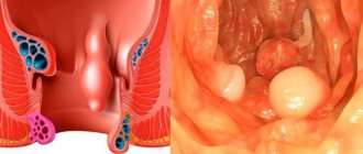
- Gastric and intestinal types of hemorrhages. Possible due to ulcerative processes in the stomach and duodenum, gastritis changes, inflammation of the intestines, also if the mucous membrane is cracked and there are newly formed structures. If esophageal hemorrhages are observed, the cause is liver dysfunction. In case of intestinal hemorrhages or intraperitoneal bleeding, the cause is hemorrhoidal changes or rectal fissures.
- Hemorrhages localized in the cavity spaces of the pleura due to torn vascular tissues between the ribs (hemothorax). This condition occurs when there is a closed injury to the chest.
- Hemorrhages that flow into the pericardium (pericardial sac), which compresses the heart muscle (hemipericardium). If this condition is ignored, it can manifest itself as heart failure followed by death. Such hemorrhage can occur with a mechanically damaged chest in the front, or surgical intervention on the heart muscle.
- Intra-articular hemorrhages (hemarthrosis). This type of bleeding often occurs when joint tissues are damaged (usually the knee joints).
According to the location where blood accumulates, there are:
- Hemorrhages in cavities. They are divided into bleeding of the abdominal cavity (if the abdominal area, chest and various organs are injured), pleural membrane (if the ribs are broken), and the skull.
- Hemorrhages within tissue structures, where blood accumulates in deep tissues that disintegrate, causing hematomas.
According to the amount of blood loss there are:
- Mild hemorrhages, where total blood loss is no more than 15% of the total blood circulation.
- Moderately severe hemorrhagic conditions, where blood is lost 20% of the total blood circulation.
- Severe bleeding, where the patient has lost approximately 1.5 liters of blood
- Massively manifested hemorrhagic condition. The total volume of blood circulation is reduced by more than 30%.
- Fatal blood loss occurs when the total circulating blood volume decreases above 60%.
Hemorrhages can occur:
- Obviously. After a certain time, the blood comes out into the external environment through the patient’s natural type of orifices.
- Is hidden. There are no main symptomatic manifestations; perhaps they are mild.
Given the time period, there are:
- Primary identified blood loss. They signal themselves after the vascular wall is ruptured.
- Secondary detected hemorrhages. They are observed a certain time after the traumatic factor that provoked them. This type of hemorrhage is divided into early, where manifestations occur after a period of 1 to 3 days due to the fact that an embolus has come out of damaged vascular tissue or the suture has been incorrectly applied. Late, secondarily detected hemorrhages appear three days after the vessel is damaged as a result of infection joining the wound.
Arterial bleeding, first aid: nuances of using a tourniquet
The tourniquet should not be kept on the damaged area for a long time. The maximum application time in warm seasons is 2 hours, in cold periods of the year – 1 hour. If the time for safe application of the tourniquet has expired, then every half hour it is loosened for 5 minutes so that the affected area is supplied with blood. When the bleeding stops, the device is untied, but the victim continues to be monitored. If the biological fluid begins to flow again, the tourniquet is reapplied above the place where it was previously located.
If for some reason the tourniquet was not loosened and was not removed on time, or was on the body for more than 3 hours, then it cannot be removed. The fact is that some of the cells died due to lack of oxygen and nutrients, and signs of tissue death appeared on the skin. When the tourniquet is removed, blood will begin to flow into this area. Toxins formed in dead tissue will enter the biological fluid. They will spread throughout the body. The spread of harmful substances will lead to the death of the entire organism.
Finger pressure
This technique should be used as a starting one when assisting a wounded person. The basic principles of digital compression depend on the anatomical region in which the artery injury occurred. The general rule is that the vessel should be pressed above the site of injury. But if bleeding occurs in the neck or head area, then the vessels are compressed downward from the wound. This is explained by the fact that the arteries in this area go upward from the heart.
Attention! When using any methods to stop bleeding, you need to lift the affected limb upward to reduce blood flow to it.
Damaged arterial vessels must be pressed against the bony protrusions, as they can slip out, and then bleeding will resume.
Places where arteries are pressed during bleeding
To better remember the method, you can use the 3D mnemonic rule:
- “Press.”
- "Ten".
- "Ten".
Also read: Applying a tourniquet for bleeding
It means that you need to press the artery by pressing with ten fingers of both hands for 10 minutes, after which it is recommended to check whether the bleeding has stopped. If it is stopped, and this happens if it is not the main arterial vessel that is damaged, then you can limit yourself to applying a pressure aseptic dressing to the wound.
Since the blood pressure in the arteries is very high, it will take a lot of effort to apply pressure to the vessel and stop the bleeding. Finger pressure is a method of temporarily stopping bleeding, so while one person is pressing the artery, the second should already be looking for a tourniquet and dressing material. There should be no time wasted taking off clothes or freeing limbs. At the same time, one of the eyewitnesses must immediately call an ambulance to provide first aid and transport the victim to a hospital.
The biggest disadvantages of the finger pressing technique are:
- significant pain for the injured person;
- physical fatigue of the person providing emergency assistance.
Speed of execution is considered the most important advantage of temporarily stopping external arterial bleeding using finger pressure.
Intracavity
A significant proportion of the total number of internal bleedings are those that occur inside the cavity of any organ and arise as a result of chronic pathologies of the gastrointestinal tract: peptic ulcers of the intestines and stomach, malignant tumors, varicose vessels of the esophagus with cirrhosis of the liver, erosive gastritis, etc. d. Gastrointestinal bleeding according to ICD-10 has a code of K92.2.
In addition, in surgical practice, Mallory-Weiss syndrome is often encountered, when a patient develops cracks in the esophagus as a result of alcohol abuse or a single large meal.
Another common cause of bleeding into the abdominal cavity is gynecological pathologies: ectopic pregnancy, ovarian rupture, etc. In gynecological practice, bleeding after abortion is often observed. This pathology may also occur with premature placental abruption or presentation, rupture of the birth canal and uterus during the birth process.
Below we will consider the types of bleeding and ways to stop them. This information is useful for everyone to have.
General principles of first aid for arterial damage
Providing first aid for arterial bleeding is based on several principles:
- Stop the bleeding and call an ambulance.
- Disinfection of the wound. To prevent the entry of pathogenic microorganisms, work with clean hands whenever possible. It is necessary to treat the edges of the wound with alcohol, vodka, cologne, lotion or other available means; apply sterile material to the damaged surface before bandaging; bandage the wound.
- Fixation of the injured limb with a scarf or splint.
- Anesthesia. If a person has a serious injury, then to prevent pain shock it is recommended to use painkillers (analgin tablets, tramadol capsules, ice).
- Safe transportation to a medical facility or awaiting the arrival of emergency doctors.
How to distinguish arterial bleeding from capillary and venous?
First aid for capillary, venous, and arterial bleeding is different. The following types of vascular lesions have different signs:
- with venous bleeding, cherry-colored biological fluid flows evenly from the wound;
- in the capillary form, blood flows evenly from the entire surface of the wound, as if permeating those tissues that are damaged.
Since during arterial bleeding, blood quickly flows out of the body, the likelihood of shock and death is high. To prevent this, you need to begin providing first aid to the injured person at the first sign.
Cauterization of blood vessels
Sometimes internal bleeding is stopped by cauterizing the bleeding vessel or tamponade. But in most cases, the patient needs emergency surgery under general anesthesia. If there are signs of hemorrhagic shock or the threat of its occurrence at all stages (preparation, surgery, postoperative period), transfusion measures are carried out. We looked at how to identify internal bleeding. The main thing is to do everything quickly and correctly, then a person’s life can be saved.
Intestinal bleeding
Intestinal bleeding can occur with various diseases of the intestine (both large and small). Most often it is caused by a duodenal ulcer. Bleeding can also be caused by tumor rupture and simple trauma. The patient may feel dizzy, spots flashing before the eyes, and sticky cold sweat. If bleeding is in the upper intestines, you may vomit coffee grounds and have black or dark cherry-colored stools. Intestinal bleeding most often requires urgent surgical intervention. In any case, stopping internal bleeding and treating its consequences are carried out in a hospital under the supervision of medical personnel.
Causes of pathology formation
The following diseases and conditions can lead to internal bleeding:
- Closed and open abdominal injuries with damage to internal organs (liver, spleen, mesentery of the small intestine);
- Ovarian apoplexy (ovarian ruptures);
- Rupture of an ovarian or pelvic cyst (most often occurs during sexual intercourse);
- Active physical activity (can provoke ruptures of cystic ovaries or enlarged internal organs);
- Injuries of the lumbar region;
- Aortic aneurysms that begin to dissect;
- Peptic ulcer of the duodenum and stomach;
- Ruptures of the mucous membrane of the esophagus (Malory-Weiss syndrome);
- Varicose veins of the esophagus and stomach;
- Disintegrating tumors of the gastrointestinal tract, abdominal cavity and retroperitoneal space.
The following circumstances are typical for internal bleeding:
- As a result of a head injury, a cerebral type of hemorrhage occurs.
- Rib fractures, both multiple and single, where the arterial vessels between the ribs and the pleural membrane are affected.
- Severe trauma to the abdominal region, leading to destruction of organs located in the abdominal area, which will cause intra-abdominal bleeding.
- Intra-articular tissues are fractured. These hemorrhages are not dangerous, but treatment must be carried out, otherwise aggravated conditions will arise.
- Severe dysfunction of the stomach and intestines in the form of ulcerative processes, malignant tumors, which will cause bleeding into the abdominal cavity.
- Cirrhotic changes in the liver also contribute to the fact that the patient may experience intra-abdominal bleeding.
- An aneurysmal vascular change that causes the aorta to dissect.
- The presence of esophageal cracks in the mucous tissue.
- Defective conditions of the reproductive system: cystic and polyposis changes. An intense regime of physical activity and active sports contribute to the rupture of the cyst. Hemorrhages can be due to the fact that the pregnancy occurs outside the uterus, in case of abortion, premature placental abruption, or the placenta is retained upon delivery. Sometimes women experience bleeding after childbirth.
Internal bleeding can never be an independent disease. They are always either the main manifestation or a complication of a number of pathological conditions.
Each type of bleeding has its own causes, the most common of which are injuries and illnesses in acute or chronic form.
- Open and closed injuries of the abdomen and lumbar region with damage or rupture of internal organs, most often the spleen or liver, less often the intestines and pancreas. Massive bleeding into the abdominal cavity is caused by a blow during a fight or during a car accident, compression - when pressed with a heavy object, etc.
- A rib fracture leads to bleeding into the pleural cavity.
- Traumatic brain injury. Bleeding inside the skull is life-threatening because the volume of the skull is limited. Any hematoma leads to compression of brain structures and serious consequences. It can develop not only immediately after an injury, but also several hours or days after it.
- Chronic diseases of the digestive tract. Bleeding into the cavity of the corresponding organ occurs with varicose veins of the esophagus, erosive gastritis, cirrhosis of the liver, peptic ulcer, malignant tumor process, the formation of a through hole in an ulcer of the duodenum or stomach.
- Gynecological diseases and pathological conditions - apoplexy (rupture) of the ovary, ectopic pregnancy, malignant neoplasm, rupture of an ovarian cyst. In obstetrics and gynecology, uterine bleeding can provoke abortion, previa or premature placental abruption. It can begin after childbirth due to rupture of the birth canal or uterus, late release of the placenta.
- Ruptured aortic aneurysm.
- Hemophilia is a hereditary disease in men with a malfunction of the blood clotting process.
- Hemorrhage in the brain as a consequence of traumatic brain injury.
- Multiple/single rib fractures affecting the intercostal arteries and pleura. Blood accumulates in the pleural cavity.
- Severe injury to the abdomen (impact, fall, car accident, etc.), which leads to damage to the internal organs located directly in the abdominal cavity. Most often in such situations, the spleen, liver, and intestines suffer.
- Fractures inside joints. In terms of their manifestations and consequences, intra-articular bleeding does not pose a great danger. But therapeutic measures must be carried out in any case: ignoring the pathology can cause exacerbations in the future.
- Serious disruptions in the gastrointestinal tract: ulcers, malignant neoplasms.
- Cirrhosis of the liver.
- Aneurysm, which is accompanied by dissection of the vascular aorta.
- Cracks in the mucous membrane of the esophagus.
- Reproductive system defects: cysts, polyps. Intense physical activity and active sports can contribute to the rupture of the cyst. Bleeding can also occur with an ectopic pregnancy, abortion, untimely abruption of the placenta or a delay in its release. Sometimes during the postpartum period, women may complain of bleeding.
We suggest you read How to determine the freshness of an egg
The cause of internal bleeding can be either injury or some chronic diseases. Massive, life-threatening post-traumatic bleeding into the abdominal cavity can develop as a result of blunt trauma to the abdomen with damage to the spleen and liver, less commonly the pancreas, intestines or mesentery (due to a blow, a fall from a height, a car accident, etc.).
Bleeding into the cranial cavity is one of the dangerous complications of traumatic brain injury. Since the skull, unlike other natural cavities, has a rigidly fixed volume, even a small amount of spilled blood causes compression of the brain structures and poses a threat to the patient’s life.
Bleeding into the joint cavity can be caused by both an intra-articular fracture and a bruise. It does not pose an immediate threat to life, but if left untreated it can lead to serious complications.
A significant proportion of the total amount of internal bleeding consists of bleeding into the cavity of any organ, developing as a result of chronic diseases of the gastrointestinal tract: malignant tumors, peptic ulcers of the stomach and intestines, erosive gastritis, varicose veins of the esophagus with cirrhosis of the liver, etc.
Another fairly common cause of internal bleeding is gynecological diseases: ovarian ruptures, ectopic pregnancy, etc. In gynecological practice, internal bleeding occurs after abortion.
There are many reasons why this pathological process is formed. As a rule, internal bleeding appears as one of the secondary symptoms of a serious disease.
The most common causes of internal bleeding include the following:
- Varicose veins of the esophagus or stomach.
- Tumors that have begun to disintegrate in the organs of the gastrointestinal tract and retroperitoneal spaces.
- Internal or external injuries in the abdominal area with damage to organs.
- Rupture of the hand (ovarian or pelvic).
- Physical exercise. Active sports can provoke rupture of cysts of various origins.
- Injuries in the lumbar region.
- Aortic aneurysms.
- Ulcers.
- Rupture or damage to the mucous membrane of the esophagus and intestines.
The reasons that cause bleeding from the esophagus are known to doctors. Common factors are:
- stomach ulcer;
- cirrhosis of the liver;
- pathological formations in the esophagus;
- varicose veins of the digestive system;
- neoplasms;
- burns;
- diseases associated with dysfunction of the cardiovascular system;
- constant uncontrolled use of medications;
- hemophilia;
- chronic stressful experiences;
- poisoning with alcohol-containing products.
In some cases, lymphogranulomatosis is mistaken for a malignant tumor. Only with surgery is a non-cancerous tumor diagnosed. The most common organs of the digestive system affected by lymphogranulomatosis are the stomach and small intestine.
Some medications taken by a person, if not properly monitored by medical personnel, can cause pathological conditions in the mucous membrane of the stomach and intestines. Damage leads to slow bleeding. The condition affects a person’s functioning.
In case of alcohol abuse, vomiting that occurs in this condition can lead to tearing of the longitudinal lining of the esophagus. Intoxication is accompanied by blood loss.
A fairly wide range of pathologies can be accompanied by blood loss in the intestines.
Causes of gastrointestinal bleeding
They are usually divided into 4 groups:
- Diseases of ulcerative and non-ulcerative nature. Ulcerative lesions are the most common cause of intestinal bleeding (about 75% of all cases, and the figure is higher in men).
- Duodenal ulcer that occurred after gastrectomy and other surgical interventions in the gastrointestinal tract;
- Nonspecific ulcerative colitis;
- Multiple ulcers of the large intestine accompanying Crohn's disease;
- Bleeding ulcers resulting from burns of the mucous membrane (due to poisoning with concentrated acid, mercury, lead, etc., long-term use of medications);
- Ulcers in places of mechanical trauma to the gastrointestinal tract;
- Formed due to stress or physical strain.