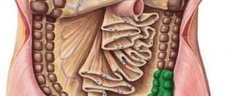
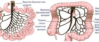
The large intestine is the final segment of the digestive tract and is divided into the colon and rectum. The colon is located along the perimeter of the abdominal cavity and has the following sections: cecum, ascending colon, transverse colon, descending colon and sigmoid colon. The length of the large intestine is 1.5 - 2 meters.
Colorectal cancer ranks third in frequency among all cancers. The number of patients with this diagnosis increases every year.
Colon tumors develop rather slowly and metastasize late. If detected in the early stages, they have a high chance of being completely cured. However, the initial stages of development are asymptomatic, as a result of which patients seek medical help late. It is quite difficult to motivate a person who is not worried about anything to undergo such an unpleasant examination as a colonoscopy.
This problem is relevant all over the world. There are recommendations for screening the population for colorectal cancer.
Clinical picture
With malignant neoplasms of the colon, patients most often complain of the following symptoms: stool disorders (diarrhea, constipation), periodic abdominal pain, bleeding, mucus during bowel movements. With carcinoma of the right half of the colon, anemic and intoxication syndromes come to the fore: weakness, shortness of breath, dizziness, nausea, weight loss.
Age matters a lot. The incidence increases sharply after 50 years.
There may be a history of intestinal cancer in blood relatives (in such individuals, the chance of having the same diagnosis increases by 3-4 times).
In some cases, colon tumors can be palpated through the anterior abdominal wall. If the tumor is localized in the rectum, the tumor can be detected during a rectal examination.
When do doctors refer for tests for colon cancer?
Severe diarrhea
In each patient, developing malignant pathologies are accompanied by various signs. In order to promptly identify problems and go to the doctor for a consultation, patients should know what symptoms indicate the possible development of a malignant tumor in the large intestine.
Indications for tests:
- severe constipation or diarrhea;
- the abdomen is swollen, there is partial or complete intestinal obstruction;
- a large amount of gases accumulates;
- disturbing painful sensations in the abdomen of a cramping nature;
- the patient vomits;
- the patient feels weak in the body;
- signs of anemia develop;
- weight loss quickly;
- nutrients have become worse absorbed;
- colic bothers you;
- bleeding appeared.
It’s not just men over 45 or women with menopause who are at risk. The same applies to people who abuse bad habits, in particular, heavy smokers. Mutation of colon cells occurs under the influence of fatty, salty, spicy foods that a person abuses. Drinking alcoholic beverages is also a provoking factor.
Sometimes inflammatory processes in the intestinal area develop against the background of constant constipation or the cause is a lack of vitamin B6. Any manifestations and alarming symptoms should force the patient to undergo a medical examination and tests. This is the only way qualified specialists will help establish an accurate diagnosis and the true cause of the development of pathological processes.
Laboratory diagnostics
- General blood analysis. Characteristic changes: decrease in erythrocytes, hemoglobin, increase in the number of leukocytes, erythrocyte sedimentation rate (ESR).
- Fecal occult blood test, benzidine test and guaiac test. A group of studies aimed at detecting macroscopically invisible bleeding, to which colorectal tumors are prone. In some cases, it serves as a method for identifying asymptomatic malignant and benign neoplasms of colorectal localization. The disadvantages of these studies are: low specificity:
1. there is a significant risk of obtaining false-positive results (for example, in the presence of gingival bleeding);
2. the need to follow a diet excluding animal products before the examination.
- Immunochemical test for occult blood. It is the most advanced laboratory research method, which is free from the above-described disadvantages.
- Intestinal tumor markers. A group of protein compounds that are formed during tumor metabolism or produced by the body in response to neoplasia. They can be detected in the blood by immunochemical methods. There are no 100% specific tumor markers for the intestine and rectum. Their determination is not recommended as a reliable method for diagnosing early forms of cancer. However, the study is valuable in determining the prognosis of the disease, monitoring the effectiveness of treatment, and identifying relapses. For intestinal cancer, CEA (carcinoembryonic antigen), CA 19.9, CA 242, TIMP-1 are most often determined.
- In the process of experimental development, large centers are introducing the so-called liquid biopsy: an innovative genetic method in which fragments of tumor DNA are detected in the blood, which is detected already in the early, asymptomatic stages of carcinoma growth.
CEA in blood analysis
The antigen is contained in the intestinal glands in small quantities and does not have any particular effect on the functioning of the body. If a tumor develops in the intestine, the concentration of CEA increases significantly. In patients suffering from cancer, the antigen level is increased to 90%. The normal value is 3 ng/ml.
In smoking patients, the marker value is 5.5 ng/ml, and for patients with a benign intestinal tumor this figure is 40.0 ng/ml.
The test for determining CEA is highly sensitive and depends on the size of the tumor. The occurrence of a relapse of the disease is indicated by a marker level above 25 ng/ml. In cancer patients, a decrease in the amount of antigen is detected after a 6-week course of treatment.
If the marker value remains constant, the presence of metastases should be assumed. CEA is determined at intervals of several months and great attention is paid to a sudden rise in the concentration of antigen in the blood. In this case, the presence of an extensive tumor process in the intestine should be assumed.
Chemotherapy treatment does not affect the value of CEA in serum.
https://youtu.be/4E_TPlGoUDs
Instrumental examination methods
- Fibercolonoscopy (FCS) is the “gold standard” in the diagnosis of colon tumors. An apparatus is inserted through the anus - an endoscope, with the help of which the doctor sequentially, as he moves from the rectum to the cecum, examines all parts of the intestine. If a polyp or tumor is detected, the endoscopist has the opportunity to immediately take tissue for examination (biopsy). In preparation for the procedure, it is necessary to follow a slag-free diet for 2-3 days, and 1 day before - cleanse with enemas or special laxatives.
- Irrigoscopy and irrigography. This is a method of X-ray contrast examination of the intestine. Using an enema, the colon is filled with a barium suspension and several images are taken. Irrigoscopy is less informative than colonoscopy. The indication for use is the presence of stenotic (narrowing the intestinal lumen) tumors that are impenetrable to the endoscope, in order to assess the extent of the tumor and the degree of stenosis.
- Virtual colonoscopy. A research method that is carried out using a computed tomograph. The device takes pictures of the intestines in three projections and is able to detect both benign and malignant neoplasms larger than 0.5 cm. This method can become an alternative to classic colonoscopy. If a pathology is detected, the patient is still sent to the FCS with a biopsy.
- Ultrasound of the abdominal cavity and pelvis. It is carried out to identify pathology in parenchymal organs and lymph nodes. Ultrasound can detect tumor invasion into adjacent structures and metastases to lymph nodes.
- CT scan of the abdominal cavity with contrast is an imaging method that allows highly informative diagnosis of metastases in the liver and lymph nodes, tumor growth into surrounding tissues. To study pelvic pathology (for rectal cancer), MRI is preferable.
Cancer interferes with blood clotting
Blood cancer is one of the main causes of blood clotting disorders.
The coagulation process when stopping bleeding is a complex of complex sequences that are determined by dozens of active physiological substances. The cause of deviations in the process of normal blood clotting is an imbalance of factors that ensure its normal clotting. The presence of platelets in the blood, which are synthesized by bone marrow cells, is also very important.
Oncological diseases contribute to a decrease in the overall level of platelet and fibrinogen concentration in the blood. In addition, damage to bone marrow stem cells is noted. All this negatively affects blood clotting. With cancer, bleeding can be very difficult to stop. The result is often a high mortality rate of patients from blood loss due to cancer.
Morphological methods
A final conclusion is impossible without pathological confirmation. It is at this stage that the diagnosis of a malignant neoplasm is made or refuted. For analysis, a scraping is taken or a piece of tissue is pinched off.
- Cytological examination is a method for identifying atypical cells. Material is collected from a surface suspicious for carcinoma and applied to a glass slide, which is subsequently examined by cytologists for the presence of malignant cells.
- Histological method. For this study, during colonoscopy, tissue samples (at least 3-5) are taken with forceps and sent to the pathology laboratory. Polyps excised during endoscopy, as well as tissues and organs removed during surgery, are sent for histological examination. The answer comes in 7-10 days.
- Immunohistochemical study (IHC). A clarifying method for diagnosing malignant tumors, which involves analyzing the antigenic properties of the sent tissue. Performed when there is doubt about the histological identity of the tumor.
Symptoms of cancer bleeding (symptoms of coagulopathy)
In cases of cancer, the patient's blood clotting disorders are often observed. This may be due to various pathologies of blood clotting or the structure of the vessels themselves. The following symptoms indicate coagulopathy:
- excessive bleeding;
- unusual speed of formation of hematomas on the skin;
- prolonged nosebleeds;
- excessively heavy periods in women.
It is possible to develop complications of bleeding in cancer, as evidenced by damage to the joints, various mental and neurological disorders, and partial loss of vision by the patient due to bleeding in the intraocular space. In such cases, we can talk about a critical stage in the development of the situation, which requires taking decisive measures to correct it.
Schedule of follow-up examinations in the postoperative period
Any malignant neoplasm is dangerous due to its progression and relapses. After radical treatment, the patient is examined by an oncologist in the first year - every 3 months, then - six months. After 3 years, the frequency of examinations is annually.
Colonoscopy after surgery is performed after a year and after 3 years, then every 5 years. If polyps are detected, this examination is performed annually.
The study of tumor markers for intestinal cancer CEA and C 19.9 is carried out every 3 months for the first 2 years, and every 6 months for the next 3 years. An increase in the level of tumor markers is a reason for a thorough examination.
CT or MRI of the chest and abdominal cavity is performed 1-1.5 years after surgery.
If within 5 years the patient has no evidence of progression or relapse, then he is considered completely cured and is removed from the dispensary register.
Symptoms of small intestine cancer
The formation of cancerous tumors in the small intestine is quite rare, but such cases do occur. In such cases, the disease manifests itself with the following symptoms:
- dyspepsia: nausea, vomiting, intestinal spasms, pain in the epigastric region;
- lack of appetite, aversion to food, weight loss;
- intestinal bleeding, the main manifestation of which is the dark color of stool;
- intestinal obstruction – in the later stages of the disease;
- compression of nearby organs with the subsequent development of many severe symptoms: jaundice, ascites, pancreatitis, peritonitis.
Symptoms of colon cancer
When the tumor is localized in one of the parts of the large intestine, the patient develops symptoms similar to small intestine cancer, but there are some differences. The development of colon cancer is characterized by the following manifestations:
- pain in the abdominal area: they can be quite long-lasting, dull and aching in nature;
- the presence of blood in the stool (as a rule, these are scarlet streaks, often mistaken by the patient for symptoms of hemorrhoids);
- loss of appetite, weakness, apathy, weight loss;
- periodic bloating, increased peristalsis;
- the presence of mucus and pus in the stool.
Colon cancer in the early stages most often occurs without characteristic signs or with vague symptoms. As the tumor develops, the symptoms worsen, and severe intestinal disorders occur.