Ultrasound of the spleen is a procedure that takes place without instrumental intervention in the abdominal cavity, which allows you to determine with a high degree of reliability the size and condition of the organ being studied. In addition, ultrasound examination is not replaceable in childhood. This is due to the fact that it is impossible to palpate a child’s healthy, non-enlarged spleen.
The purpose of an ultrasound examination is to confirm/refute the presence of injuries to the organ being examined, neoplasms, inflammatory processes and developmental anomalies. For diseases of the hematopoietic system, chronic damage to the liver and lymphatic system, it is recommended to undergo ultrasound of the spleen systematically.
General information
One of the main functions of the spleen is the dissolution of red blood cells
The spleen has auxiliary functions. During intrauterine development, it takes part in hematopoiesis. When the baby is born, this function stops.
The main functions of the spleen include:
- dissolution of red blood cells;
- absorption of bacteria and other foreign substances that enter the bloodstream;
- the production of antibacterial bodies that increase the body's resistance to infections.
Spleen dimensions
Information about the size of the spleen is provided in the tablet.
Table 1. Normal boundaries of the spleen.
| Age | L (mm) | D (mm) |
| 40 | 38 |
| 68 | 50 |
| 80 | 60 |
| 90 | 60 |
| 100 | 60 |
| 120 | 60 |
The normal diameter of the splenic vein is 5-8 mm, the artery is 1-2 mm. The normal size of the spleen according to Kurlov is 4/8 cm, the spleen is not palpable.
Main splenic pathologies
The main splenic pathologies include:
- calcification;
- enlarged spleen;
- thrombosis of the splenic vein.
Causes of calcification
Calcifications refer to areas with calcium accumulation. Their size varies.
Sometimes the central part of the calcification becomes softer and thinner. The result of this is the formation of a cyst.
Note! Against the background of the formation of multiple areas of calcification, there is a risk of necrosis of areas of the spleen.
The main cause of calcium accumulation inside the spleen is inflammation. The percentage of occurrence of other causes is indicated in the table.
Table 2. Main causes of calcification.
| Cause | % occurrence |
| 92 |
| 47 |
| 31 |
| 14 |
Causes of enlarged spleen
The causes of diffuse changes in the spleen parenchyma are presented in the table.
Table 3. Causes of enlarged spleen.
| Cause | Description |
| The disease is diagnosed immediately after birth. |
| The reason for the enlargement of the spleen is a reduction in the flow of blood from other organs. |
| The spleen becomes thicker, there are changes in the blood. |
| An increase in organ size in 73% of cases is a consequence of oncology. |
| The main provocateur of size changes is low hemoglobin. |
| Occurs against the background of anemia. |
| The root cause is chronic hepatitis. |
| The risk group includes children and young people under 20 years of age. |
Causes of splenic vein thrombosis
This pathology is infectious in nature. Another reason for the development of splenic vein thrombosis is intoxication of the body.
Table 4. Occurrence of other causes.
| Cause | % occurrence |
| 76 |
| 53 |
| 37 |
| 28 |
| 19 |
| 15 |
| 8 |
| 5 |
| 3 |
| 1,8 |
https://youtu.be/zUxeU7XfjZg
Preparation and methodology for the procedure
The purpose of preparation for the examination is to create an acoustic window and reduce the volume of gases in the colon, which greatly complicate the examination. Lack of preliminary preparation complicates ultrasound scanning, as a result of which the organ may not be fully examined. In some cases, this leads to unreliable results and sizes.
Preparation for the procedure includes nutritional correction for 3 days (before the study). The patient should exclude the following foods from his diet:
- confectionery, baked goods, fresh bread;
- dishes made from peas, beans and other legumes;
- fresh vegetables, carbonated drinks;
- fast food.
Before the examination, the last meal should be eaten at least 8 hours before the appointed time. People who suffer from flatulence are recommended to take sorbents ( Smecta , Laktofiltrum ) or carminatives ( Espumizan ) in the evening before the procedure. The ultrasound itself is performed in the morning on an empty stomach (persons with diabetes are allowed to take a cracker or a small mug of tea).
Ultrasound technique:
- as a rule, the examination is carried out in a lying position on the right side, facing the doctor;
- a special transparent gel is applied to the projection area of the organ, which removes the air “cushion”;
- the sensor is applied to the left costal arch, the study is carried out polypositionally;
- the organ is displayed on the screen in full, the structure of its parenchyma, echogenicity, evenness and clarity of contours, and the nature of blood flow are assessed;
- measurements are taken: length and width, diameter of the lumen of the splenic vein and additional lobules, if any.
In some cases, the scan is performed while lying on your back, sitting or standing (especially if there is an abdominal injury). When formations are detected, the doctor evaluates their size, structure (homogeneous or not), echogenicity, location and blood supply.
When to see a doctor
The sign lists symptoms that indicate the development of a serious disease.
Table 5. Warning signs.
| Symptom | Description |
| They occur regardless of food intake and radiate to the left rib. |
| The man turns pale. Sometimes the skin becomes bluish or greenish. |
| Leads to vomiting reactions. This symptom can be confused with a sign of rotavirus. |
| Increases to 37-38 degrees. |
| The person may vomit blood. |
| Has different intensity. Most often there is a dull nagging pain. |
| The symptom may be combined with constant fatigue and signs of intoxication. |
| Occurs with calcification. The nature of the pain ranges from dull to sharp. |
Who treats the spleen?
Detailed information about doctors who treat spleen is provided in the tablet.
Table 6. Who should I contact?
| Cause | Which doctor treats you? |
| Therapist. Depending on the specifics of the clinical picture, the doctor gives a referral to specialists. |
| Surgeon. |
Establishing a diagnosis
The most informative diagnostic method is radionuclide scanning of the liver and spleen
It helps to identify the disease at an early stage. The study involves intravenous administration of a radioactive substance. Together with the blood, it moves to the liver and spleen.
By the location of isotopes in the liver and spleen, the presence of abscesses and cysts can be determined.
Note! Both organs are scanned simultaneously.
What needs to be done to ensure that the size of the organ remains normal
Restoring the organ to normal size is possible only with treatment, which can be aimed at combating oncology, anemia, and inflammation.
In the article we will consider what the norms of the splenic vein are.
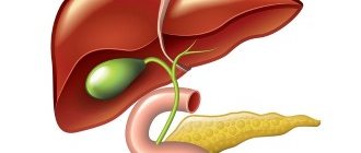
The spleen is a lymphoreticular organ located in the circulatory system. It is located in the hypochondrium on the left.
The splenic vein leaves the spleen and receives blood from the veins of the stomach and pancreas.
How can you help?
Therapeutic tactics depend on the diagnosis. In less complex cases, drug therapy is prescribed. More serious diagnoses require hospitalization and surgery.
The prognosis depends on the brightness of the clinical picture, the stage of the disease, the timeliness of diagnosis and the correctness of treatment.
Treatment of splenic calcifications
In the absence of complications and patient complaints of malaise, treatment is not carried out. If the calcifications are small in size, it is allowed to resort to traditional therapy.
Table 7. Application of folk recipes.
| Means | How to cook? | How to use (24 hours)? |
| 25 grams of the product are poured into 180 ml of just boiled liquid and infused for 40 minutes. Then the product is cooled and well filtered. | 3, 40 ml each. |
| 15 grams of the product is brewed in 170 ml of very cool boiling water and kept under the lid for half an hour. Then the product is filtered and combined with 200 ml of boiled cool water. | 3. |
| Squeeze the juice from ½ pomegranate and warm to room temperature. Persons who have gastrointestinal pathologies are recommended to first dilute the juice with water. | 3, half an hour before meals. |
| Warm freshly squeezed juice to room temperature. | 3, 20-30 minutes before meals. |
Treatment of enlarged spleen
The instructions look like this:
- If the cause of an enlarged spleen was an infection, the patient is prescribed antibiotics. At the same time, restorative medications are taken.
- If parasites are detected, anthelmintic medications are prescribed.
- Regardless of the provocateur, the patient is advised to avoid physical activity. General restorative therapy is carried out.
If the patient's condition does not cause concern, he is treated at home. In case of serious violations, the person is urgently hospitalized.
Table 8. Use of home remedies.
| Means | How to cook? | How to use (24 hours)? |
| 20 grams of the product are combined with 200 ml of boiled liquid, wrapped up, and chilled. After half an hour, the same amount of water is added to the broth. Then the broth is re-chilled and well filtered. Before use, you can add ½ tsp. honey | 3, 1/3 cup each. |
| 30 grams of finely chopped fruits are combined with 170 ml of very cool boiling water and infused for 45 minutes. Then the product is well filtered. | 3-4. |
| 40 grams of crushed horseradish roots are placed in the middle of the radish and filled with honey. Next, the radish is placed in the oven for 20-25 minutes. You need to eat the drug entirely. | 3-4, 2 tablespoons each. |
| Dry the seeds of overripe cucumbers and chop them thoroughly. The resulting powder (the color should be yellow) should be washed down with water. | 3, 10 grams each. |
Normal pancreas on ultrasound in adults
Ultrasound of the pancreas: the norm in adults - what is it? It is difficult to obtain a complete picture using ultrasound, but indirect signs can be detected, for example, narrowing of one of the ducts or the presence of an additional duct. In order to confirm the detected pathology, the doctor usually sends the patient for a series of additional examinations.
A normally functioning gland has the shape of the Latin letter S. Pancreas: dimensions (normal for adults), according to ultrasound, should be as follows:
- Weight ranges from 70 to 80 grams;
- The anteroposterior length of the head part is from 2.5 to 3 cm. The head must also have a hook-shaped process;
- The tail usually does not exceed 3 cm. The body is about 21 mm.
The gland should have an even contour and a uniform structure. The width of the Wirsung duct cannot be more than 1.5 - 1.9 mm.
The ultrasound examination procedure is carried out in case of constant pain in the left side, newly discovered diabetes mellitus, pathological changes in the duodenum that were discovered during an x-ray. Also, an ultrasound examination of the pancreas is indicated if deformation of the posterior gastric wall is confirmed, as well as if it is necessary to diagnose acute or chronic pancreatitis. In addition, an ultrasound examination of an organ is extremely necessary if surgical intervention is planned on it or nearby organs.
“Pancreas: size (normal in adults with ultrasound)” - you need to remember that this information can only be interpreted by a doctor, since he takes into account additional factors, such as the patient’s age and gender.
The spleen is a fairly large organ, which, although not vital, still performs a number of functions. This organ is unpaired and is localized in the abdominal cavity to the left and behind the stomach. In its shape it looks like a slightly flattened elongated hemisphere.
Treatment of splenic vein thrombosis
The main goals of therapy include:
- reducing the risk of consequences;
- restoration of blood flow;
- preventing further blockage of blood vessels.
Note! First, intravenous administration of heparin anticoagulants is prescribed. Then indirect-acting medications are prescribed. The dosage is reduced gradually.
Heparin anticoagulants
Information about recommended medications is presented in the tablet.
Table 9. Heparin anticoagulants.
| A drug | Description | Price |
| Direct anticoagulant. Affects platelet adhesion. Affects the vascular wall and the fibrinolysis system. Prescribed for acute thrombosis. | From 323 rubles. |
| The drug has a powerful antithrombotic and weak anticoagulant effect. Does not change general blood clotting tests. | From 187 rubles. |
| Enoxaparin | It is a low molecular weight heparin preparation. Has a powerful antithrombotic effect. Duration of exposure – up to 12 hours. | 258 rubles. |
| Low molecular weight heparin with antithrombotic and anticoagulant effects. Helps reduce blood viscosity, increases the permeability of membranes of granulocyte and platelet cells. | From 198 rubles. |
Indirect anticoagulants
The effect of drugs in this group is opposite to the effect of vitamin K. They help reduce the formation of proteins and suppress coagulation factors.
Table 10. Recommended indirect anticoagulants.
| Medicine | Description | Price |
| Suppresses the synthesis of vitamin K-dependent blood clotting factors. Reduces the risk of developing new blood clots. It has no effect on formed blood clots, but prevents the process of their enlargement. | 101 rubles. |
| Blocks the synthesis of vitamin K-dependent blood coagulation factors in the liver and has an anticoagulant effect. The result occurs 2-7 days after taking the drug. | 98 rubles. |
| Powerful anticoagulant. The effect occurs 3-5 days from the moment you start taking the medicine. | 77 rubles. |
| The medicine disrupts the synthesis of prothrombin and proconvertin. The maximum effect occurs within 12-24 hours. | 626 rubles. |
Note! Independent selection of drugs and dosage carries the risk of severe bleeding. Only a doctor can prescribe and discontinue medications.
Splenectomy
The table lists the main reasons why a patient is prescribed surgery.
Table 11. When is spleen removal indicated?
| Cause | Description |
| The spleen is removed due to its rupture. Usually, surgery is resorted to due to a fall on the left side, a strong blow to the stomach, or a car accident. |
| People at risk include people with malaria and some forms of liver cirrhosis. |
| The indication for surgery is bleeding of the organ. After removal of the spleen, the patient feels much better. |
| Removal of the spleen is resorted to only when the size of the tumors increases. |
If the surgery goes well, there are no restrictions on the regimen, and no special diet is prescribed.
Structural features
The organ is located in the left hypochondrium and extends from 9 to 11 ribs. The spleen is covered with a serous membrane that lines the walls of the abdominal cavity on all sides, except for a small area in the area of its gate.
The organ has the following surfaces:
- external – adjacent to the serous membrane, has a convex shape;
- internal - the surface has a concave shape and is represented by three parts, each of which is in contact with nearby organs (kidney, stomach, pancreas).
In addition to the serous cover, the gland has its own capsule, which is formed from connective tissue elements, and also has parts of non-striated muscle fibers.
This capsule seems to enter the cavity of the spleen, forming its trunk. Here there is a burgundy-colored pulp in which particles of lymphoid tissue are located. They have a round shape, adjacent to the walls of the branches of the arteries. The pulp is represented by reticular tissue filled with decaying blood cells.
Disability after removal
The final decision regarding disability is made by members of the medical and labor expert commission
Radical surgical intervention does not entail any disturbances in the vital functions of the body. The functions of the spleen are compensated by the lymph nodes.
Note! Disability is established not by a specific diagnosis, but only if there are 2 conditions for recognizing a citizen as disabled.
These conditions include:
- a health disorder accompanied by a persistent disorder of body functions;
- complete or partial loss of ability to self-care;
- the need for social protection measures, including rehabilitation.
If removal of the spleen does not result in renal failure, then disability is usually not given. But the final decision remains with the medical labor expert commission.
Comprehensive examination
Ultrasound examination allows you to evaluate the structure of organs and their functional features.
Reference! An ultrasound examination of the spleen is often performed during an ultrasound of other internal organs.
An ultrasound scan of the spleen is necessary if:
- pain in the abdomen;
- there is a bitter taste in the mouth;
- there is a feeling of a “bloated” stomach;
- Frequent hiccups bother me;
- unexpected weight loss;
- hypertension occurs;
- the patient suffers from hepatitis;
- there is a feeling of heaviness under the ribs;
- The attending physician suspects cancer.