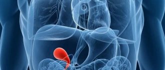
The gallbladder is a small, hollow organ located under the lower part of the liver. It serves to temporarily store bile produced in the liver.
As soon as a person begins to eat, the digestion process begins, in which bile gradually passes from the bladder into the intestines. However, part of the bile, bypassing the gallbladder, enters directly into the intestines, and only the excess is sent for temporary storage.
Bile not only participates in the digestion process, but also regulates peristalsis, removes steroids, cholesterol, and glutathione from the body. In addition, bile breaks down fats, neutralizes the harmful effects of hydrochloric acid on the intestines, and prevents fermentation and putrefactive processes in the gastrointestinal tract.
At most, the bladder contains about 200 ml of bile fluid, and in its normal state is not palpable. The shape, size of the organ and the development of pathology can only be determined using ultrasound.
Features of the structure of the gallbladder
The bile bladder is pear-shaped and consists of three main parts: the bottom, the organ itself and its neck. The walls consist of a mucous, muscular and outer layer. In the body of the liver there are small ducts that form large ducts at the exit on the right and left sides of the organ.
Further, both ducts join into one, which is called the hepatic duct and connects the gallbladder with the liver. And the outflow of bile into the intestines occurs through the bile duct. The hepatic and cystic ducts are transformed into one common duct, which flows into the intestine.
At the beginning of the duct, at the exit from the gallbladder, there is a sphincter that prevents the free circulation of bile. The sphincter opens and releases another portion of bile only when food enters the stomach.
Normally, bile is secreted in small quantities to aid digestion, break down food in the small intestine and create conditions for the formation of enzymes in the pancreas. Any pathologies and diseases of the gastrointestinal tract can be easily determined using ultrasound.
What does an ultrasound show?
At the slightest suspicion or discomfort, you should immediately consult a doctor. For a more accurate examination and diagnosis, an ultrasound examination is prescribed, and the liver is examined along with the gallbladder.
Using an ultrasound, the doctor will accurately determine the presence of pathology, focusing on the length and width of the organ itself, the length of the neck, the thickness of the walls and the diameter of the ducts. The norm of these indicators directly depends on the age of the patient and may vary slightly.
It is necessary to seek medical help in the following cases:
- bitterness in the mouth;
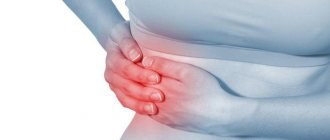
- pain in the hypochondrium on the right;
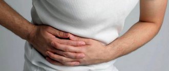
- heaviness in the side;
- bad tests;
- jaundice;
- severe poisoning, intoxication.
As a rule, the gallbladder is affected by alcohol abuse and taking a large number of medications. Ultrasound helps to timely determine the presence of acute or chronic cholecystitis, cholelithiasis, pancreatitis and congenital anomalies.
Watch the video of an ultrasound of the gallbladder:
Diagnostics with function determination
This type of diagnosis is carried out for dyskinesia (impaired wall motility with a hypotonic or hypertensive type) and inflammatory diseases, which include cholecystitis (inflammation mainly in the bladder) and cholangitis (inflamed ducts), as well as cholelithiasis.
Ultrasound is performed non-invasively; it is the fastest, most informative, absolutely painless and safe examination method. In just a few minutes, the doctor receives data on the parameters and shape of the organ being checked, the thickness of the walls, existing defects, and the presence and parameters of stones.
Ultrasound of the gallbladder to determine function is performed with a choleretic breakfast. It allows you to track the dynamics of the contractile-evacuatory function.
The doctor performing the ultrasound focuses on three main indicators: the time of the contraction period, the efficiency of bile secretion, and the tone of the sphincter of Oddi. This procedure takes approximately an hour. Decryption is carried out immediately. Initially, the examination is carried out on an empty stomach in a lying position; you cannot even drink water. After the first scan, the subject should have breakfast with two chicken yolks, or 250 ml of heavy cream or sour cream. A sorbitol solution can also be used for these purposes. An echography is done after breakfast 3 times: after 5-10 minutes, after 20 minutes and after 40-45 minutes.
Readings are taken in different positions (lying on your back, on your side, standing, sitting, etc.). The dimensions of the bladder are measured in longitudinal and cross sections, which makes it possible to assess the ducts and walls in more detail and reliably. If the bladder decreases in size by 60-70% of its original size during the examination, then it is considered that there is no impairment of contractile function. It should also be taken into account that with age there is a natural decrease in contractile function. Ultrasound of the gallbladder with determination of function shows the most accurate information about the condition of the cavity and ducts. To obtain reliable readings, the patient only needs to accurately follow all the doctor’s instructions.
Loading…
Share with friends!
Preparing for an ultrasound
In order for the study to be successful and show reliable results, it is necessary to properly prepare for it. 10 days before the procedure, you should completely stop drinking alcohol, fatty and fried foods.
5 days before the ultrasound, patients are advised to refrain from eating foods that cause increased gas formation (milk, peas or beans, cabbage), and should not drink soda or natural juices.
A couple of days before the examination, you can start taking enzyme medications. You will have to fast for 10-14 hours before the test, and smoking is prohibited immediately before the procedure.
First, an ultrasound is performed on an empty stomach to determine the contractility of the gallbladder. After this, the patient is given a little fat sour cream or cream to eat, and after 40 minutes the examination is repeated.
A healthy organ should shrink by no less than 60% and no more than 80%. Any deviations from these indicators are pathology. Using an ultrasound, the doctor determines the size of the gallbladder, its structure and wall thickness, and the presence of an inflammatory process.
How are indicators measured?
The exact size of the gallbladder is normally determined by ultrasound in adults and children. For the results of the study to be reliable, you need to prepare for it:
- 8-12 hours before the examination, you should have a light dinner (preferably before 7 pm the day before the ultrasound). At the same time, give up coffee and tea, alcohol and smoking. You can't use chewing gum.
- During the examination you will need to lie on your side, then on your back. A change in body position is necessary when searching for stones in the ducts.
- Sometimes you may need to eat a so-called choleretic breakfast. It could be a piece of butter, chocolate, or a raw egg. Such products stimulate the release of bile into the duodenum, and the uzologist will be able to assess the motility of the biliary tract.
After taking readings from the device, an ultrasound diagnostic specialist calculates the size of the gall bladder.
Decoding the results
It is important to consider that the normal size of the organ may vary slightly depending on age. They will differ in older people and children, and the average volume of the gallbladder in an adult is about 40-80 ml.
Normal for adults
A healthy adult patient must have certain organ parameters, which include width, diameter, wall thickness and length.
The following are considered normal indicators:
- volume about 70 ml;
- wall thickness no more than 0.4 ml;
- organ length, about 6-10 cm;
- the width should not exceed 5 cm;
- the diameter of paired ducts is normally about 0.3 cm;
- the diameter of the main duct cannot exceed 0.7 cm.
Interestingly, in women over 40 years of age, gallbladder lesions are much more common than in men. Moreover, naturally overweight blondes often suffer from such diseases. In men, such pathologies are observed much later, and only under conditions of alcohol abuse and poor nutrition.
Find out the norms of the gallbladder from the video:
Norm for children
The normal size of the gallbladder in children varies depending on age. So, for newborns, the norm is considered to be a length of 3.4 cm and a width of 1.08 cm. From a month to five, the length should be 4 cm and the width should be 1.02.
A one-year-old child has a bladder length of 5.5 cm and a width of up to 1.07 cm. At 3 years old, these figures increase to 5 cm and 1.60 cm, respectively. For a 7-year-old child, the norm will be a length of about 7 cm and a width of no more than 3.70 cm. Children aged 10 years and older should have a length of the gallbladder of 7.7 cm, a width of about 3.7, and a diameter of up to 1. 4 cm.
Deviation from the norm
Any deviations from normal values are a signal of the presence of a serious pathology. A large organ size indicates the development of cholelithiasis or acute cholecystitis, and a small one indicates hepatitis.
A sign of chronic cholecystitis is significant thickening of the walls of the organ. As a rule, all pathologies of the liver and gallbladder can be diagnosed using ultrasound, but in addition, the doctor prescribes a blood test, FGDS and a coprogram.
In addition, an upward deviation in size may indicate the development of oncology, dyskinesia, or liver damage. A deviation from the norm is considered to be an irregular shape of the bladder, an increase or decrease in size, scars and adhesions on the walls of the organ, and an uneven neck of the bladder.
Gallbladder diseases
With the help of ultrasound, any diseases not only of the gallbladder, but also of the liver and bile ducts are easily diagnosed. There are several diseases that occur most often.
For example, dyskinesia, which manifests itself in the form of thickening of the walls of the organ and disruption of its contractile functions. The cause of the disease is considered to be a hormonal imbalance that prevents the normal flow of bile. On ultrasound, the bubble has uneven outlines and kinks that disrupt the functioning of the organ.
An equally common pathology is cholesterosis, which occurs against the background of diabetes mellitus and other endocrine disorders. A common disease is cholecystitis, which is caused by an intestinal infection that has penetrated the gallbladder.
Severe inflammation of the walls occurs, accompanied by nausea, flatulence, and pain in the hypochondrium. The acute form of the disease can be recognized not only by thickening of the walls, but also by an increase in the size of the organ. The chronic course is characterized by a decrease in the bubble and the presence of small foreign inclusions.
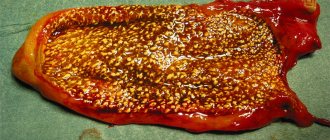
With the help of such a study, cholelithiasis (cholelithiasis), the cause of which is a hereditary predisposition and hormonal problems, is well determined.
Adenomyomatosis is considered a dangerous disease, which is characterized by benign thickening and compaction of the walls of the bladder, reaching 10-12 mm. This almost completely blocks the lumen inside the organ and is often accompanied by the appearance of polyps.
Sometimes, polyps can grow on the walls on their own, without accompanying pathologies. They look like small round growths and can be either benign or malignant. In the latter case, their rapid growth and the manifestation of accompanying metabolic disorders occur.
The most dangerous, incurable and severe pathology of the bladder is cancer. Determine it by an increase in volume, thickening of the walls and the presence of metastases.
At an early stage, it is almost impossible to make a diagnosis, because the disease is initially asymptomatic. The progression of the pathology is accompanied by severe pain, weakness, weight loss and bloating of the peritoneum.
In addition, congenital developmental anomalies occasionally occur, which include uncharacteristic protrusions of the walls, incorrect location or complete absence of the gallbladder.
Why do you need to know the diameter of an organ?
Our internal organs should normally be of a certain size. Disturbances caused by infection or functional disorders are immediately reflected in ultrasound - the shape and volume change. For example, when pus accumulates inside an organ, the pathogenic contents begin to stretch the walls of the organ. This is clearly visible to the diagnostician on an ultrasound, so he will immediately refer you for additional examinations.
The following diseases entail changes in the size of the gallbladder:
- cholelithiasis;
- acute cholecystitis;
- obstruction of the biliary tract and hepatic ducts.
Sometimes a change in size in children is a consequence of a congenital anomaly in the structure of the gall bladder. Such an anomaly may not manifest itself in any way until the occurrence of provoking factors. These include various kinks that arise due to non-compliance with the normal sizes of different parts of the cholecystis. If the corresponding anomalies were detected in children on ultrasound, you need to pay great attention to nutrition, since an unhealthy lifestyle will lead to the occurrence of pathologies (cholecystitis, hepatitis, blockage of the bile ducts, colic).
Sometimes in children and adults the following structural anomaly can be seen on ultrasound: double gallstone. These rare anatomical changes occur approximately once in 4 thousand people. This structure also weakens biliary function, requiring more careful care of the body.
If the size of cholecystis has not changed on ultrasound, but jaundice is present among the symptoms, this gives the doctor reason to understand that the problem lies not in the gall bladder, but in the liver. Dystrophic changes in this organ of the biliary system led to yellowing of the skin, and not stagnation of fluid inside the bladder. Among such diseases: hepatitis, cirrhosis of the liver.