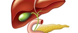
The term “obstructive jaundice” refers to a symptom complex that occurs when the outflow of bile through the bile ducts of the liver is disrupted. Its main symptoms are yellow discoloration of the skin, sclera and visible mucous membranes, which is associated with an increase in the level of bilirubin in the blood (hyperbilirubinemia).
Bilirubin is the end product of hemoglobin metabolism, which is contained in red blood cells. When aged red blood cells break down, the hemoglobin they contain is eventually converted into bilirubin through several transformation processes.
Normally, bilirubin should be excreted with bile into the lumen of the duodenum, but if there is an obstacle to such outflow, it enters the blood and has a toxic effect on the entire body.
In oncological practice, the cause of obstructive jaundice, as a rule, is compression of the bile ducts by primary or metastatic tumors of the liver, pancreas, and retroperitoneum.
Why is obstructive jaundice dangerous?
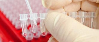
Bilirubin is found in the blood and is normal. However, its maximum concentrations, as a rule, do not exceed 20.5 µmol/l.
With obstructive jaundice, its concentration in the blood begins to constantly increase and can reach several hundred micromol/l. Such a high level of bilirubin has a pronounced toxic effect on almost all biochemical processes, organs and systems of the body. In addition, in the presence of obstructive jaundice, neither surgical nor chemotherapy treatment of the underlying disease is possible. A further increase in bilirubin concentration leads to the death of the patient.
Although infusion therapy can somewhat “dilute” the concentration in the blood for a short time, the only way to reduce its concentration is to restore the outflow of bile from the liver.
Indications for organ drainage
Surgery is a risky business. Its consequences are sometimes difficult to predict. Therefore, surgical intervention is resorted to in emergency cases.
Drainage of the biliary tract and gallbladder is done for the following indications:
- With obstructive jaundice. With a large amount of bilirubin in the blood, the skin and sclera of the eyes acquire a yellowish tint. The manifestation is not an independent disease - it is a symptom of bile stagnation. Bilirubin is the pigment that colors it.
- Gallstone disease. Conglomerates form in the bladder, less often in the ducts. Gradually increasing in size, the stones block the path of bile movement.
- Neoplasms of both benign and malignant nature. As the tumor grows, it compresses the ducts, which leads to the cessation of bile circulation.
The main indication for the use of drainage is obstruction of the bile ducts. The attending physician decides how to cope with the problem. The appointment is made only after examination and testing.
There are additional indications for the procedure. Thus, gallbladder drainage is used in diagnosing purulent cholangitis and acute pancreatitis. Damage to the common bile duct is also an indication. This is what doctors call the main duct. In fact, there are dozens of them. Some of the channels are located in the liver, and the other part is outside it.
https://youtu.be/uNtDxvkykVg
How is obstructive jaundice of tumor etiology treated?
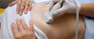
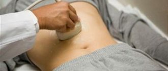
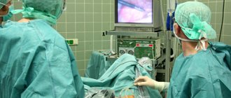
Currently, the most effective tactics to combat obstructive jaundice caused by compression of the bile ducts by tumors are drainage interventions on the bile ducts. As a rule, they are performed under the control of X-ray television and (or) ultrasound.
Most often, at the first stage, the bile ducts are punctured using a thin and long needle (Chiba needle) through the intercostal space. A special contrast agent is injected through a needle, which allows you to see the bile ducts themselves on an x-ray, as well as determine the level at which they are blocked. This intervention is called puncture cholangiography.
Then, using a special tool, it is possible to install a special drainage into the bile ducts. Drainage is distinguished:
- external - in which all bile is discharged only outside;
- and external-internal - in which the drainage is placed in such a way that the bile is evacuated both outward and in the natural direction, into the intestine.
As a rule, external-internal drainage is more physiological, since many important substances contained in bile, which are normally absorbed back in the intestine, are not lost.
In some cases, when tumor compression isolates several different segments of the biliary tree, multiple drains may be required.
Although drainage allows you to cope with the symptom itself, it significantly limits the patient’s quality of life - the drainage constantly irritates the abdominal wall, requires constant care, there is a risk of its displacement or even accidental removal, as well as the development of infectious complications at the site of erection.
To overcome these drawbacks of drainage, it was recently proposed to complete drainage interventions with bile duct stenting. The essence of stenting is to install a special (usually metal) endoprosthesis in the area of compression of the bile duct - a stent, which maintains the bile duct in an open state due to its high radial rigidity. In this case, the drainage tube can be removed completely, the outflow of bile will be carried out in the normal direction along the stent, or rather along the bile duct restored with the help of a stent.
Types of gallbladder drainage
Modern medicine has developed various methods to solve the problem of duct obstruction.
There are several options for draining the gallbladder:
- External. An external bile receptacle is installed. Serves as a temporary measure in preparing the patient for surgery. External drainage of the biliary tract allows you to record the amount of secretion secreted, the presence of pus and other foreign liquids in it. This helps to better control the patient's condition. The disadvantage of the method is the need to forcefully inject bile into the duodenum.
- External-internal method. In this case, most of the bile enters directly into the duodenum. A smaller part is diverted through special tubes to an external receiver.
- Internal method. Percutaneous transhepatic drainage of the bile ducts is used. An endoprosthesis is installed in the area of the damaged structures. The device ensures the outflow of fluid into the intestine. In this case, a drainage tube is left in the ducts for two or three days. This is necessary for decompressive changes in pathways. When the transformation occurs, the tube is removed.
The first two drainage options are used when preparing patients for surgery to remove stones and tumors. The methods allow us to ensure the normal functioning of the gastrointestinal tract, as well as monitor the condition of the gallbladder.
Internal drainage is intended for inoperable patients. The intervention is resorted to after the elimination of jaundice. For patients with neoplasms, internal drainage is the final stage in normalizing the functioning of the bile outflow system.
Feedback on the treatment of obstructive jaundice at the European Oncology Clinic
Compression of the biliary tract by malignant neoplasms leads to the development of obstructive jaundice. In patients, bilirubin levels increase and intoxication develops. Oncologists perform drainage of the bile ducts in case of obstructive jaundice. The Yusupov Hospital employs professors and doctors of the highest category. Drainage of the biliary tract is performed by leading oncologists. All complex cases of the disease are discussed at a meeting of the expert council.
Drainage of the bile ducts is performed to create an anastomosis between the bile ducts and the small intestine. Surgery can prolong the life of patients suffering from gallbladder or liver cancer. Oncologists perform drainage of the gallbladder in patients with neoplasms of the bladder and bile ducts, tumors in the area of the major duodenal papilla, and cancer of the head of the pancreas.
The following types of bile duct drainage are distinguished:
- external - the outflow of bile occurs through a specially installed external receiver;
- external-internal - most of the bile enters the intestine, flowing through the near channel, and the smaller part collects in the receiver located outside;
- internal drainage - requires endoprosthetics (biliary drainage is used as a palliative method for the treatment of inoperable cancer).
Indications
The need for drainage of the bile ducts may be associated with the severity of tissue strictures. The list of indications includes:
- cholangitis, which is accompanied by purulent inflammation;
- acute form of pancreatitis;
- lack of positive results from surgical intervention on the biliary tract;
- damage to the common bile duct area (expansion of the bile duct, taking the form of a sac).
On this topic
Can a stomach ulcer turn into cancer?
- Editorial board of Oncology.ru
- October 16, 2020
Another indication is impacted stones, which are difficult to remove. Medical drainage of the bile ducts is carried out in the presence of tumor formations.
Intervention can be carried out if there are changes due to ductal hypertension, if intraoperative cholangiography (a technique for examining the common passage in the bladder area) is necessary in the future. And finally, another indication is to monitor the restoration of bile flow into the duodenum.
Methods of drainage of the biliary tract in cancer patients
Oncologists prefer to perform external internal or, if technically feasible, external drainage of the bile ducts in patients with obstructive jaundice of tumor origin. Both methods are quite effective in preoperative preparation for radical surgery or as a final treatment method. Their advantage is:
- constant monitoring of bile flow;
- the possibility of active removal of blood, pus, and clots from the bile ducts;
- washing the ducts with aseptic solutions;
- dynamic x-ray monitoring of the location of the drainage tube.
Unlike external-internal drainage, bile completely flows out through the external drainage of the gallbladder. The disadvantage of external drainage of the bile ducts compared to external internal drainage is the complete flow of bile through the drainage to the outside. To compensate for the vital substances contained in bile, patients are forced to drink their own bile or medical personnel administer it through a nasogastric drainage. With external internal drainage, the distal end of the tube is located further than the narrowing site and most of the bile flows directly into the intestine. It remains possible to control the patency and flush the drainage and replace it with an internal transpapillary endoprosthesis.
Internal endoprosthetics of the bile ducts is performed after the elimination of jaundice. This is the final stage of treatment for inoperable patients. To successfully perform external or external internal cholangiostomy, oncologists use a set of special instruments: wire guides, special puncture needles, bougies and catheters.
Under local anesthesia using a Shiba needle, the surgeon tightly fills the bile ducts with a contrast agent. A long needle with a diameter of 1.5-1.7 mm performs a puncture of one of the segmental ducts. A conductor wire is then passed through it. The end of the conductor is passed beyond the narrowing, the narrowed area is widened with bougies, and a drainage tube is installed. It is fixed to the skin and the bile ducts are washed with sterile solutions.
This drainage method has disadvantages: there is a risk of bile and blood leaking into the abdominal cavity when the needle is removed, when passing a guidewire or bougienage the canal. Additionally, this complication may be encountered if the outer diameter of the needle is larger than the outer diameter of the guidewire. In order to reduce the number of complications associated with liver puncture, oncologists use the technique of installing a cholangiostomy using a stylet catheter.
Types of drainage
Obstructive jaundice is a serious pathological process that requires immediate medical intervention. Only some diseases that cause this complication can be treated conservatively. These include parasitic diseases, helminthic infestations and pancreatitis. As therapy, anthelmintic drugs, antibiotics, enzymes and other drugs are used, selected by the doctor individually.
In the vast majority of cases, a special type of surgical intervention is used to temporarily or permanently eliminate symptoms - drainage of the biliary tract and bladder.
There are the following types of this surgical intervention:
- External - the outflow of cystic contents occurs through specially installed conductors to an external receiver.
- External-internal - most of the bile enters the intestine through a channel formed by the doctor, and the remaining contents go into the external receiver.
- Internal drainage - with it, a duct endoprosthesis is surgically formed, ensuring normal passage of bile.
The choice of treatment method depends on the nature of the pathological process, age, concomitant diseases and the state of the patient’s cardiovascular system.
External drainage
It is one of the methods of preparing patients for further surgery. The intervention is low-traumatic, does not require special training and can be performed on any group of patients.
The advantages of this technique include the ability to control the flow of bladder contents, pus and blood. Through the catheter, the cystic cavity and ducts can be washed with antiseptic solutions in order to eliminate the inflammatory process. Through surgical access, carried out to install drainage, it is also possible to remove stones, as well as excise scars that narrow the lumen of the ducts.
Contraindications to the method are:
- Blood clotting disorders, decreased platelet levels below 50 G/l.
- Ascites, severe liver failure.
- Extensive, multiple metastases of a malignant neoplasm.
- Presence of hypervascular liver tumors along the path of catheter placement.
After surgery, constant monitoring of the drainage tube is necessary. The catheter should be washed with a mixture of saline solution with novocaine and heparin on the first day.
In the following days, 20 ml of saline solution is injected into the drainage lumen daily to remove clots and prevent obstruction. After the acute period has stopped and the general condition has improved, doctors can carry out the next stage of the operation, aimed at restoring the normal passage of bladder contents into the intestine.
Patients who have undergone external drainage of the gallbladder should undergo periodic examinations to determine the level of bilirubin and blood electrolytes. Removing large amounts of bile from the body can lead to hyponatremia and worsening general condition.
External-internal and internal drainage
Internal drainage of the bile ducts is performed as palliative treatment for patients with advanced cancer pathology. In this case, a permanent endoprosthesis is installed to ensure normal flow of bile into the intestinal cavity.
The external-internal type of drainage is recognized as the most effective. With this type of operation, it is possible to control the patency of the tube and flush the drainage with antiseptic solutions. In addition, most of the bile is not excreted, but enters the duodenum through a special anastomosis, thereby preventing the occurrence of electrolyte disturbances.
The catheter can be placed endoscopically, as well as using percutaneous transhepatic drainage. The choice of technique depends on the localization of the pathological process that caused the disruption of bile outflow.
Thanks to this surgical intervention, it is possible to better prepare the patient for the main operation (removal of stones, tumors), and in the case of palliative treatment, to extend the life of patients up to 1 year.
How is the operation performed?
Surgical external drainage of the common bile duct is a simple procedure, the duration of which is on average 1.5-2 hours. No special preparation is required; the procedure can be performed either as an emergency or as planned.
The operation is performed in several stages:
- On the eve of the planned intervention, it is necessary to take a general blood test and a coagulogram to assess the blood coagulation system.
- On the day of surgery, elective patients are prescribed antibiotic therapy to prevent infectious complications. During the intervention itself, the patient remains conscious. Painkillers and sedatives enter the bloodstream through an installed venous catheter.
- Drainage installation is carried out in an X-ray room. Once the patient is on the operating table, they will be connected to a machine that monitors blood pressure, pulse and other vital signs, and will also be injected with contrast to better visualize the surgical site.
- Under the control of the X-ray image obtained on the screen after the administration of a contrast agent, the surgeon injects a solution of local anesthetic into the liver area, after which, through a small incision, he will install a catheter to the blocked bile ducts, above the site of obstruction.
- The catheter is washed with sterile saline, its free end is brought out, sutured to the skin and connected to a special bag for receiving bile.
- After this procedure, the patient is transferred to a ward for further observation.
If there are indications for installing internal drainage during the intervention, in addition to the drainage tube that removes bile out, an endoprosthesis is installed in the duodenum to ensure the outflow of bile from the common bile duct, above the site of blockage with a stone. Subsequently, the temporary tube through which the bile flows out is removed, and the outflow of the contents occurs through the established endoprosthesis.
Permanent drains are made of metal, polyethylene and other non-reactive polymers. Experts prefer metal prostheses because they have a longer service life.
The success of the operation largely depends on the diagnosed pathology, the location of the narrowing of the common bile duct and on average is 90%. The rest of the patients achieve partial decompression, which also significantly improves their quality of life.
Endoscopic method of drainage of bile ducts
Using an endoscope, doctors perform nasobiliary drainage of the biliary tract. Indications for endoscopic drainage of the biliary tract are:
- obstructive jaundice caused by malignant and benign neoplasms;
- acute purulent cholangitis;
- external biliary fistulas;
- damage to the walls of the extrahepatic ducts, retroduodenal perforations;
- acute cholecystitis.
There are no contraindications to endoscopic drainage, except in cases where the tube for drainage of the biliary tract cannot be passed through the area of tumor narrowing. The endoscopic kit for drainage of the biliary tract through the nose includes:
- conductor wire;
- drainages of various shapes;
- a connecting tube for collecting bile and flushing the drainage;
- nasal tube, clamp and spatula.
The operation of endoscopic drainage of the biliary tract includes the following steps:
- cholangiography to determine the level and location of drainage;
- introduction of drainage with a metal guide-conductor;
- removal of the guidewire and endoscope;
- control cholangiography;
- assessment of drainage position;
- transferring the drainage from the mouth to the nose and fixing it on the head.
After using the endoscopic method of drainage of the bile ducts, complications do not develop. They may occur due to the progression of the disease.
How is drainage performed?
The operation takes one and a half to two hours. No special patient preparation is required. Therefore, the intervention is carried out both planned and emergency.
External drainage of the bile ducts is carried out in a certain sequence:
- Blood is taken for analysis. In addition to the clinical one, a clotting test is carried out.
- On the day of surgery, the patient takes antibacterial agents. Infection may occur during surgery. It is important to prevent its development.
- The patient is not put to sleep during the operation. The patient is conscious. Through a venous catheter, sedatives and painkillers enter the bloodstream. Equipment is also connected to the patient to monitor blood pressure and other vital parameters.
- Surgery is performed in an operating room with X-ray equipment. This need is associated with ensuring control over the manipulations being carried out. For the same purpose, the patient is injected with a special solution – contrast.
- Using X-ray equipment, the doctor injects local anesthetics into the liver. An incision is then made.
- The next step is the introduction of external bile duct drainage. Using a guide, the tube is brought to a place located above the “blockage” area. Next, an “insertion” is made into the bile ducts. Tubes, the diameter of which is 2-3 millimeters, are brought out and sutured to the skin. The system is then connected to a special bag for collecting bile.
- After the operation, the patient is transferred to the ward.
Inoperable patients undergo internal drainage. It requires an endoprosthesis. It is made of metal, polyethylene and other polymers. Metal endoprostheses last longer.
The sequence of the internal drainage operation is similar to the external one. The exception is the installation of an endoprosthesis. The device is connected to the ducts along with a tube for external removal of bile. The other end of the endoprosthesis is brought out into the intestine. After carrying out control measures and identifying positive dynamics, the outer tube is removed.
Drainage after gallbladder removal
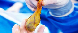
After cholecystectomy, surgeons often install a gallbladder drain. Indications for cholecystectomy are:
- cholelithiasis;
- acute cholecystitis;
- gallbladder carcinoma.
The operation is performed laparotomy or laparoscopically. Drains from the abdominal cavity after open cholecystectomy are removed on the eighth day, and in weakened and oncological patients - on the twelfth day. To prevent the skin around the drainage from becoming inflamed, the bile is drained into a special vessel. The skin around the wound is lubricated with zinc ointment or Lassar paste. Drains are changed no earlier than the twelfth postoperative day. In this case, fistulography is performed through the drainage to ensure the free patency of the bile ducts. Removal of drains after cholecystectomy is not performed on the fourteenth day, and when draining the bile ducts, no earlier than the twenty-first day after removal of the gallbladder.
Make an appointment by calling. Doctors at the Yusupov Hospital use various methods of draining the bile ducts for obstructive jaundice. Medical staff care for the drainage of the gallbladder and bile ducts.
Video
Gallbladder drainage.
- 4 minutes to read
Malignant neoplasms, chronic or acute forms of pathologies inevitably lead to narrowing of the bile ducts. This is a prerequisite for the occurrence of a mechanical form of jaundice, the characteristic feature of which is an increase in bile pigment (bilirubin) and intoxication. The only effective way to solve this problem is to carry out drainage in order to normalize bile outflow. Also, this is the main and only way to eliminate obstructive jaundice.
Content
Forecast
Mortality with obstructive jaundice depends on the cause of obstruction of the bile ducts, the duration of the disease and the severity of the patient’s condition. Modern technical conditions make it possible to eliminate cholestasis in more than 90% of patients in a minimally invasive way and, if necessary, to operate on patients in “favorable” conditions after the resolution of jaundice. If this step-by-step approach is followed, mortality due to the benign nature of bile duct obstruction does not exceed 2-5%.
The most difficult group consists of patients in whom it is not possible to eliminate biliary hypertension using minimally invasive approaches, and surgical intervention is forced to be performed at the height of cholestasis. Surgeries for increasing obstructive jaundice, cholangitis, and liver failure pose the greatest danger and are accompanied by high mortality.
Gallbladder drainage is
Drainage is a system for draining liquids. It is for these purposes that it is used in medicine. Drainage is installed on the bile ducts.
The systems for removing bile from the bladder to the duodenum may not function.
There are several reasons leading to pathology:
- conglomerates that have moved from the bubble where they formed into the ducts and clogged them;
- narrowing of bile flow due to a number of diseases, for example, dyskinesia;
- tumors blocking the flow of bile;
- cancer metastases in the liver, preventing its cells from producing digestive secretions.
There are many reasons for system failures. A significant part of them cannot be solved with the help of medications alone. In this case, drainage is carried out. With the help of surgery, an alternative path for the outflow of bile is provided. Most often, the procedure is necessary as a temporary measure. The intervention is used to prevent stagnation of bile during the period of preparation for surgery to remove a tumor, stones or the bladder itself.
Tumors in the bladder and bile ducts are inoperable. In this case, drainage allows you to prolong the patient’s life and is done on an ongoing basis. The tubes through which liver secretions are now directed leave the body or are located inside. Containers for collecting bile are attached to the ends of the drains.
Contraindications
Installation of a drainage tube into the gallbladder is indicated for:
- eliminating stagnation of bile due to narrowing of the bile duct;
- increasing the lumen of the common bile duct (bile duct) to transport cystic secretions into the duodenum;
- antimicrobial therapy: medicinal effects with antibiotics or antiseptic solutions;
- prevention of scarring and fistula formation in the postoperative period;
- carrying out a liver cleansing procedure mechanically.
The decision on drainage is made by the doctor when diagnosing the following diseases:
- Gallstone disease (cholelithiasis) - installation of drainage creates conditions for the timely outflow of bile, as well as small stones (stones) and sand. Drainage is included in the complex of preoperative treatment.
- The acute course of cholecystitis is the prevention of infection when fluid stagnates as a result of inflammation.
- Mechanical jaundice - removal of active enzymes from liver cells, decrease in bilirubin levels.
- Malignant and benign neoplasms – to eliminate blockage of the bile ducts by tumor-like growth.
- Pancreatitis - inflammation provokes swelling of the head of the pancreas, which creates an obstacle to the flow of bile into the initial part of the small intestine.
- Inflammation of the walls of the bile duct (cholangitis), cystic formations.
- Improper development of the biliary tract at the genetic level.
Bile duct drainage is not performed if there is a history of the following abnormalities:
- ascites – accumulation of fluid in the abdominal cavity;
- pregnancy complicated by severe gestational diabetes;
- germination of cancerous tumors blocking the duct;
- blood clotting disorder;
- obesity;
- respiratory and heart failure in the stage of decompensation.
To treat mechanical jaundice, an external drainage procedure is performed. The method eliminates bilirubin stagnation during therapeutic measures, and after recovery the catheter is easily removed.
The decision on the need for drainage is made by the attending physician when the following indications appear:
- acute inflammatory process in the gallbladder or pancreas;
- damage to the bile duct;
- formation of pus in the biliary tract;
- the appearance of neoplasms;
- presence of stones.
After drainage for obstructive jaundice, the patient's condition improves due to the normalization of bile pigment (bilirubin) and the resumption of fluid outflow from the liver.
Drainage in medical terminology is an invasive process to eliminate blockage of the duct with stagnant bile. During the procedure, sedatives or painkillers are administered at the initial stage, and then the necessary anesthesia is administered.
The procedure, if the process is inoperable, is carried out using radiography, since this technique allows you to prolong the patient’s life by 1.5–2 years if he is diagnosed with severe cancer.
The need for drainage occurs when the normal flow of bile into the duodenum is disrupted. With this pathology, the patient feels a pronounced feeling of discomfort in the right hypochondrium, nausea, vomiting with an advanced course of the disease.
Drainage of the gallbladder is a difficult surgical operation, which is performed when there are clear indications:
- Obstructive jaundice is characterized by a high presence of bilirubin in the blood, which gives yellowness to the skin and pupils. Pathology indicates severe stagnation of bile and requires surgical intervention;
- Stones in the gall bladder, since the formed stones block the normal outflow of bile, and a complication of the disease is blockage of the conglomerate in the bile duct;
- Neoplasms of malignant and benign origin, since with increasing size there is a lack of normal bile circulation.
The main indication for drainage is obstruction of the bile ducts, since without surgical intervention death can occur. The operation is carried out only after a thorough examination and the appointment of the necessary tests. Manipulation can be prescribed for acute pancreatitis and purulent cholangitis.
Before performing drainage, it is necessary to exclude possible contraindications:
- A history of hepatitis;
- Late pregnancy;
- Obesity of the second and third degree;
- The occurrence of diseases of the cardiovascular system and respiratory organs.
The possibility of surgical intervention is decided by the attending physician after studying the presented clinical picture and passing the necessary list of tests.
Drainage does not require preliminary preparation, so the manipulation is carried out according to the following algorithm:
- Taking a general and biochemical blood test to study general indicators (coagulability must be checked);
- Before the operation, the patient is prescribed antibacterial therapy, since involuntary infection may occur during surgery;
- Drainage does not imply the administration of general anesthesia, since during the operation painkillers and sedatives are infused intravenously, and the patient is connected to equipment to monitor the general condition of vital organs;
- The surgical operation is performed using radiography with the introduction of a contrast agent, which allows you to monitor the overall progress of the surgical intervention;
- Direct drainage is based on the introduction of tubes into an area located slightly above the area of blockage of the bile duct and subsequent insertion into the organ. The tubes are brought out and sutured to the skin, and a container for collecting bile is connected to the system;
- After all necessary manipulations have been completed, the patient is transferred to the ward for further recovery.
When carrying out internal drainage, a special endoprosthesis is used, which is connected to the bile duct and the second end is connected to the intestine. The device is made of metal or polymer material.
If positive dynamics and favorable recovery of the body are observed, then the outer tube is subsequently removed.
The procedure for cleaning the gallbladder at home is called tubage. The process requires compliance with certain rules and can be carried out using a duodenal tube or taking special choleretic drugs.
This technique can be easily implemented at home, as it has no serious contraindications and does not require special medical equipment.
The process must be carried out in the following cases:
- Prevention and treatment of persistent bile stagnation;
- Preventing the development of gallstone disease;
- Improving the functioning of the digestive system;
- Stimulating intestinal functionality.
Drainage at home has contraindications that must be taken into account before performing the procedure:
- Gallstones;
- Acute cholecystitis;
- Obstruction of the bile ducts;
- Acute inflammation of the duodenum;
- Stomach or intestinal ulcer;
- Period of menstruation;
- Pregnancy;
- Course of acute infectious diseases;
- Oncological processes in the body.
In the presence of the pathologies described above, active production and promotion of bile can cause additional complications in the body. It is best to consult your doctor before performing the procedure yourself.
As preparatory measures, a few days before tubage, it is necessary to establish proper nutrition with maximum fiber consumption and the exclusion of heavy foods (fried, salted, pickled dishes) from the general diet.
To carry out the procedure, it is necessary to first cleanse the intestines and it is preferable to take a free day, since bed rest will be required. The process can be carried out using alkaline mineral water (Essentuki, Borjomi, Slavyanskaya, Moskovskaya).
You must first release all the gases, and in the morning drink 1–2 glasses of heated water (temperature 40–50 degrees) on an empty stomach. In the future, you need to lie down in bed and apply a heating pad to your right side to activate the liver.
The patient should take a comfortable position that will not cause unnecessary discomfort (on the right or left side or on the back). The amount of warm water can be increased to 500 ml. At the same time, it must be at a certain temperature, since cold liquid can cause spasms of the gallbladder.
The second way to activate the functioning of the gallbladder is based on drinking warm mineral water and performing special abdominal exercises (bending and twisting). If necessary, drink more fluid and repeat the exercises.
After the actual procedure, it is recommended to follow the basics of proper nutrition and lead a moderately active lifestyle, since violation of these rules can lead to re-inflammation.
Cleansing the gallbladder makes it possible to reduce heaviness in the right hypochondrium, improve the digestion process and prevent the development of constipation. As a preventative measure, this procedure can be performed once a month.
When performing tubage, in addition to mineral water, you can use sorbitol, xylitol or magnesium sulfate. This technique is effective as a preventive measure to stabilize the functioning of the digestive system.
Mechanical or in other words subhepatic jaundice is a serious complication of diseases of the digestive tract and abdominal cavity and a direct indication for drainage of the gallbladder. The pathological process consists of a violation of the outflow of bile from the bladder into the lumen of the duodenum. This leads to an increase in the content of bilirubin in the blood and, as a consequence, to intoxication of the body.
Subhepatic jaundice can occur at any age and can be either benign or malignant.
The main reasons leading to its formation are:
- Gallstone disease (in this case, the outflow of bile is obstructed by stones in the cystic cavity and in the excretory ducts).
- Tumors (neoplasms of the bladder, ducts, tumors in the area of the major duodenal papilla, cancer of the head of the pancreas).
- Pancreatitis, accompanied by swelling and an increase in the size of the head of the pancreas.
- Inflammatory processes, scars on the duodenal mucosa.
- Cholangitis and common bile duct cysts.
- Parasitic diseases - opisthorchiasis, giardiasis.
- Congenital obstruction, atresia of the excretory ducts.
Obstructive jaundice – what are the diagnostic and treatment options?
Obstructive jaundice is dangerous for the body, causing severe intoxication and dysfunction of all organs. In oncology, it is more often diagnosed during the treatment of metastatic cancer abroad, and such patients are often given up as not promising for treatment.
This formulation of the question is typical for medical institutions, the level of which leaves much to be desired, where there are no new technologies for examining and eliminating this complication.
However, in modern clinics and centers, both foreign and domestic, methods are used that allow:
- pinpoint the causes;
- jaundice level;
- take measures to restore normal bile circulation.
Next, we will look at the clinical nature of the development of this condition, and also find out how long obstructive jaundice lasts.
Carrying out drainage at home
The procedure for cleaning the gallbladder at home is called tubage. The process requires compliance with certain rules and can be carried out using a duodenal tube or taking special choleretic drugs.
This technique can be easily implemented at home, as it has no serious contraindications and does not require special medical equipment.
The process must be carried out in the following cases:
- Prevention and treatment of persistent bile stagnation;
- Preventing the development of gallstone disease;
- Improving the functioning of the digestive system;
- Stimulating intestinal functionality.
Drainage at home has contraindications that must be taken into account before performing the procedure:
- Gallstones;
- Acute cholecystitis;
- Obstruction of the bile ducts;
- Acute inflammation of the duodenum;
- Stomach or intestinal ulcer;
- Period of menstruation;
- Pregnancy;
- Course of acute infectious diseases;
- Oncological processes in the body.
In the presence of the pathologies described above, active production and promotion of bile can cause additional complications in the body. It is best to consult your doctor before performing the procedure yourself.
As preparatory measures, a few days before tubage, it is necessary to establish proper nutrition with maximum fiber consumption and the exclusion of heavy foods (fried, salted, pickled dishes) from the general diet.
To carry out the procedure, it is necessary to first cleanse the intestines and it is preferable to take a free day, since bed rest will be required. The process can be carried out using alkaline mineral water (Essentuki, Borjomi, Slavyanskaya, Moskovskaya).
You must first release all the gases, and in the morning drink 1–2 glasses of heated water (temperature 40–50 degrees) on an empty stomach. In the future, you need to lie down in bed and apply a heating pad to your right side to activate the liver.
The patient should take a comfortable position that will not cause unnecessary discomfort (on the right or left side or on the back). The amount of warm water can be increased to 500 ml. At the same time, it must be at a certain temperature, since cold liquid can cause spasms of the gallbladder.
Currently reading: Symptoms of gallbladder inflammation and how to treat them at home
The second tubage technique when using mineral water
The second way to activate the functioning of the gallbladder is based on drinking warm mineral water and performing special abdominal exercises (bending and twisting). If necessary, drink more fluid and repeat the exercises.
After the actual procedure, it is recommended to follow the basics of proper nutrition and lead a moderately active lifestyle, since violation of these rules can lead to re-inflammation.
Cleansing the gallbladder makes it possible to reduce heaviness in the right hypochondrium, improve the digestion process and prevent the development of constipation. As a preventative measure, this procedure can be performed once a month.
When performing tubage, in addition to mineral water, you can use sorbitol, xylitol or magnesium sulfate. This technique is effective as a preventive measure to stabilize the functioning of the digestive system.
How to care for drains after the procedure
The result of the intervention largely depends on the further actions of both the patient and medical personnel.
For example, the following procedures for caring for drainage tubes are important:
- the tube coming out is placed in a special sterile container without any foreign liquid inside;
- The patient's blood is periodically taken to analyze the level of bilirubin and electrolytes;
- caring for drains for external bile drainage also includes avoiding kinking of the outer tube;
- the catheter must be washed daily to prevent infection of the ileostomy (excreting opening), and subsequently the bile ducts;
- Daily monitoring of the volume and quality of the exiting liquid.
On the first day after drainage, the bile has a dark tint. After the inflammatory processes subside, the liquid becomes lighter. This is a reason to shut off the tubes. Initially, the release of bile to the outside is stopped for one hour. If the patient does not experience pressing pain in the right hypochondrium, the duration of tube closure is increased. If the prognosis is favorable, the drainage is removed two weeks after the intervention.
A test is performed before the drain is removed. The goal is to identify the degree of patency of the bile ducts. For testing, a water-soluble contrast agent containing iodine is injected into the tubes.
In what cases is it necessary to install such drainage?
The biliary tract is a special system of channels, the main task of which is to drain bile produced by the liver into the digestive system (more precisely, into the duodenum). The main factors leading to disruption of the normal functioning of this channel system are various pathologies of the biliary system (liver and gallbladder), although strictures can also cause diseases of other internal organs.
The main pathologies for which such drainage is used are:
- the presence of concretions (stones) in the gallbladder and its ducts. This pathology is called cholelithiasis. The main risk group is patients who are overweight. The main factor leading to stone formation is stagnation of bile in the gallbladder, which leads to disruption of the normal metabolic process. The basis of such stones is the precipitated components of bile, such as cholesterol, bilirubin and others. The danger of this pathology lies in the fact that often the process of stone formation is asymptomatic for a long time, does not cause discomfort to the patient, and manifests itself only in the later stages of the development of the disease. In some other cases, stones block the bile ducts and damage their walls. The onset of inflammation manifests itself in acute and sharp pain, called hepatic colic. The pain is localized in the right hypochondrium and can radiate to the back and under the shoulder blade. In some cases, this pathological process is accompanied by fever, attacks of nausea and vomiting. In such cases, solving this problem is possible only after removing the gallbladder;
- Also, narrowing of the bile ducts is provoked by various serious diseases (such as malignant tumors). It is possible to stop the obstructive jaundice that occurs against their background just by installing a special drainage to normalize the process of bile outflow.
Read also: How long does it take for a suture to heal after laparoscopy of the gallbladder?
Methods for removing the gallbladder
Prices for treatment after gallbladder drainage
Conservative treatment of obstructive jaundice - following a diet, taking medications. Surgical treatment in surgery depends on the disease that caused the syndrome. Stenting and drainage using endoscopic or percutaneous methods are used. The latter include: percutaneous transhepatic cholangiostomy (PTCHS) or percutaneous transhepatic cholecystostomy under ultrasound and CT control. Among the types of endoscopic drainage are: papillosphincterotomy (EPST), duodenobiliary drainage.
The choice of treatment for obstructive jaundice with medications depends on the pathogenesis of the disease. Antispasmodics are used to reduce pain and increase the lumen of the ducts. For cholelithiasis, cholangitis, and chronic cholecystitis, ursodeoxycholic acid is indicated. To protect the liver parenchyma, hepatoprotectors are needed, and for Klatskin tumor, gastric and pancreatic carcinoma, chemotherapy is needed. Along with these medications, your doctor may prescribe:
- B vitamins;
- lipocaine;
- Vikasol;
- methionine;
- Trental;
- administration of glucose.
Transhepatic percutaneous drainage (TPD) promotes the flow of bile. External drainage directs the discharge of bile through a catheter into the digestive system so that losses do not lead to stomach upsets. Drainage of the gallbladder with obstructive jaundice (external-internal) requires the installation of a catheter from the outside.
Nutrition eliminates excess stress on liver parenchyma cells. The diet for obstructive jaundice should include: plenty of fluids, carbohydrate-rich foods, low-fat dairy products, day-old or dried bread, fruits, vegetables, cooked meats and steamed fish. All products and food should be at room temperature, boiled and pureed. To obtain fat, you can consume a small amount of butter and vegetable oil.
Conservative treatment for obstructive jaundice of tumor origin is ineffective, since it does not eliminate its main cause. Moreover, jaundice can progress, as over time the patency of the biliary tract becomes worse. Infusion therapy can reduce the level of bilirubin in the blood, but the primary goal in the treatment of liver tumors is to restore the outflow of bile.
Since patients with already formed obstructive jaundice are admitted for treatment quite late, treatment is divided into 2 stages - temporary bile diversion followed by radical surgery. This approach reduces mortality and the number of postoperative complications.
Surgical techniques allow not only to get rid of obstructive jaundice, but also to prepare for radical surgery to remove the tumor. Several approaches have been developed - endoscopic, percutaneous, intraoperative (during another operation). Before any intervention, careful visualization of the lesion is carried out using ultrasound and x-ray techniques.
Drainage using a special catheter helps to relieve the bile ducts well, as well as wash them, which is necessary for purulent inflammation. The catheter is a polyethylene tube with many holes at the end. This drainage is well tolerated by patients and can be installed for up to several weeks.
If the tumor affects the extrahepatic ducts, when it is impossible to perform a radical operation, the patient undergoes stenting - installation of a thin elastic tube that supports the bile duct in a straightened state. Thus, the stent allows bile to freely exit the liver. It is placed over a guidewire after an endoscopic examination of the patient.
Stenting is performed after the inflammation has been eliminated. Stents maintain patency for 3 to 6 months (metal stents last longer), after which they require replacement. The disadvantages of plastic prostheses include the possibility of their movement into the intestines, as well as clogging of the lumen with bile or germination by a tumor.
Percutaneous drainage (that is, ensuring the outflow of bile) can be:
- external;
- external-internal;
- performed using arthroplasty techniques.
External drainage ensures the discharge of bile through the catheter from the body. In this case, however, the loss of bile leads to disorders of the gastrointestinal tract, so the secreted bile is usually sent back to the digestive system. External drainage is used in extreme cases when it is impossible to insert the catheter further than the tumor narrowing.
With external-internal drainage, an external catheter is installed in the patient for several days, during which time the swelling in the narrowing zone subsides, thanks to which the catheter can be passed further into the duodenum. Thus, bile is sent to the gastrointestinal tract, where it performs its function.
During percutaneous endoprosthetics of the bile ducts, a plastic prosthesis is inserted into the place where they are narrowed by the tumor, replacing part of their wall. The disadvantages of the technique include high trauma when carried out through the liver, the possibility of migration of the prosthesis into the intestine, and rapid blockage.
In half of the cases, radical surgery for obstructive jaundice ends with the formation of an opening for the drainage of bile into the gastrointestinal tract or drainage of the ducts. When forming an opening to drain bile, the gallbladder is most often connected to the small intestine.
Experts try to give preference to minimally invasive methods of correcting obstructive jaundice in oncology; in each case, the choice of one or another method of operation is made individually.
What happens if you refuse to drink bile?
Bile plays several roles in the body.
These include:
- neutralization of the negative effect of pepsin from gastric juice on the walls of the digestive tract;
- participation in the process of fat emulsification;
- participation in the synthesis of micelles;
- normalization of mucus production;
- participation in the process of producing intestinal hormones;
- antibacterial effect;
- preventing the development of putrefactive processes in the intestines;
- normalization of digestion processes and intestinal motility;
- participation in the synthesis of enzymes that are important for the complete digestion of food.
If there is not enough bile in the body, digestion, absorption of nutrients from food and the process of cleansing the body of toxins are disrupted.
Disturbances caused by a deficiency of secretions lead to poisoning of the body and a deterioration in the general condition of the patient.
Without liver secretions:
- The intestines are unprotected from pathogenic microorganisms.
- As a result of impaired peristalsis of the organ, constipation or, conversely, diarrhea may occur.
- Due to poor emulsification of fats in the intestines, the body begins to suffer from a deficiency of fat-soluble vitamins.
Owners of patent RU 2532404:
The invention relates to medicine, namely to surgery, and describes a method for compensating for disturbances in the flow of bile into the intestines in patients with external drainage of the biliary tract. The method of replenishing bile deficiency during its external diversion consists, in addition to the main treatment, in prescribing a medicinal composition consisting of the following ingredients: ursodeoxycholic acid 12-15 mg/kg/day (up to 20 mg/kg) in 2-3 doses, eslidin (phospholipids from soy lecithin 300 mg; methionine 100 mg, soybean oil up to 550 mg) 1 capsule 3 times a day with meals; milife 0.2 g 3 times a day. The invention makes it possible to improve the general condition of the patient and relieve pain in a safe and technically simple way. 1 ave.