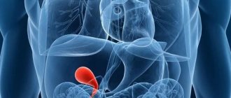
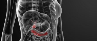
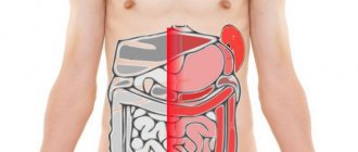
The gallbladder is a small, hollow, pear-shaped organ located under the liver. It concentrates and stores bile, a liquid necessary for the digestion of fats in the small intestine. For gallbladder cancer, it is important to start treatment on time, since the disease can be asymptomatic and is often detected too late. Cancer can affect nearby organs and tissues, making the disease much more difficult to cure.
Malignant tumors of the gallbladder are a fairly rare phenomenon; they most often develop in women, in 7 out of 10 cases.
Our company, medical service "Tlv.Hospital", offers services for organizing the diagnosis and treatment of gallbladder cancer in Israel:
1. Selection of doctors and clinics, individual approach taking into account all wishes, comfortable conditions of stay.
2. Carrying out all procedures as quickly as possible.
3. Moderate prices thanks to contracts with medical centers for the provision of medical care at wholesale prices.
4. Providing a “second opinion” (getting advice from another specialist), after completing treatment, maintaining contact with the doctor.
Causes of gallbladder cancer
There are several known risk factors associated with this type of cancer.
- Like most types of malignant diseases, gallbladder cancer occurs more often in older people.
- A family history of gallbladder cancer increases the risk of the disease by 5 times, especially if there is a genetic mutation known as BRCA2.
- Risk factors are gallstones and inflammation (cholecystitis). 8 out of 10 people with gallbladder cancer are diagnosed with these disorders. A family history of stones increases the risk of cancer, according to research.
- “Porcelain” gallbladder—calcium deposits on the inner wall of the organ—is one potential factor.
- Primary sclerosing cholangitis (PSC), a type of inflammation of the bile ducts, can increase the likelihood of cancer.
- Smoking and some industrial chemicals contain nitrosamines, chemicals that damage DNA and increase the risk of cancer.
- Certain abnormalities of the pancreas and bile ducts increase the likelihood of this disease. These are congenital disorders - growths along the bile duct, an abnormal connection of the bile ducts and the pancreas.
- Gallbladder polyps are small growths that appear on the mucous membrane of the organ. The larger the size of the polyp, the higher the risk that it will become malignant.
- Excess weight increases the likelihood of many types of cancer, including gallbladder cancer. Obesity means being more than 40% above the maximum desirable weight for a given height. Excess weight causes changes in the hormonal balance in the body, which increases the risk of this type of cancer.
- According to several studies, diabetes increases the likelihood of gallbladder cancer and bile duct tumors.
- There is some evidence that diet affects the risk of developing this disease.
- Ethnicity is one factor. The highest incidence of gallbladder cancer is in North India.
- Salmonella infection increases the risk of this disease. Some studies suggest that the bacteria Helicobacter Pylori may also have an effect.
Signs and symptoms of gallbladder cancer
In the early stages, the disease does not cause any symptoms. When the disease is diagnosed, it often already extends beyond the primary focus and affects surrounding tissues and organs.
Most of the symptoms are typical for the later stages of the disease. It is worth knowing that other pathologies can also cause these symptoms.
- Abdominal pain may occur on the right side of the abdomen. Some people describe her as dragging. If cancer or stones are blocking the bile duct, the pain will be severe.
- Nausea is very common in advanced stages of gallbladder cancer. It is easily controlled with antiemetic drugs.
- Jaundice means that the liver is not functioning properly. Symptoms include: yellowing of the skin and whites of the eyes; darkening of urine; white color of stool. Jaundice is associated with the accumulation of bile salts in the blood. Half of people diagnosed with gallbladder cancer have jaundice, a sign of advanced stages of cancer. It is worth remembering that hepatitis is a more common cause of jaundice.
- Enlargement of the gallbladder occurs due to blockage of the bile duct and bile filling the organ.
- Other less common signs of the disease include loss of appetite, weight loss, and constipation.
Types of Gallbladder Cancer
In 85 out of 100 cases of this disease, adenocarcinoma is diagnosed. This type of cancer develops in the glandular cells of the organ that produce mucus. Andenocarcinoma is divided into papillary, non-papillary and colloid.
Less common types of gallbladder cancer include squamous cell, small cell and sarcoma.
Rare types of gallbladder cancer include neuroendocrine tumors, lymphomas, and melanoma.
Diagnosis of gallbladder cancer in Israel
The oncologist collects anamnesis and asks about symptoms. Examines the patient, palpating the abdomen for signs of enlargement. Checks the whites of the eyes and skin color for the symptom of yellowing. Examines the lymph nodes in the neck and groin area.
Depending on the results, the following types of examinations may be prescribed:
- Blood tests called liver tests. This is a series of tests that check the functioning of the liver and gallbladder. It also includes a test for bilirubin, a chemical in bile. A small amount of bilirubin in the blood is completely normal. But high levels usually mean there are problems with the gallbladder or liver.
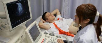
- Ultrasound. If a tumor is found in the gallbladder, ultrasound can determine whether the cancer has spread to the walls of the organ.
- A CT scan will show tumor growth in and around the gallbladder and whether the common bile duct, lymph nodes or liver are affected.
If the scan shows abnormal areas around or inside the gallbladder, the following may be done:
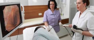
- ERCP - x-ray of an organ using an endoscope (endoscopic retrograde cholangiopancreatography - ERCP). The patient swallows a flexible tube, which the doctor uses to examine the inside of the small intestine and take a biopsy of areas that appear abnormal. This test shows narrowing or blockage of the bile ducts of the pancreas and helps plan surgery. Takes from 30 minutes to 2 hours.
- MRCP is a type of MRI of the gallbladder, pancreas and bile ducts. MRCP stands for magnetic resonance cholangiopancreatography. The procedure requires preparation; you must stop eating and drinking 2 hours before. MRCP is less uncomfortable than ERCP and does not require painkillers or other medications, but does not allow tissue samples to be removed.
- Biopsy and fine needle aspiration. A biopsy means taking a sample of tissue and examining it using a microscope. This is the only way to determine whether a tumor is cancerous. But if the doctor is absolutely sure, based on the results of other tests, that it is cancer, a biopsy is not needed. The gallbladder will be removed in any case.
If a biopsy is necessary, it can be performed in different ways: during laparoscopy for ERCP or using fine-needle aspiration biopsy.
To monitor and further treat gallbladder cancer, as well as to perform aspiration biopsies, the doctor uses CT or ultrasound to guide the needle to the right place. Takes a sample of cells and sends it to the laboratory for further study. A tissue sample may be taken from the liver or lymph nodes to see if the cancer has spread to them. After the gallbladder biopsy, the patient remains in the clinic for several hours or overnight. This is necessary because there is a risk of bleeding.
If tests indicate gallbladder cancer, further tests may be needed to determine the extent of the cancer's spread. Cancer most often affects the liver - in 8 out of 10 people. It can also penetrate the lymph nodes in the abdomen. To treat gallbladder cancer, the following types of diagnostics may be performed.
- MRI shows soft tissue more accurately than CT. The use of MRI with cholangiography can show blockage of bile flow by the tumor, as well as the spread of cancer to the portal vein. If there is metal in the body (for example, a pacemaker), this test is contraindicated.
- Endoscopic ultrasound uses an ultrasound scanner and an endoscope to help determine the stage of cancer, whether the tumor has grown into the wall of an organ or whether it has spread to the liver. All this facilitates the process of planning the operation.
- Cholangiography examines the bile ducts using dye, X-rays, and an endoscope. The procedure lasts 30-60 minutes, with its help you can find out whether there is a tumor in the gallbladder or whether the duct is blocked. If there is a blockage, a stent may be placed.
- Laparoscopy is a minor operation. A laparoscope equipped with a camera and light is inserted into the abdominal cavity through small incisions and examined for signs of cancer. Using a laparoscope, the surgeon is able to look inside the gallbladder. Laparoscopy helps in planning surgery and choosing other cancer treatments for gallbladder cancer. The procedure requires general anesthesia and overnight hospitalization will be required. During this procedure, a biopsy may be performed. If there are stones or inflammation in the gallbladder, the surgeon will immediately remove the organ. This operation is called cholecystectomy. The advantage of this type of treatment is a shorter recovery period.
Gallbladder cancer stages
The stages indicate the growth and spread of the malignant process. Determining the stage of the disease is necessary to select the optimal treatment option for gallbladder cancer.
The TNM system is used to classify gallbladder cancer.
- T – indicates the size and spread of the gallbladder tumor.
- N – for damage to lymph nodes.
- M – for the penetration of the tumor process into other parts of the body.
According to this classification, there are 5 stages - T1 - T4 and a very early stage called Tis or carcinoma in situ.
Tis (cancer in situ) – the tumor is located inside an organ. At this stage, the disease is diagnosed extremely rarely. It occurs more often when the gallbladder is removed for other reasons, such as stones.
- T1 tumor began to grow into the wall of the gallbladder. The stage is divided into T1a and T1b. T1a indicates damage to the connective layer under the inner lining of the organ wall, T1b indicates penetration of cancer into the muscle layer located behind the connective layer.
- T2 tumor is localized in the gallbladder, but has grown through the muscle layer into the next layer of connective tissue.
- T3 tumor has gone beyond the boundaries of the organ, spreading to the liver or another nearby organ - the stomach, intestines or pancreas.
- T4—The cancer has invaded the portal vein or hepatic artery and has developed secondary lesions in two or more organs outside the liver.
There are three stages of damage to the lymph nodes in malignant tumors of the gallbladder
- N0 – lymph nodes are healthy.
- N1 – the tumor process has affected one or more neighboring lymph nodes, for example, along the bile duct or the main arteries of the liver.
- N 2 - pathological cells have spread to the lymph nodes located further than the gallbladder.
M indicates the penetration of the tumor process into other organs and tissues.
- M0 – the malignant process has not affected distant organs or structures.
- M1 – secondary lesions have arisen in other organs, for example, in the brain or lungs.
The combination of T, N and M gives a complete description of the stage of the disease.
Stages of gallbladder cancer according to another classification
There are 4 main stages, some doctors also talk about stage 0.
Stage 0 or carcinoma in situ. This cancer is at a very early stage. Malignant cells are found only in the layer of tissue lining the gallbladder. There is a low risk of spreading the disease.
Stage 1: The earliest stage of invasive cancer. It means that the malignant process is located only in the inner layers lining the tissue of the gallbladder. There is no penetration into nearby tissues or organs. Stage 1 is identical to T1, N0, M0 according to the TNM classification.
Stage 2. Cancer grows through the muscle layer of the gallbladder wall into the connective tissue that follows it, remaining localized within the organ. Stage 2 corresponds to T2, N0, M0 according to TNM.
Stage 3 is divided into 3A and 3B.
- 3A – cancer has grown through the walls of the gallbladder, there are no malignant cells in the lymph nodes. Corresponds to 3, N0, M0.
- 3B – the tumor is located within the boundaries of the gallbladder or has grown through the outer layer and affected the nearest lymph nodes. Identical to T1, T2 or T3, N1 or M0.
Stage 4 indicates metastatic cancer, divided into 4A and 4B.
4A - the tumor process has affected the artery leading to the liver, or has spread to 2 or more organs outside the liver. Adjacent lymph nodes (T4, N0 or N1, M0) may be affected.
4B indicates cancer of any size that:
- affected lymph nodes located further than the gallbladder, but did not penetrate distant organs (any T, N2, M0);
- gave metastases to structures or organs located further from the gallbladder (any T, any N, M1).
Simple staging system
Doctors sometimes use a simplified staging system to decide what treatment is needed for gallbladder cancer. There are three stages:
- Localized gallbladder cancer (1 and 2) – the tumor is located within the boundaries of the gallbladder and can be removed surgically.
- Inoperable gallbladder cancer (3 and 4) – the malignant process has spread beyond the primary tumor and cannot be removed surgically. Sometimes it is possible to resect a stage 3 tumor.
- Relapse – the disease returned after therapy. A secondary tumor may appear in the gallbladder or in another area.
Treatment
Surgery
Treatment of gallbladder cancer with surgery remains the most effective method. The type of surgery will depend on the size and location of the tumor and whether the cancer has spread.
If the cancer is only near the area where it started, it may be possible to remove the tumor completely. If the cancer has not spread beyond the bladder, it is removed (cholecystectomy). If the cancer has spread to other tissues around the organ, the gallbladder and other tissues will be removed.
If the cancer has spread to other parts of the body, it may not be possible to completely remove the cancer.
Surgery may also be done to relieve symptoms of advanced breast cancer, such as a blocked bile duct. This may involve inserting a stent to open a blocked duct or creating a shunt to drain bile blocked by a tumor.
Radiation therapy
Radiation therapy may be used to relieve symptoms of advanced gastric cancer. Sometimes radiation therapy may be given after surgery (called adjuvant radiation therapy).
Chemotherapy
Chemotherapy uses drugs to kill cancer cells. This may be given after surgery (called adjuvant chemotherapy treatment). If surgery cannot be performed or the cancer has spread to distant parts of the body, you may receive chemotherapy. Can be used alone or in combination with radiation therapy.