Bile duct stones are a common condition. Stones obstruct the flow of bile from the bladder, which causes pain. The pathology affects 25% of women and 10% of men. Most often, gallstone disease is diagnosed in people over 40 years of age and prone to obesity. According to research, in 15% of people with stones in the ducts, stones are also found in the gallbladder (it is from there that they penetrate into the bile ducts). In addition, stones can form in the ducts even after the bladder is removed, which is caused by metabolic disorders.
Cause of the disease
The most common reason for the formation of stones in the bile ducts is that they are brought there from the gallbladder. There are different types of stones that enter the bile duct - small and large; some can completely block the opening of the duct and block the movement of bile. Large stones are rare because they are much more difficult to remove than small stones a few millimeters in diameter. Such small stones subsequently even enter the lumen of the duodenum. Stones can also form after removal of the gallstone, if there are predisposing factors.
The second reason, more rare, is the appearance of stones in the bile ducts themselves after removal of the gallbladder. The fact is that removing the bladder does not always solve the main problem - metabolic disorders. Therefore, patients may repeatedly experience small bilirubin stones localized in the hepatic ducts and common duct. Their occurrence is provoked by sclerosing cholangitis, congenital anomalies of the ducts, and traumatic stricture.
Often, even before surgery to remove the bladder, a stone can be caused by an infection (Klebsiella, streptococcus, clostridia) that has settled in the biliary tract. And even after the bubble is removed, the stone still continues to form in the duct.
Typically, bile duct stones are small, they have a smooth structure and oval shape, are located along the duct, pushed by the movement of bile. They get stuck in narrow places, most often in the Vater nipple.
Examination methods
Diagnosis of diseases of the gallbladder and ducts always begins with ultrasound and obtaining blood biochemistry results. Specialists should be alert to characteristic changes in laboratory tests: increased bilirubin and alkaline phosphatase. Sometimes stones in the ducts do not cause serious problems, but changes in biochemical liver tests may indicate serious problems in the functioning of the biliary tract.
Using ultrasound of the gallbladder and liver, you can detect signs of bile stagnation, stones, and dilated ducts. Diagnosis is often complemented by endoscopic retrograde cholangiopancreatography (ERCP). This method allows not only to detect stones, but also to remove them with minimal health risks.
To select the most effective method of treating and eliminating the symptoms of stones in the bile ducts, the diagnosis is supplemented with magnetic resonance cholangiopancreatography - a method of computer scanning of the ducts. The doctor receives a three-dimensional model of the biliary system, on which stones, tumors, cysts, and foci of obstruction are clearly visible.
It is also possible to conduct an X-ray contrast study - percutaneous transhepatic cholangiography (PTCHG). The method allows you to assess the degree of stone blockage of the jelly ducts. A contrast agent is administered through a puncture needle, which the doctor places in the area of the hepatic hilum, and then a series of x-rays are taken.
Signs and diagnosis of the disease
Formations in the bile ducts may not produce symptoms for a long time if their size is small and they do not block the lumen of the duct. Most often, their presence is established after an ultrasound examination of the gallbladder, when the patient went to a medical facility with symptoms of cholelithiasis. If the gallbladder is removed, the stones will make themselves known when they begin to block the duct.
Symptoms of the disease in eighty percent of patients give a clear picture, while in the rest the signs can be erased, the so-called “silent” course of the disease. With obvious signs of stones in the ducts, patients experience sharp pain in the right hypochondrium, which occurs in attacks and is relieved with analgesics. In advanced cases, the attacks are protracted, prolonged, the pain radiates to the back, under the shoulder blade, and patients begin to vomit with bile. The feces become discolored, but the urine, on the contrary, darkens and acquires the color of strong tea; its color depends on the degree of blockage of the bile duct.
Making a diagnosis in such patients is not difficult, especially if the classic symptoms appear: hepatic colic and fever. When jaundice appears, it is necessary to carry out a differential diagnosis with cholestasis caused by the presence of a tumor and acute viral hepatitis.
When analyzing bile and blood, the presence of infection is confirmed, and sensitivity to antibiotics is determined at the same time. An x-ray of the abdominal organs shows the presence of stones and their location in the duct.
Ultrasound examination allows you to determine the degree of enlargement of the intrahepatic ducts. If it is necessary to make a more accurate diagnosis, endoscopic cholangiography is performed to determine the number of stones, their size, and shape.
Can stones form after removal of an unhealthy gallbladder?
Due to metabolic disorders, stones can form, a disease called choledocholithiasis. Causes of the condition:
- unhealthy diet, eating fried and fatty foods;
- violation of meal frequency (long breaks);
- chronic inflammatory processes;
- formation of cysts and strictures;
- worm infection.
Stones in the bile ducts are formed from cholesterol crystals or calcium lime salts. The formation takes place in the bladder itself, then they move along the duct.
If you violate your diet or do not follow your doctor’s instructions, stones may appear in the ducts.
Bile is involved in the breakdown of fats along with intestinal and pancreatic enzymes. After removal of the painful gallstone, a deficiency of the enzyme occurs. The system cannot cope with large amounts of fat. If you neglect your diet, after removing unhealthy bile, cholesterol stones can form in the ducts, leading to blockage and the formation of jaundice.
Treatment of the disease
Today, the choice of optimal treatment for patients with choledocholithiasis largely depends on the specifics of the disease, the severity of symptoms, the presence of complications, and contraindications to a particular type of intervention. Let's look at the most commonly used methods for removing stones from the ducts.
Laparoscopic stone removal
The method has been used not so long ago, but a certain number of operations performed have allowed us to gain experience and improve the technique of surgical intervention. Treatment is carried out through access through the gallbladder duct using special instruments. All manipulations are carried out under visual x-ray control, which allows you to obtain an image even with angular displacement. During laparoscopy, the duct is opened and stones are removed through the opening in the duct. The stones end up in a special basket, which is evacuated after the operation is completed.
Choledochotomy
Opening the bile duct is performed if other treatment for the disease has not brought the desired result. Statistics show that in thirty-five percent of cases, stone removal during laparoscopy fails, so surgeons resort to choledochotomy. This type of surgical intervention is used if:
- the size of the stones is much larger than the size of the duct (stuck with stretching of the ducts);
- it was not possible to evacuate stones using access through the bladder duct;
- several large stones were diagnosed in the ducts;
- so-called “impacted” (strangulated) stones were discovered;
- There are contraindications to the use of other treatment methods.
During the intervention, access to the bile ducts is opened, the duct is dissected at the location of the stone, and the stones are removed from the ducts using special instruments (a spoon, probe or forceps). After the operation, surgeons ensure the patency of the duodenal nipple so that stenosis does not occur. A drainage is placed for two weeks to allow the infected bile to leave the body.
Contact lithotripsy
It is carried out if the bubble has already been removed. The process occurs by making five incisions on the abdominal wall, into which special instruments are inserted for the operation (trocar, optical system, and others). Stone crushing is carried out with a solid-pulse holmium laser - the most effective device for crushing stones today. The entire treatment takes about half an hour, and after a week the patient can go home. For some time after surgery, monitoring of the ducts for the presence of residual elements is indicated.
Ultrasonic stone removal
Ultrasound removal treatment is performed without any incisions. When performing ultrasonic lithotripsy, waves are transmitted to the stone, which not only affect it from different sides, but also accumulate as much as possible at a certain point, ultimately leading to the destruction of the stone. The operation takes about forty minutes; if there are no complications after the procedure, the patient can be discharged home on the same day or the next. After a few weeks, it is necessary to undergo diagnostics again to ensure the result of the procedure.
Prerequisites for the development of the disease
Based on the mechanism of appearance, there are two types of stones: true and false. True stones appear with the development of scars and narrowing of the bile ducts. The cause may be complications of choledocholithiasis associated with the progression of cholelithiasis. The composition of the liver secretion changes, it itself becomes stone-forming. Crystals are usually located along the inner walls of the channel, and they can also be floating. In this case, the stone moves freely in the gallbladder duct.
If the patient has had a cholecystectomy, residual or false stones may appear.
Sometimes a calculus or its fragment is difficult to detect inside the hepatic ducts. Removing a stone from the bile duct can be difficult. False stones have a mixed composition (cholesterol and pigment). These are yellow stones. True stones are soft, dark in color, no more than 3 cm, oblong. Composed mainly of bilirubin, they may grow some time after surgery.
How to recognize?
Choledocholithiasis may not bother a person for a long time: many months or even years. Problems begin when a stone gets stuck in a duct and blocks the flow of bile. The disease develops rapidly. When the first signs appear, urgent consultation with a doctor is necessary. Symptoms indicating pathology:
- obstructive jaundice;
- sharp, paroxysmal pain in the upper and upper right parts of the abdomen;
- nausea, vomiting;
- chills, fever;
- elevated temperature;
- decreased appetite;
- prostration.
Return to contents
Consequences of gallstones entering the ducts
The formation of conglomerates in any organ significantly complicates the patient’s life.
The first thing that bothers me is the pain. But this symptom can be relieved by taking painkillers and antispasmodics. Stones in the gallbladder ducts are fraught with more dangerous complications:
- when a stone enters the bile duct, the channel is blocked, and liver secretions stop moving through the system;
- the risk of cholangitis, that is, inflammation of the ducts, increases;
- Acute biliary pancreatitis develops, accompanied by nocturnal vomiting, diarrhea, flatulence, bitter taste in the mouth, and fever;
- cholecystitis occurs - inflammation of the walls of the gallbladder;
- Obstructive jaundice appears on the skin, mucous membranes, and sclera of the eyes;
- due to insufficient supply of bile to the digestive system, intestinal obstruction is diagnosed;
- dropsy develops, characterized by the accumulation of fluid in the subcutaneous fat and organs.
If left untreated, a blocked bile duct increases not only the risk of complications, but also rupture of the duct.
The duct can break due to excessive pressure on its walls from the bile or the stone itself. Sometimes, stones pierce the walls of organs, since they are needle-shaped.
Damage to the walls of the bile duct threatens infection of the abdominal cavity. The condition is dangerous, and without urgent surgical intervention it can be fatal.
Reasons for appearance
Cholesterol stones have a yellowish-mustard color and consist mainly of cholesterol, which in the body of a healthy person is found in dissolved form in bile. In overweight people, patients with diabetes, hypertension, and atherosclerosis, increased secretion of cholesterol occurs, its crystallization and fusion into larger structures. If the contractility of the gallbladder is impaired, small forms do not have time to move into the intestine and grow to a size that can block the bile duct. The reason for the onset of the stone formation process is:
- hereditary predisposition;
- regular use of contraceptives;
- injury to the bile ducts during surgery;
- fat-rich foods and long-term strict fasting.
People who abuse alcoholic beverages are at risk.
Pigment stones are:
- Black in color and composed of bilirubin. Formed when the liver overproduces the substance. Occurs in patients with alcoholic cirrhosis, with a long period of hemolysis (destruction of red blood cells with the release of hemoglobin). They make up almost 50% of gallstones in children.
- Brown color. Contains calcium salts of fatty acids and calcium bilirubinate. They occur directly in the ducts due to infection by parasites or bacteria. They are more common in regions with poor sanitation and hygiene.
Return to contents
Types of stones and their interaction with the bile ducts
Formations are divided into primary and secondary.
In the first case, conglomerates are formed due to a violation of the composition of bile. Secondary ones are less common. Conglomerates of this group are formed due to stagnation of bile. Doctors distinguish several types of conglomerates depending on their composition.
Stones in the bile ducts are:
- Cholesterol. Conglomerates are formed due to a decrease in the level of bile acids. Eighty percent of the stone's composition is cholesterol. This substance, with a reduced level of bile acids and phospholipids, is prone to crystallization. Stones with cholesterol composition are formed in diabetes mellitus, chronic diseases of the liver and thyroid gland. Conglomerates also occur due to poor diet and use of hormonal contraceptives. The size of the stones varies from a millimeter to 3-4 centimeters.
- Bilirubin formations. Small stones with sizes up to ten millimeters. The emergence of conglomerates is promoted by infectious and autoimmune diseases. Chronic intoxication and hemolytic anemia also lead to the problem, since bilirubin is designed to utilize destroyed red blood cells. In some cases, the cause of stones is the use of medications.
- Calcareous formations. We are talking about conglomerates that contain calcium salts. The core of the stones is a cholesterol crystal, microorganism or epithelial cells. The formation of calcareous conglomerates is promoted by inflammatory processes in the walls of the gallbladder.
- Mixed type. In eighty percent of cases, stones of this category are found in patients. Conglomerates are a multilayer structure. The composition contains bilirubin and cholesterol as a basis. Lime deposits are layered on top.
It is the composition of stones in the bile ducts that is the main indication of treatment regimens. This applies to non-surgical methods. Knowing the composition of the conglomerate, the doctor can determine the methods by which the formations are dissolved and removed.
Why is it dangerous?
The most dangerous stones are those whose size is comparable to the width of the bile duct. A large formation will not be able to move out of the bladder, and very small ones will exit into the intestines with little pain. If the bile duct is partially or completely blocked, bile cannot move freely, stagnates and, under the influence of intestinal microflora, becomes infected. When infected, the liquid changes color to dark brown and becomes cloudy. The bacteria travel to the liver and contribute to the development of cirrhosis. If part of the duct from the pancreas to the duodenum is blocked, pancreatitis occurs, the acute forms of which require rapid surgical intervention to prevent death.
Types of stones
Liver stones have their own classification. Cholesterol is present in cholesterol formations; when an excess of it appears in the bile, this substance accumulates in the body. Bilirubin liver stones are also isolated (photo). Their main characteristic is their slightly greenish color. The diameter of the stones and their shape are also important.
These formations also differ in consistency. In some cases they crumble, they can also be soft or dense. Their surface can be smooth or rough.
Diagnostics
Jaundice in combination with hepatic colic makes it possible to make a preliminary diagnosis quite accurately. Chemical tests make it possible to determine some features of the course of the disease, but do not determine their location or size. Therefore, the doctor must give a referral for one of the instrumental examination methods:
- Ultrasound;
- plain radiography;
- direct cholecystocholangiography;
- endoscopic ultrasound;
- indirect cholecystocholangiography;
- ERCP;
- hepatoscintigraphy;
- cholescintigraphy;
- CT and MRI.
The most common are 2 methods:
- Ultrasound of the abdominal cavity. This relatively inexpensive test, when properly configured, allows you to determine the presence of stones larger than 2 mm. But not all parts of the biliary system are well accessible due to the depth of their location.
- Endoscopic retrograde cholangiopancreatography. In addition to diagnosis, ERCP allows you to consolidate the result of a surgical operation, sometimes even avoid it. Disadvantage: complicated by pancreatitis.
The disease is more common in women and older people.
Why do they appear?
The formation of stones in the liver is due to the physicochemical and physiological properties of the environment. If it is impossible to dissolve organic or inorganic compounds that are in bile, suspended substances precipitate. It is believed that the formation of sediment occurs as a result of metabolic disorders, which can be congenital or acquired (poor nutrition, destructive processes, poisoning).
Stones can form in the liver and bile ducts due to a sedentary lifestyle, due to dietary habits, fever, and climatic conditions. A prerequisite for the formation of a calculus is a violation of the secretory and resorptive activity of the organ, stagnation of bile or inflammatory processes.
It is believed that due to strictures of the bile ducts, bile stagnation occurs in the gland, the motility of the ducts is disrupted and atony develops, which leads to the formation of an abscess, biliary cirrhosis, and sepsis. When bile secretion is impaired, the concentration of dissolved substances in the bile increases, but they cannot be excreted because there is no component that should bind them, as a result, they precipitate.
With inflammation, stagnation of liver secretions occurs, and this also leads to sedimentation, changes in salt solubility and deterioration in the functioning of protective colloids. The stones are divided into a core and a layer. The core of any stone is an organic or colloidal substance (desquamated epithelium, leukocytes, thick mucus, fibrin, protein sediment, bacteria) on which inorganic compounds are layered over time.
At first, the stones are small, the size of crystals or grains of sand, but gradually they become covered with salts and increase in size. Even small single formations can disrupt the flow of bile, and once they get into the gland duct, they can clog it, which will provoke a disruption of the digestive process and an inflammatory reaction.
Stones in the human liver can be formed from bile pigment, calcium salts, and cholesterol. They can be round, oval, or polygonal. Their color depends on their composition, for example, cholesterol stones are yellow, and bilirubin stones are brown or black (as in the photo below). The size of the formation can be up to 3 cm. Stones in the liver grow relatively slowly, increasing by about 10 mm in six months.
Most often, cholesterol stones are formed; stones from phosphorus or calcium carbonate are extremely rarely formed. For the formation of cholesterol stones, three factors must be met: excess cholesterol in the bile, its precipitation in the form of crystals and impaired liver function.
Thanks to bile acids and phospholipids, cholesterol in bile is in a dissolved state; if cholesterol-retaining factors are insufficient, it precipitates. Sometimes bile becomes lithogenic due to increased secretion of cholesterol, which occurs with obesity, hypothyroidism, diabetes, pregnancy, and fatty hepatosis.
Any pathology that leads to metabolic disorders in the liver can trigger the process of stone formation in the liver. This may be obesity, inflammatory processes both in the liver itself and in other organs, for example, the bladder, urinary tract, pancreas. Pigment stones appear in the gland if the patient's red blood cells are rapidly destroyed (thalassemia or sickle cell anemia).
Treatment of choledocholithiasis
Treatment of cholangiolithiasis is impossible only with the use of medications; inclusions that interfere with the free flow of bile must be removed surgically. Several options are possible:
- Endoscopic biliary sphincterotomy is a common method where the stone is removed through a small puncture. If necessary, a stent is installed.
- Lithotripsy is the removal of gallstones by crushing. Fragments are removed or they come out on their own.
- Direct removal. It has to be done when other methods are impossible. This is the most difficult option due to the configuration of the biliary excretory system.
Return to contents
How often do stones appear?
Diseases of the gallbladder (GB) and biliary tract (cholelithiasis, chronic cholecystitis, cholangitis, etc.) are detected in 10-20% of the population of developed countries. In women they occur approximately 2-3 times more often than in men. With age, the incidence of pathological formations becomes higher. So, in children this figure is less than 5%, and at the age of 60–70 years, the frequency of carriage of stones is 30–40%.
What diseases or conditions can cause stones? In people who are obese, have a sedentary lifestyle, hereditary predisposition, or abnormalities in the anatomical structure of the gallbladder, the risk of developing gallstones is greater. Alcoholic liver disease, chronic biliary tract infections, helminthiases, diabetes mellitus, and pregnancy can also be the source of these formations. In addition, the formation of stones is influenced by a high-calorie, high-cholesterol diet and the use of certain medications (oral contraceptives, clofibrate, etc.)
Prevention measures and forecasts
Unfortunately, treatment of choledocholithiasis does not mean getting rid of the problem forever; about 14% of people with stones removed from the bile ducts experience symptoms again within 15 years. But it is possible to prevent stone formation. First of all, they get rid of excess weight and parasites in the body. It is recommended to change your diet, consume plant fiber, eat less fatty foods, and also move more and include walking in your daily schedule.
Gallstones (gallstones) are a disease associated with impaired metabolism of cholesterol and bilirubin, resulting in the formation of stones (stones) in the gall bladder (cholecystolithiasis) and/or in the bile ducts (choledocholithiasis).
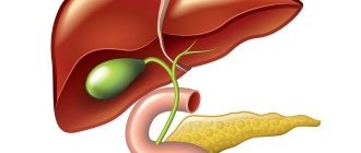
Gallstones are crystalline formations located in the gallbladder or bile ducts. The gallbladder is a reservoir for bile produced by the liver. The movement of bile along the biliary tract is ensured by the coordinated activity of the liver, gallbladder, common bile duct, pancreas, and duodenum. This ensures the timely entry of bile into the intestines during digestion and its accumulation in the gallbladder on an empty stomach.
The formation of stones in it occurs due to changes in the composition and stagnation of bile (dyscholia), inflammatory processes, motor-tonic disorders of bile secretion (dyskinesia).
Gallstones lead to the development of cholecystitis. Stones in the bile ducts contribute to the development of inflammation (cholangitis), which, when combined with inflammation of the pancreas (pancreatitis) or liver, can be life-threatening. When the bile system is blocked, favorable conditions are created for the proliferation of bacteria, and then inflammation of the ducts quickly occurs. Bacteria can enter the bloodstream and infect other organs.
Preventing stones from entering the ducts
The disease is easier to prevent than to treat.
If there is an increased risk of stone formation in the gallbladder or ducts, the patient must take preventive measures:
- First of all, this concerns nutrition. In medicine, patients with stones in the gallbladder or ducts are prescribed “table No. 5.” It is necessary to avoid foods high in fat and cholesterol. It is not recommended to eat fried, salted, smoked foods. You should eat in small portions, 5-6 times a day in small portions.
- To prevent stones from appearing, you need to monitor your weight. This means not only reducing the calorie content of food consumed, but also exercising.
- You should give up bad habits - smoking and drinking alcohol.
- Patients at increased risk of conglomerate formation should closely monitor their health. If diseases of the gastrointestinal tract are detected, timely treatment is necessary. Therapy cannot be delayed. It is chronic diseases of the digestive system that most often lead to the appearance of conglomerates in the gallbladder and further transition into the ducts.
Periodic scheduled examination is also included in the list of preventive measures. With regular visits to the doctor, the disease is detected at an early stage of development, which facilitates and speeds up treatment.