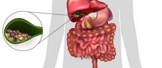
The gallbladder is a small, hollow organ located under the lower part of the liver. It serves to temporarily store bile produced in the liver.
As soon as a person begins to eat, the digestion process begins, in which bile gradually passes from the bladder into the intestines. However, part of the bile, bypassing the gallbladder, enters directly into the intestines, and only the excess is sent for temporary storage.
Bile not only participates in the digestion process, but also regulates peristalsis, removes steroids, cholesterol, and glutathione from the body. In addition, bile breaks down fats, neutralizes the harmful effects of hydrochloric acid on the intestines, and prevents fermentation and putrefactive processes in the gastrointestinal tract.
At most, the bladder contains about 200 ml of bile fluid, and in its normal state is not palpable. The shape, size of the organ and the development of pathology can only be determined using ultrasound.
Features of the structure of the gallbladder
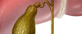
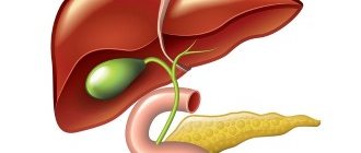
The bile bladder is pear-shaped and consists of three main parts: the bottom, the organ itself and its neck. The walls consist of a mucous, muscular and outer layer. In the body of the liver there are small ducts that form large ducts at the exit on the right and left sides of the organ.
Further, both ducts join into one, which is called the hepatic duct and connects the gallbladder with the liver. And the outflow of bile into the intestines occurs through the bile duct. The hepatic and cystic ducts are transformed into one common duct, which flows into the intestine.
At the beginning of the duct, at the exit from the gallbladder, there is a sphincter that prevents the free circulation of bile. The sphincter opens and releases another portion of bile only when food enters the stomach.
Normally, bile is secreted in small quantities to aid digestion, break down food in the small intestine and create conditions for the formation of enzymes in the pancreas. Any pathologies and diseases of the gastrointestinal tract can be easily determined using ultrasound.
What does an ultrasound show?
At the slightest suspicion or discomfort, you should immediately consult a doctor. For a more accurate examination and diagnosis, an ultrasound examination is prescribed, and the liver is examined along with the gallbladder.
Using an ultrasound, the doctor will accurately determine the presence of pathology, focusing on the length and width of the organ itself, the length of the neck, the thickness of the walls and the diameter of the ducts. The norm of these indicators directly depends on the age of the patient and may vary slightly.
It is necessary to seek medical help in the following cases:
- bitterness in the mouth;
- pain in the hypochondrium on the right;
- heaviness in the side;
- bad tests;
- jaundice;
- severe poisoning, intoxication.
As a rule, the gallbladder is affected by alcohol abuse and taking a large number of medications. Ultrasound helps to timely determine the presence of acute or chronic cholecystitis, cholelithiasis, pancreatitis and congenital anomalies.
Watch the video of an ultrasound of the gallbladder:
Postscript with Inspector Warnicke
For those who want to relax and at the same time activate cerebral circulation, which is no less important for our health than the bile ducts, I suggest trying to solve detective mystery stories together with Inspector Warnicke. Meet me.
I started publishing these stories on July 04, 2020 “HERE” and continued on July 06, 2020 “HERE”
The gallbladder is part of the biliary system of the liver, a connecting link with the intestines. It has the shape of a pear-shaped reservoir with a capacity of 70 ml. The structure includes the body of the bladder, the bottom, the neck, and the duct with a valve. The body is responsible for its functioning; stones and deposits can accumulate at the bottom if there is a malfunction; the neck and duct serve to remove bile and enter the reservoir.
The gallbladder (hereinafter referred to as the gallbladder) is an organ responsible for the quality of the digestive system. Unconcentrated bile is synthesized by liver cells with the participation of enzymes. It enters the bladder through a duct.
Before digestion has begun, secretion water is removed. The liquid acquires the concentration necessary for normal operation. While the bile is stored, the channel is shortened. The walls of the organ will begin to work when digestion is activated. The secretion enters the lumen of the duodenum and participates in the breakdown of substances.
The composition of bile from the bladder and liver is the same, but differs in concentration. The main component is acids, synthesized from cholesterol, improve metabolism, help in lipid hydrolysis, and participate in liver restoration.
Bile is designed to solve the body's problems:
- The breakdown of fats, which helps the body absorb.
- Strengthening the absorption of proteins and carbohydrates through the intestines.
- Neutralization of gastric juice.
- Sterilization of food in the intestines.
- Removing toxins.
- Stimulation of duodenal receptors.
- Participation in the creation of joint fluid.
Disturbances in the functioning of the bladder will lead to a number of diseases. For the normal functioning of the organ, you will need to eat right and give up bad habits. Prevention of diseases is better than cure.
https://youtu.be/ZsyESVYCkho
Preparation" for ultrasound
In order for the study to be successful and show reliable results, it is necessary to properly prepare for it. 10 days before the procedure, you should completely stop drinking alcohol, fatty and fried foods.
5 days before the ultrasound, patients are advised to refrain from eating foods that cause increased gas formation (milk, peas or beans, cabbage), and should not drink soda or natural juices.
A couple of days before the examination, you can start taking enzyme medications. You will have to fast for 10-14 hours before the test, and smoking is prohibited immediately before the procedure.
First, an ultrasound is performed on an empty stomach to determine the contractility of the gallbladder. After this, the patient is given a little fat sour cream or cream to eat, and after 40 minutes the examination is repeated.
A healthy organ should shrink by no less than 60% and no more than 80%. Any deviations from these indicators are pathology. Using an ultrasound, the doctor determines the size of the gallbladder, its structure and wall thickness, and the presence of an inflammatory process.
Functions of the biliary tract
Normally, the biliary system functions as a single whole. Bile passes through the ducts throughout the biliary chain: it is synthesized in the liver, stored and concentrated in the gallbladder, and then enters the intestinal space.
The bile ducts perform the following functions:
- Muscle bundles of smooth muscles keep the walls of the ducts in tone, which contribute to the active promotion of bile secretion.
- The biliary microflora of the bile ducts fights pathogenic microorganisms that have penetrated the system.
- Participates in the preparation of the bile-pancreatic mixture for normal digestion.
A decrease in the functionality of the bile ducts leads to a deterioration in the transportation of bile, which entails a chain of pathological processes in the gastrointestinal tract.
Decoding the results
It is important to consider that the normal size of the organ may vary slightly depending on age. They will differ in older people and children, and the average volume of the gallbladder in an adult is about 40-80 ml.
Normal for adults
Normal liver and gallbladder for an adult
A healthy adult patient must have certain parameters of the organ, which include width, diameter, wall thickness and length.
The following are considered normal indicators:
- volume about 70 ml;
- wall thickness no more than 0.4 ml;
- organ length, about 6-10 cm;
- the width should not exceed 5 cm;
- the diameter of paired ducts is normally about 0.3 cm;
- the diameter of the main duct cannot exceed 0.7 cm.
Interestingly, in women over 40 years of age, gallbladder lesions are much more common than in men. Moreover, naturally overweight blondes often suffer from such diseases. In men, such pathologies are observed much later, and only under conditions of alcohol abuse and poor nutrition.
Find out the norms of the gallbladder from the video:
Norm for children
The normal size of the gallbladder in children varies depending on age. So, for newborns, the norm is considered to be a length of 3.4 cm and a width of 1.08 cm. From a month to five, the length should be 4 cm and the width should be 1.02.
A one-year-old child has a bladder length of 5.5 cm and a width of up to 1.07 cm. At 3 years old, these figures increase to 5 cm and 1.60 cm, respectively. For a 7-year-old child, the norm will be a length of about 7 cm and a width of no more than 3.70 cm. Children aged 10 years and older should have a length of the gallbladder of 7.7 cm, a width of about 3.7, and a diameter of up to 1. 4 cm.
Organ health
Cleaning of the gallbladder and bile ducts is carried out as prescribed by a gastroenterologist. The procedure has contraindications. Self-prescription may cause pancreatitis, peritonitis and other complications! Depending on the examination performed, treatment methods are prescribed.
Reasons for cleaning procedures
If the body gives signals indicating problems with the gallbladder, cleaning the organ and bile ducts is indicated. The method helps remove toxic compounds, cholesterol deposits, and regulates the functioning of the bladder. For children and the elderly, cleansing is prescribed exclusively under the supervision of a gastroenterologist.
The cleaning procedure solves the problems:
- Inflammation, infections of the digestive system;
- Obesity;
- The appearance of stones in the bladder;
- Dyskinesia GB;
- Stagnation of bile;
- Removing toxins;
- Reducing the adverse effects of medications;
- Neoplasms in the liver and gallbladder.
For prevention purposes, the procedure is performed by people with a sedentary lifestyle, lovers of spicy, fried and fatty foods, fast food, and women before conceiving a child.
Proper nutrition and therapeutic exercises often help in solving problems with gastrointestinal tract. Cleansing will affect the general condition of the body. Sleep and appetite will improve, and the syndrome of constant fatigue will disappear.
In the absence of contraindications, cleaning is carried out on an outpatient basis or at home. There are different methods in traditional and folk medicine. Chemical or natural remedies, probing, gymnastics and therapeutic massage are used.
Situations in which it is impossible to cleanse the gallbladder
Before cleaning the bladder, you will need to conduct a series of examinations to detect other diseases. There are reasons limiting the use of the procedure. Techniques that may seem harmless to a non-specialist can cause serious complications.
This treatment method should be abandoned in the following cases:
- Colds;
- Infectious and chronic diseases of the gastrointestinal tract;
- Problems of the cardiovascular system;
- Oncology;
- Diseases in the acute stage;
- Stone in the ducts;
- Exhaustion of the body;
- Childhood;
- Pregnancy.
The list goes on. Body systems are interconnected. Disruption of one organ is associated with problems in other systems. To avoid the situation of “we treat one thing and cripple another,” we need a competent approach.
Gallbladder diseases
Using ultrasound, any diseases not only of the gallbladder, but also of the liver and bile ducts are easily diagnosed.
With the help of ultrasound, any diseases of not only the gallbladder, but also of the liver and bile ducts are easily diagnosed. There are several diseases that occur most often.
For example, dyskinesia, which manifests itself in the form of thickening of the walls of the organ and disruption of its contractile functions. The cause of the disease is considered to be a hormonal imbalance that prevents the normal flow of bile. On ultrasound, the bubble has uneven outlines and kinks that disrupt the functioning of the organ.
An equally common pathology is cholesterosis, which occurs against the background of diabetes mellitus and other endocrine disorders. A common disease is cholecystitis, which is caused by an intestinal infection that has penetrated the gallbladder.
Severe inflammation of the walls occurs, accompanied by nausea, flatulence, and pain in the hypochondrium. The acute form of the disease can be recognized not only by thickening of the walls, but also by an increase in the size of the organ. The chronic course is characterized by a decrease in the bubble and the presence of small foreign inclusions.
With the help of such a study, cholelithiasis (cholelithiasis), the cause of which is a hereditary predisposition and hormonal problems, is well determined.
Adenomyomatosis is considered a dangerous disease, which is characterized by benign thickening and compaction of the walls of the bladder, reaching 10-12 mm. This almost completely blocks the lumen inside the organ and is often accompanied by the appearance of polyps.
Sometimes, polyps can grow on the walls on their own, without accompanying pathologies. They look like small round growths and can be either benign or malignant. In the latter case, their rapid growth and the manifestation of accompanying metabolic disorders occur.
The most dangerous, incurable and severe pathology of the bladder is cancer. Determine it by an increase in volume, thickening of the walls and the presence of metastases.
At an early stage, it is almost impossible to make a diagnosis, because the disease is initially asymptomatic. The progression of the pathology is accompanied by severe pain, weakness, weight loss and bloating of the peritoneum.
In addition, congenital developmental anomalies occasionally occur, which include uncharacteristic protrusions of the walls, incorrect location or complete absence of the gallbladder.
The gallbladder is an important organ. His health and performance must be carefully monitored. People can be helped with this by deciphering the results of an ultrasound scan of the gallbladder to determine its function. Thanks to ultrasound, everyone can verify the health of their gallbladder and the ducts that connect to it.
Pathologies
There are many diseases and formations that can be seen in the gallbladder using ultrasound. Each of them has symptoms and developmental characteristics. Let's take a closer look at the most common pathologies.
Cholesterosis
This disease is characterized by the process of cholesterol deposition in the organ. These pathological actions lead to inflammation. Factors that can influence the onset of cholesterol formation are diseases: diabetes mellitus, obesity, thyroid disease and others.
Using ultrasound, it is possible to characterize the anterolateral wall. In an ultrasound image, this disease is visible as an uneven thickening of the walls of the bladder. In the diffuse form, individual areas similar to a dotted line are observed.
Dyskinesia
This disease means that there is a disturbance in the process of bile outflow due to incorrect contractions of the gallbladder. It often occurs due to a hormonal, hypotonic or hyperkinetic disorder. To improve the flow of bile, you should adjust your diet and avoid stress.
It is with the help of ultrasound that this disease can be detected. Dyskinesia is visible in the image as uneven outlines of the organ, the presence of constrictions and kinks that have formed. The walls are thickened and are in strong tone. Also, during this procedure, it is possible to track the contractility of the bladder walls, and their incorrectness is the main sign of dyskinesia.
Cancer is one of the most serious and dangerous pathologies.
There are many causes of the disease.
These include genetic predispositions, smoking and addiction to alcohol and heavy food.
In the first stages there are almost no symptoms.
Over time, the cancer begins to manifest itself, leading to general weakness, bloating, and loss of kilograms.
First of all, to determine the disease, they resort to ultrasound of the gallbladder, MT (ureters) and liver. If there are other questions, an ultrasound scan of the abdominal cavity is also performed. Cancer signals on ultrasound are: unevenly dense and thickened walls of the organ, its enlargement. It is possible to detect metastases.
Tumor
A gallbladder tumor is the growth of cells atypical for the body on the tissue of the organ. It can be benign or malignant. Often this malignant tumor occurs in conjunction with gallstone disease. In case of a malignant tumor, immediate surgery is required.
Norms
The normal parameters (size and volume) for the gallbladder of an adult are:
- length – 6-10 cm, width – 3-5 cm;
- volume ranging from 30 to 70 cm3;
- organ wall thickness – up to 4 mm;
- pear-shaped or cylindrical shape;
- size in diameter – 3-3.5 cm;
- lobar bile ducts no more than 3 mm;
- the bottom protrudes no more than 1.5 cm from the bottom of the liver;
- reduction to 70% of the volume measured on an empty stomach and 15 minutes after a choleretic breakfast;
- The diameter of the bile duct is 6-8 mm.
Any disease detected on ultrasound requires clarification and observation.
Protocol
the transcript of an ultrasound scan of the gallbladder and other abdominal organs looks like
Video
The video below gives an idea of many issues related to ultrasound of the gallbladder, including normal indicators of the organ’s condition.
Bile duct tumors
Among benign tumors, adenomas, papillomas, fibroids, lipomas, adenofibromas, etc. can be found. An echogram can reveal a tumor-like formation of different sizes and echogenicity with localization in the projection of the extrahepatic bile ducts, but more often in the projection of the common bile duct, without specifying the histological forms, the differentiation of which is carried out by using a targeted biopsy of the tumor site.
Anatomy of the gallbladder
This is a pear-shaped organ connected to the liver and located in its lower part. It is attached to the liver with the help of connective tissue and accumulates bile, which is formed during the work of this largest human gland. After filling the bladder, its walls relax, the accumulated bile flows through the duct into the duodenum.
The design of the walls is the main feature of the gallbladder; it is thanks to it that it carries out its functions. The bladder wall consists of three layers:
- The mucous membrane that forms folds in the cervical area. The tubular-collar glands are hidden in these folds.
- Muscle tissue consisting of smooth fibers. It smoothly passes into the layer of the cystic duct, and near the neck it forms a closure that prevents the arbitrary outflow of bile.
- Dense tissue that forms the adventitia. It consists of elastic fibers.
Normal sizes
The size of the gallbladder in women and men should be within the following standards:
- width 3-5 cm;
- length 6-10 cm;
- wall thickness up to 3 mm;
- the internal size of the common duct is up to 6-8 mm, the lobar duct is up to 2-3 mm.
For children, there are no clear parameters that the bubble must meet. This is due to constant growth, to which the gall bladder may not adapt immediately.
Problems can only be indicated by the results of tests performed by a gastroenterologist. If a pathology is suspected, an ultrasound will be performed.
Biliary tract: structure
The structure of the canal is not very complicated. All small ducts originate in the liver. The fusion of the left and right canals (both located in the liver) forms the common hepatic canal. The channels carry the burn formed by the hepatic lobes. The bile duct is formed in the bladder, then it connects with the common hepatic duct and forms the common bile duct. A bend in the gallbladder may indicate abnormalities in its development. Strictures of the common hepatic duct are not normal. Occurs due to strong blows to the liver area.
Return to contents
How are indicators measured?
The exact size of the gallbladder is normally determined by ultrasound in adults and children. For the results of the study to be reliable, you need to prepare for it:
- 8-12 hours before the examination, you should have a light dinner (preferably before 7 pm the day before the ultrasound). At the same time, give up coffee and tea, alcohol and smoking. You can't use chewing gum.
- During the examination you will need to lie on your side, then on your back. A change in body position is necessary when searching for stones in the ducts.
- Sometimes you may need to eat a so-called choleretic breakfast. It could be a piece of butter, chocolate, or a raw egg. Such products stimulate the release of bile into the duodenum, and the uzologist will be able to assess the motility of the biliary tract.
After taking readings from the device, an ultrasound diagnostic specialist calculates the size of the gall bladder.
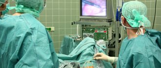
Contraindications to cholecystectomy
Usually there are absolute and relative indications for any operation. The doctor must assess the risk to the patient before and during organ removal.
- Surgery is absolutely contraindicated for people with severe acute and chronic diseases in which removal of the gallbladder would lead to death.
- Relative contraindications include those health problems that can make the postoperative period more difficult and prolonged, and cause complications. However, the consequences of these diseases can be foreseen and influenced: for example, normalizing blood pressure in patients with hypertension, glucose levels in people with diabetes.
What to do if surgery is contraindicated and stones need to be removed? For such cases, there is a procedure for non-surgical stone crushing. Why, then, is this procedure not performed on everyone who suffers from cholelithiasis? At the moment, it shows little effectiveness, it has many contraindications and risks, and usually it ends with the patient having the entire bladder removed anyway. For more information about how this procedure is carried out and why doctors do not favor it, read the article about crushing gallstones.
In some cases, you can try to dissolve the stones using Ursosan or other ursodeoxycholic acid preparations. Not all stones can be dissolved: this depends on their composition, size and other factors. Read about the cases in which it is possible to use tablets, what the disadvantages of this method are, and whether it is possible to dissolve stones using traditional methods in the article about litholytic therapy.
An expert on the website Pokhmelye.rf, gastroenterologist Daniela Purgina, warns about a common mistake.
What I strongly do not recommend to you is to try to dissolve the stones yourself using folk remedies. It just doesn't work! If it worked, we would prescribe this treatment to patients ourselves. The most common recipe is lemon juice with olive oil; as a result of taking them together, you may see stone-like formations in the stool, and you will think that they came out; but in fact, in the body, under the influence of enzymes, a saponification process simply occurred and you produced soap within yourself. Only very small stones or sand can come out, but even trying to expel small stones is dangerous, because they can get stuck in the bile duct or get into the pancreatic duct, and this can lead to the development of pancreatitis.
Possible deviations from the norm
Deviations manifest themselves in the form of changes in the shape and size of the gall bladder. It can increase or decrease; when kinked, the contours of the organ change. Additionally, changes in the condition of the bile ducts are diagnosed, which can expand or narrow depending on the existing pathologies.
Increase
The norm of the gallbladder is disrupted to a greater extent when the outflow of bile becomes difficult. The reason for the increase in bile volume is:
- Inflammatory processes. Most often this is cholecystitis. The disease is accompanied by fever, pain in the right hypochondrium, nausea and a feeling of weakness.
- Infections caused by parasites, bacteria and viruses.
- Bubble bend. Often appears when gallstones form.
- Diseases of the liver and digestive system. The bladder enlarges if the pancreas is affected by a malignant tumor - carcinoma. The gallbladder also reacts to diseases such as cirrhosis of the liver and intestinal obstruction.
After ultrasound diagnostics, it will be necessary to establish the cause of the enlarged gallstone. This can be done using laboratory tests that will identify diseases that have caused a change in the size of the organ.
Decrease
Downward size changes are rare. Most often, it indicates improper preparation for the examination. Organ shrinkage occurs in the following conditions:
- Hypogenesis. This is an anomaly in which a small gallbladder is a congenital characteristic of a person. Such an organ functions properly, the bile ducts remain of normal diameter, and there are no obstacles to the accumulation and removal of bile.
- Wall perforation. This occurs when an organ is damaged from the inside by a stone, as well as in some injuries. The pathology is identified by ultrasound results and persistent severe pain in the right side. Perforation can only be treated surgically.
- Chronic cholecystitis in the later stages. The examination will show a decrease in the structure of the organ, as well as foreign bodies inside the gall bladder.
Wall thickening
The cause of changes in the thickness of the walls of the gallbladder is swelling. It is caused by inflammatory processes, such as cholecystitis, as well as toxic and infectious lesions.
Wall thickening is most often diagnosed along with a change in the size of the bladder.
Change in duct diameter
Narrowing of the bile ducts is not a sign of disease. This is an anatomical feature that does not interfere with the normal flow of bile and does not cause problems to humans. If the bile ducts have a larger diameter than usual, this indicates inhibition of the outflow mechanism. The cause of the violation may be stones, kinks in the body, bottom or neck.
These pathologies cause severe pain in the right side, which radiates to other parts of the body - under the right shoulder blade, in the arm and leg. The patient's body temperature rises, vomiting and weakness appear. The skin may become yellow.
When diagnosing, it is necessary to exclude liver diseases - hepatitis, steatosis, cirrhosis and oncology, since they give similar symptoms.
Pathologies and methods of their treatment
According to statistics, 50% of patients who sought medical help were diagnosed with diseases of the digestive system, of which more than half are pathological changes in the biliary system. The common bile duct interacts with the liver and gallbladder, so stones or bacteria easily penetrate into it. This leads to damage to the walls and its involvement in the general pathological process.
The most common diseases include:
- stricture (stenosis) of the bile ducts;
- cholangitis – inflammatory pathology;
- Choledocholithiasis is the presence of stones in the ductal system.
Impaired functioning can be caused by injuries, the presence of a foreign body in the abdominal cavity, a hematoma in the area of the bile ducts, or dissection of the common bile duct during surgery. Sometimes tumor growths are diagnosed in the canal. Most often these are metastases; common bile duct cancer practically does not occur.
Biliary strictures
Scar growths that lead to narrowing (stricture) of the duct mainly appear after surgery to remove the gallbladder. But the incidence of injury after laparoscopic cholecystectomy does not exceed 1%, so this is a rare cause. Most often, strictures appear:
- after chronic inflammation of the gallbladder (cholecystitis) or pancreas (pancreatitis);
- often a decrease in the size of the common bile duct causes sclerosing cholangitis, opisthorchiasis (helminthic infestation);
- tumor-like growths of benign and oncological origin.
Strictures obstruct the flow of bile, but this condition may not appear for years. When the contraction is critical, the patient is bothered by causeless itching of the skin and body temperature rises. Over time, signs of obstructive jaundice appear. The skin becomes yellow, the stool is light, and the urine is dark.
It will not be possible to restore the lumen using conservative methods. Dilation of the duct requires surgical treatment, which is carried out in the following ways:
- reconstruction by classical surgery;
- percutaneous puncture;
- retrograde intervention.
The patient should contact a hepatologist surgeon at the first suspicion of narrowing of the ductal system. Delay is fraught with the development of dangerous consequences. Long-term retention of bile in the liver leads to damage to hepatocytes and the development of biliary cirrhosis. When stagnant secretions become infected, cholangitis develops. An acute form of inflammation can cause liver abscess and blood poisoning (sepsis).
Choledocholithiasis
The cause of the appearance of stones in the bile ducts may be the movement of stones along with bile from the affected gallbladder. In some patients, stones form in the common bile duct. Why this happens is explained by the lithogenicity of the bile, which persists even after the bladder has been removed. Strictures, cysts, polyps, and helminthic infestations can provoke pathology.
With choledocholithiasis, an increase in pressure is observed in the common bile duct, as a result of which a small stone can migrate along the duct. The sharp edges of the stone irritate the walls, causing inflammation, swelling, and sometimes complete blockage. As a result, the common bile duct is dilated and bile evacuation is difficult. This means that an environment favorable for the growth of bacteria appears.
If the stone in the bile duct is small, it can move freely into the duodenum and not cause discomfort. The presence of large stones is accompanied by the following manifestations:
- First, pain appears, which is localized in the right hypochondrium and spreads to the back;
- the pain syndrome is dull or aching in nature, and when the stone moves to the area of the papilla of Vater, it becomes encircling;
- after 12–24 hours, jaundice appears, and the pain gradually decreases;
- yellowness of the skin and sclera intensifies, then weakens, such “swings” are a characteristic sign of choledocholithiasis;
- with prolonged blockage, the feces become white and the urine dark.
If choledocholithiasis is suspected, ultrasound diagnostics and transhepatic cholangiography are performed. Using these methods, the diagnosis is confirmed, the number, size and location of stones are revealed.
Treatment consists of removing the stones. This can be done during surgery - laparotomy or laparoscopy. For large solid formations, choledochotomy is prescribed. This is an opening of the bile duct followed by cleansing of stones. If the disease recurs, the gallbladder is removed, after which the main duct returns to normal.
Therapeutic manipulations will not bring the desired result without following a diet and limiting physical activity. Patients are advised to exclude fatty, spicy, fried foods from the diet, limit salt, sour fruits, and raw vegetables. Make a daily menu, adhering to the list of products recommended by diet No. 5 according to Pevzner.
Cholangitis
The inflammatory process in the common bile duct appears for the following reasons:
- bile stagnation;
- infections caused by staphylococci, enterococci, E. coli;
- the presence of tumor growths, cysts;
- In childhood, the pathology can be caused by helminthiasis.
Acute inflammation is accompanied by a vivid clinical picture. The patient's temperature rises sharply to 40 degrees, severe pain appears in the right hypochondrium, similar in intensity to hepatic colic. After a while, jaundice and headaches develop. Nausea, vomiting, and itchy skin are possible. The chronic form of cholangitis is characterized by a long course with alternating periods of remissions and exacerbations.
Patients with cholangitis are treated in a hospital setting. Therapy includes prescription of medications:
- antibiotics - to destroy the infectious agent;
- anthelmintics - to get rid of parasites;
- anti-inflammatory – eliminate inflammation;
- antispasmodics - to relax the walls of the bladder and ducts;
- hepatoprotective agents – for the restoration of liver cells.
If conservative methods do not produce positive results, surgical treatment is performed. During surgery, the common bile duct is widened and the stones are removed. If the course is prolonged, abdominal surgery is performed to remove dead sections of the biliary tract.
Why does the size change?
The size of the gallbladder may change due to a disruption in the natural processes of accumulation and outflow of bile. The normal size of the gallbladder allows it to carry out its functions without problems. Any deviation from the norm, not due to a congenital anatomical feature, is a sign of serious diseases of the gall and other organs: liver, pancreas.
Untimely treatment can lead to serious consequences - worsening of the disease. Some of them can subsequently be cured only by removing the gallbladder, for example, calculous cholecystitis. Timely diagnosis will allow you to get rid of diseases without serious consequences for the body and return to a full life. Therefore, it is necessary to pay attention to preparing for the studies prescribed by the doctor.
>
Prevention of consequences
It should be understood that any surgical intervention is traumatic for the body, and it needs time to adapt and recover. Therefore, you should follow the recommendations of doctors.
- You should not adhere to any long-term diet after removal of the gallbladder in the absence of problems with stool and abdominal pain: a varied healthy diet is necessary.
- If you have diarrhea or bloating, you should limit your intake of fatty, spicy foods, drinks containing caffeine, and the consumption of dairy products, as well as treating bile outflow dysfunction.
- It is necessary to control weight and hormonal levels in the presence of relevant diseases.
An expert on the site Pokhmelye.rf, gastroenterologist Daniela Purgina, refutes the typical misconception regarding cholecystectomy.
It is a very common belief that after removal of the gallbladder, you need to constantly take some kind of medication to drain bile. In fact, there is no need for this. Taking choleretic drugs is completely unjustified, since the bladder has been removed and there is nowhere to drive the bile from; it will go from the liver through the ducts to the intestine. Ursodeoxycholic acid drugs are prescribed only if there is a risk or stones have been identified in the bile ducts.
You can contact the hepatologist in the comments. Don't hesitate to ask!
This article was last updated: 07/18/2019
Didn't find what you were looking for?
Try using search
doctor or administrator.
Read the dictionary of terms.
Expert author: Gastroenterologist-hepatologist Ekaterina Kashukh