What is an intestinal tumor?
An intestinal tumor is an abnormal proliferation of normal (benign neoplasia) or abnormal (malignant neoplasia) cells to form a characteristic structure in the lumen or wall of the intestine.
Benign tumors are characterized by slow growth. They do not grow into surrounding tissues and do not metastasize to other organs and parts of the body. However, in many cases, such tumors eventually become malignant.
Malignant tumors are characterized by rapid and invasive growth with damage to the intestinal wall and invasion of neighboring organs. These tumors require immediate and early treatment.
Lump in the abdomen on the right side
If a lump is felt in the right side of the lower abdomen, then perhaps the reason lies in the formation of a formation in the area of the right kidney or ascending colon.
A lump on the right side of the lower abdomen signals the occurrence of certain diseases such as:
- appendicitis. The inflammatory process begins on the right side, but gradually moves to the navel and lower abdomen. Usually the disease is accompanied by acute pain, severe discomfort, fever and nausea. Urgent surgical intervention will help solve the problem;
- lipomas. In medicine it is also called benign wen. Occurs as a result of blockage of the sebaceous glands. On palpation, a soft tumor is felt. It can increase in size and roll under the skin. Removed by surgery;
- atheromas. This concept is usually understood as a cyst up to 3 cm in size, which is tightly attached to the skin. It is characterized by an inflammatory course, so it is hot and painful to the touch. Requires surgical removal;
- fibrosarcomas. A malignant formation, the diameter of which ranges from 2-15 mm. Resembles a smooth scar or bump on the skin. Characterized by mobility and pain. The middle part of the abdomen is tense, and tissue atrophy occurs over time. Treatment involves surgery;
- diverticulitis as a hernia with accumulation of intestinal contents as inflammation develops. Provoking factors are parasites, hereditary predisposition, poor nutrition, and decreased muscle tone. The disease is accompanied by upset stool, the passage of stool mixed with blood, an increase in temperature, and pain in the inflamed area. Treatment involves taking antibiotics. In complicated cases, surgical intervention is performed.
The painful sensation appears on the right side, but can radiate to other parts of the abdominal cavity.
What types of tumors are there by location?
The most common neoplasms of the large intestine. They account for up to 95% of all intestinal neoplasia, both benign and malignant.
This is because the small intestine is better protected from tumors due to rapid cell turnover, a general absence of bacteria, an alkaline environment, increased apoptosis, fast food transit time, a well-developed immunoglobulin A (IgA)-mediated immune system, and low levels of precarcinogenic enzymes. .
However, tumors of the small intestine do occur. And these are usually malignant tumors with a high degree of aggressiveness.
Organ Anatomy
The ileum is anatomically similar to the middle section of the small intestine - the jejunum. Its entire internal surface is a mucous membrane, covered with villi of columnar epithelium about 1 mm high.
The role of the villi is the absorption of substances. With their help, fats are sent to the lymphatic vessels, amino acids with monosaccharides are sent through the veins. In addition to villi, the mucous membrane contains cylindrical tubes (crypts) of small depth - Lieberkühn's glands.
Due to the irregularities of the ileal mucosa due to circular folds, villi, and crypt glands, its area is increased, which contributes to better and faster absorption of nutrients from the chyme.
Thanks to their contractions, food is mixed with intestinal juice enzymes and moves down the intestines. Externally, the ileum is covered with serosa along its entire length.
Signs of tumors by structure and properties
Benign tumors
Leiomyomas are tumors of one of the muscle layers of the intestinal wall. They can grow into the intestinal lumen and become damaged, becoming ulcerated. This causes bleeding and anemia, which are warning symptoms.
These neoplasias are very difficult to diagnose, especially when they occur in the small intestine. Endoscopic ultrasound is a good way to determine the location of the tumor in the intestinal wall. Some leiomyomas have malignant potential.
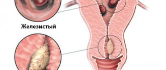
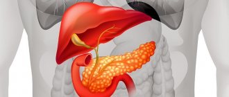
Adenomas (polyps) are benign tumors that have a high malignant potential. They cause symptoms due to blockage of the intestinal lumen. If they occur in the area of the duodenal papilla of Vater, they can cause jaundice. Because of the risk of malignant transformation, adenomas are usually removed surgically.
Lipomas are collections of fatty tissue in the intestinal wall that have a soft, yellowish appearance when examined endoscopically. These are completely benign tumors with no malignant potential. Lipomas do not need to be removed unless they grow large enough to cause obstructive symptoms (or bleeding due to ulceration).
Hemangiomas. Hemangiomas are collections of blood vessels that form a benign vascular tumor in the intestinal wall. They are benign and sometimes occur in combination with other syndromes.
Hemangiomas can cause gastrointestinal bleeding and anemia. They can be treated endoscopically using a heating probe to burn the vessels. Patients may require resection of areas of intestine associated with hemangiomas if they are causing significant bleeding.
Neurogenic tumors are benign growths that arise from neural (nervous) tissue. The most common type is neurofibroma.
Neuroendocrine tumors
Gastrointestinal neuroendocrine tumors (GI-NETs) of the intestine are intestinal carcinoid neoplasias. They are generally thought to originate from serotonin-secreting enterochromaffin cells (ECs) of the intestine (known as Kulczycki cells). But technically, intestinal carcinoid tumors can develop from any type of well-differentiated neuroendocrine cells in the digestive tract that are capable of secreting bioactive hormones and/or amines.
Historically, the most common site of gastrointestinal (GI) carcinoid tumors has been the appendix. Currently, however, the most common site of carcinoids in the gastrointestinal tract is the small intestine (30%), followed by the rectum (19.6%).
Malignant tumors
adenocarcinoma most often develops from adenomas (polyps). In 95-97% of cases, adenocarcinomas develop in the large intestine (colorectal cancer). If such a tumor forms in the small intestine, then most often it occurs in the ampulla of the duodenum.
The tumor can be diagnosed using radiological methods, endoscopy or surgery and confirmed by biopsy. Treatment depends on the location, size and shape of the tumor, and whether it has spread to nearby organs.
Options include endoscopic removal and surgery. Chemotherapy and radiation are also widely used.
Intestinal lymphoma is a recognized complication of celiac disease, but it is also possible and may occur in immunodeficiency syndromes. Symptoms include cramping abdominal pain, weight loss, signs of malabsorption, bleeding or even intestinal obstruction.
Leiomyosarcoma is a malignant tumor arising from the muscular wall of the intestine. Complete surgical resection is recommended whenever possible. Chemotherapy and radiotherapy are also used.
Metastases of malignant tumors from other parts of the body can occur in both the small and large intestines. However, if in the thick part primary malignant tumors are more common than metastases, then in the thin part the opposite is true - metastases are found more often than primary cancer of the small intestine.
Features of work
The secret secreted by the ileum, like the entire small intestine, is intestinal juice, with the help of which parietal (membrane) and cavity digestion is carried out. About two liters can be released per day.
Intestinal juice is formed due to chemical and mechanical irritation of the food lump (chyme) of the intestinal walls. The dense part of the juice are epithelial cells - special cells on the villi of which the necessary enzymes accumulate and then are released into the intestinal lumen, which promote the hydrolysis (decomposition with water) of food and the absorption of necessary substances. The main enzyme in intestinal juice is enterokinase.
Cavity and parietal digestion are closely interconnected. During the cavity, food is hydrolyzed to intermediate substances, and intermediate substances during the membrane process continue to be broken down and begin to be absorbed with the help of intestinal motility (motor function), mucosal villi and an increase in pressure inside the intestine. The environment in the ileum during the digestion process is alkaline.
Symptoms of intestinal tumors
Symptoms in the thin section
Abdominal pain, bleeding, intestinal obstruction and weight loss are the main signs of intestinal tumors in this section.
However, a palpable tumor is very rare. Perforation of the intestinal wall occurs in approximately 10% of cases. Periampullary tumors may be accompanied by obstructive jaundice, and diarrhea is more common in patients with lymphoma.
In small bowel carcinoids, carcinoid syndrome, caused by the release of serotonin and other vasoactive compounds, causes flushing, telangiectasia, diarrhea, bronchospasm, and, rarely, heart failure.
Symptoms in the thick section
A malignant intestinal tumor may not show symptoms. Colorectal cancer does not detect itself in the early stages, while the tumor is small. Symptoms often appear when the tumor grows into surrounding tissues and organs.
Other health conditions can cause the same symptoms as colorectal cancer.
For an illness such as an intestinal tumor, symptoms may include:
- diarrhea;
- constipation;
- a feeling that the rectum is not completely empty after bowel movements;
- bright or dark red blood in the stool;
- bleeding from the rectum;
- flatulence, abdominal cramps and feeling of bloating;
- pain or discomfort in the rectum;
- a lump in the stomach or rectum;
- fatigue and weakness;
- anemia, which can cause fatigue and shortness of breath;
- loss of appetite and weight;
- intestinal obstruction;
- swollen lymph nodes;
- accumulation of fluid in the abdomen (ascites);
- pain in the abdomen, back, buttocks or legs.
Forecast for life
For benign intestinal tumors, the prognosis is favorable. With timely detection and treatment, complications can be avoided.
The prognosis for malignant intestinal tumors depends on the stage of the disease. The overall five-year survival rate is 60%. With a localized process, the prognosis is better - up to 90%. In the presence of regional metastases, survival rate is only 50%, and in case of distant metastases it drops to 7%.
The prognosis also depends on the effectiveness of the therapy. To assess the patient’s condition and identify relapses of carcinoma, the following is indicated:
- Colonoscopy 1 year after completion of therapy and then every 3-5 years.
- Chest X-ray – every 5 years.
- Ultrasound of the liver every 6 months for 3 years, then annually until 5 years.
- Tumor marker assessment (CEA) every 6 months for 3 years, then annually.
If no relapse is detected within 5 years, they speak of recovery.
Causes of intestinal tumors
The pathogenesis of most intestinal neoplasias is unknown.
Risk factors that are associated with colon or small bowel cancer are:
- smoking cigarettes;
- alcohol consumption;
- dietary factors such as lack of plant fiber in food, consumption of processed red meat, etc.;
- Crohn's disease;
- celiac disease;
- peptic ulcer;
- familial adenomatous polyposis;
- cystic fibrosis;
- various types of previous operations (for example, operations to connect the urinary system to the intestines);
- neurofibromatosis.
Lumps in the lower abdomen
A pathological formation can appear in any part of the abdomen. If a lump is felt in the lower abdomen, then the cause is:
- pinched hernia;
- protrusion of the sigmoid colon;
- rupture of the walls of the diverticulum;
- inflammatory process in the pelvic organs;
- intestinal obstruction in the lower part of the organ;
- increased gas formation;
- tumors in the male genital organs.
Irritable bowel syndrome, an inflammatory process in the intestinal canal, or compression of the nerves can lead to the appearance of a lump in the lower abdomen. A lump often occurs as a stone moves down the ureter.
Diagnosis of intestinal tumors
Diagnosis of the thin section
X-ray. Enteroclis - this type of fluoroscopy is most often used abroad for diagnosing tumors of the small intestine. The tube passes through the nose or mouth into the stomach and into the small intestine. Barium is then pumped through the tube. As the barium coats and passes through the lining of the small intestine, a series of pictures are taken.
A computed tomography (CT) scan is used to look for causes of pain or swelling in the abdomen, such as a mass in the small intestine. It is also used to see if a tumor in the small intestine has spread to other organs.
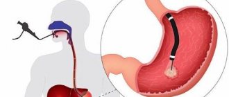
An endoscopy is done to find out what is causing bleeding in the small intestine or to find a tumor in the small intestine. The small intestine is very long and has many loops and bends that make it difficult to examine. Doctors in Belgium use different types of endoscopy to examine the entire length of the small intestine.
Push endoscopy allows doctors to examine the duodenum, which is the first part of the small intestine closest to the stomach. The doctor passes a thin endoscope through the mouth, down the throat and through the stomach. The doctor pushes it as far as possible along the duodenum and into the middle part of the small intestine (that is, the jejunum).
Balloon endoscopy uses a special balloon attached to an endoscope. The doctor may blow air into the balloon to widen the small intestine so that the endoscope can move further.
Capsule endoscopy (PillCam). This is a new and less invasive procedure. Instead of an endoscopy, the patient swallows a pill-sized capsule containing a tiny video camera and light bulb. The camera takes pictures as it moves along the gastrointestinal tract. These photos are sent to a small recording device worn on the belt. The capsule usually passes through the gastrointestinal tract in about 8 hours and is eliminated from the body through bowel movements.
Laparotomy or laparoscopy may be used to examine and collect samples from the small intestine.
Diagnosis of the thick section
Colonoscopy is the most common test used to diagnose all types of colon tumors.
A colonoscopy is performed if a patient has symptoms or as part of a screening test. A biopsy sample may be taken during a colonoscopy.
Virtual colonoscopy uses computed tomography (CT) to produce images of the colon and rectum without the use of an endoscope. If virtual colonoscopy equipment is available, you may be offered this test if you are unable to have a regular colonoscopy. CT scans are also used to check whether colorectal cancer has spread to the lymph nodes or organs of the chest, abdomen, and pelvis.
A digital rectal examination is an examination in which the doctor inserts a gloved finger into the rectum to check for lumps or swelling or to measure how far a rectal tumor is from the anus. DRE is used in conjunction with transrectal ultrasound or pelvic MRI to determine the stage of rectal cancer, including how deeply the tumor has grown into the rectal wall.
A fecal occult blood test (gFOBT) or immunochemical test (FIT) may be performed at the time of diagnosis or during screening.
Transrectal ultrasound (TRUS) is used to determine the stage of rectal cancer, including how deep the tumor has grown into the rectal wall, and to measure the distance from the rectum to the anus.
Magnetic resonance imaging (MRI) can be used to determine the stage of colon cancer and where it has spread.
Treatment methods
Based on the histological structure of the formation, its type, location in the small or large intestine and size, an intestinal tumor can be treated in different ways. The most effective is surgical removal of benign and malignant tumors. The operation is performed in cases where the tumor does not extend beyond the intestine. For cancer, such treatment has an effect in the first two stages, as well as in the third, provided there are no metastases. The tumor may require excision of part of the colon or other intestine, followed by suturing of the free ends of the intestine or a calostomy through the peritoneal wall. The operation can be performed laparoscopically or by the abdominal method.
If metastases are detected in the liver (most often colon cancer metastasizes there) or in other organs, radiation therapy is performed first, followed by surgery. Radiation can also be used in the postoperative period, which helps kill remaining cancer cells and reduce the risk of relapse. For malignant tumors, especially if they are inoperable, chemotherapy is performed. Taking chemicals improves the effectiveness of radiation, but has some side effects. To alleviate the patient’s condition after chemotherapy, he is prescribed anti-nausea drugs, vitamins, and immunomodulators.
We recommend reading Burkitt's lymphoma - causes, forms, stages
If you have intestinal tumors, you must adhere to a certain diet to regulate the functioning of the digestive system. Food should be easily digestible and digestible; food should contain proteins, fats, carbohydrates, vitamins and minerals. You should eat fractionally, that is, in small portions, but every two hours. At the initial stage of treatment, all food should be warm and puree-like in consistency, preferably steamed. You should drink enough; it is better to give preference to plain water.
It is worth excluding alcohol, coffee, strong tea and fresh milk. The patient is advised to minimize meat consumption.