Dermatomyositis is an autoimmune disease with damage to human muscle tissue, impaired motor function and the formation of edema and erythema on the skin. This is a severe progressive disease, the prognosis of which depends on the severity of the course.
The disease most often affects adults (mostly women) over the age of 40. But dermatomyositis also occurs in children aged 5–15 years. Juvenile dermatomyositis (or juvenile dermatomyositis) often affects girls during puberty.
Dermatomyositis is similar to polymyositis, in both cases the muscle tissue of the body is damaged, but dermatomyositis is also characterized by skin manifestations.
Causes of dermatomyositis
Dermatomyositis is a very complex disease, the causes of which are not fully understood, but doctors all clearly agree that many factors can provoke the development of the disease.
All of them are conditionally divided into several groups:
- The main group of causes are infectious diseases that develop in the child’s body for more than 3 months. These include diseases caused by:
- picornaviruses
- parvoviruses
- influenza viruses
- Pathogenetic group of causes - factors that provoke:
- bacteria (eg group A streptococcus)
- vaccinations
- hormonal drugs
- The trigger group of causes are factors predisposing to the onset of the disease. These include:
- hypothermia or overheating
- irradiation
- severe psychological and physical injuries
- heredity
- allergies to medications
What are the causes and risk factors?
Dermatomyositis is characterized by a rash on heliotrope. Dermatomyositis belongs to a group of muscle diseases called inflammatory myopathies or myositis (muscle inflammation).
Dermatomyositis is a chronic autoinflammatory disease, the symptoms of which may change over time. About a third of people who develop dermatomyositis as children recover recover, but the rest continue to experience symptoms.
Researchers are not sure what causes dermatomyositis. This may be due to a genetic predisposition that is caused by bacteria, viruses or even sunlight. Dermatomyositis is rare and affects fewer than 10 out of every million people.
Women are twice as likely to develop dermatomyositis as men. Although this disease can develop at any age, it is more common among children aged 5 to 10 years. Juvenile dermatomyositis affects children and can cause damage to blood vessels as well as muscle weakness.
Other risk factors for dermatomyositis and the heliotrope rash that accompanies it include:
- cancer, especially in older people (paraneoplastic syndrome)
- autoimmune disease or predisposition to autoimmune diseases
- medications, infections or injuries
- viral infection
- sunburn or severe sun exposure
Types of dermatomyositis in children
Dermatomyositis can be of 3 types:
- Idiopathic dermatomyositis - it is also called primary. With it, apart from external manifestations, no other symptoms of the disease are detected. A person develops salt deposits on the skin, which acquire a red tint and are very itchy. Most often, this symptom occurs in young children and the elderly.
- Paraneoplastic dermatomyositis is a secondary type of disease, in which, in addition to the already purple rash, the muscles begin to weaken. With this form of the disease, malignant tumors often form not only on the skin, but also on internal organs.
- Juvenile dermatomyositis is a childhood form of the disease, which we will discuss in detail in our article. It is characterized by all the symptoms of the disease that also occur in adults (most often the skin and muscles are affected). However, they appear with some features, which we will tell you about in more detail below.
Doctors also identify a fourth type of dermatomyositis. It is called polymyositis because, in addition to the symptoms of this disease, signs of other diffuse pathologies actively manifest themselves.
Lesions of the skin and underlying vessels
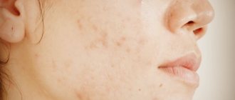
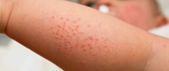
Skin lesions during illness in children are quite specific; often it is the skin and blood vessels under the epidermis that are among the first to be damaged during the formation of autoimmune aggression. Initial symptoms appear as:
- swelling of the skin,
- bluish, purple skin around the eyes, surrounding the eyes like glasses or a half mask,
- symmetrical areas of redness and areas of atrophic scars appear in the area of the affected articular surfaces,
- translucent vessels appear on the upper eyelids,
- spider veins appear in the palms and fingertips,
- marbling of the skin of the body and face is typical,
- due to malnutrition of the skin, small areas of necrosis (death) of cells appear,
- the skin is dry and very flaky, there may be areas of strong pigmentation or no pigment at all,
- it is possible to form subcutaneous calcifications with their opening to the surface of the skin with crumbly discharge,
- The mucous membranes of the mouth and respiratory tract are affected, conjunctivitis and vaginal damage occur in girls.
Degrees of manifestation of dermatomyositis
If your child has been diagnosed with dermatomyositis, you should be aware that the disease can develop in varying degrees of activity. In total, experts distinguish 3 degrees:
- I degree – primary chronic, in which only the skin and muscles are affected. With her, the child:
- body temperature does not rise
- the skin acquires a purple tint in some areas (including the eyelids)
- joints are poorly straightened
- muscles weaken only if the child begins to strain them
- the voice changes - it becomes nasal
- Myocarditis and vascular problems may develop
- Stage II – subacute, in which all organs are affected within 7 months of active development of the disease. With this form of dermatomyositis in a child:
- low-grade body temperature
- damage to muscles and skin is more severe than in grade I
- the child loses motor activity
- all internal organs begin to become inflamed, the heart and blood vessels especially suffer
- III degree – acute, in which the disease actively develops in just 1.5 months. With this form of dermatomyositis in a child:
- febrile body temperature (very high)
- The skin and muscles are very deformed (all this is accompanied by severe pain)
- blood and urine tests are always bad
- internal organs are severely inflamed
Whatever degree of disease you are diagnosed with, it must be treated urgently. Correctly selected treatment will help your child live a full life in the future.
Survival prognosis
Timely initiation of treatment for a pathological process significantly improves the prognosis for the patient. As the disease develops, several options for further developments are possible:
- the appearance of an episode of pathology as a single phenomenon, transition to the stage of inactive course (for 2 years) and absence of relapses in the future,
- alternation of constant exacerbations and remissions,
- a chronic course in which various complications are most likely.
The earlier this disease is detected, the greater the chances of a complete cure. In children, if the treatment process was started on time, this process often ends with almost complete recovery or the achievement of stable remission.
When the process is neglected, 40% of patients die within the first two years after the onset of the pathological process.
How is nasal bone repositioning done?
Dermatomyositis: symptoms
Symptoms of dermatomyositis differ depending on the degree of activity of the disease. However, there are several main signs by which doctors most often diagnose dermatomyositis:
- The first manifestations of dermatomyositis are skin. As we already mentioned, the eyelids, the area under the eyes, the places where the joints extend, become swollen and purple. At the same time, the skin peels off greatly because the dermis becomes dry.
- Calcification occurs on the elbows, knees, buttocks and shoulders - the deposition of salts on the subcutaneous tissue.
- The mucous membranes are affected. Most often the mouth, but in girls the vaginal mucosa can also be affected.
- All muscle groups are greatly weakened. The child may stop walking and have difficulty breathing and swallowing. As a result, respiratory diseases also occur as a symptom of dermatomyositis.
- Myocarditis develops. If the heart was already weak, then we may even be talking about myocardial dystrophy.
- The child’s nervous system is also severely affected. As a result, encephalitis and meningoencephalitis develop, which are accompanied by convulsions and epileptic seizures.
- The fundus of the eye changes and the optic nerve atrophies.
- The organs of the digestive tract and kidneys are affected. Ulcers and possibly even malignant tumors form on these organs. All these ailments are accompanied by severe pain.
Articular syndrome in dermatomyositis
Articular syndrome with severe dermatomyositis is not common, but it does occur. Deformation of the joint capsule may occur due to lack of treatment or factors complicating the course of the disease. In other cases, the affected area is only in small joints:
- brushes,
- wrist joints,
- ankle,
- shoulder,
- elbows,
- knee
With articular syndrome, redness of the skin, pain and swelling localized to the lesion may occur. In rare cases, deposition of calcifications is observed. With severe damage, the development of ankylosis is observed - the inability to move the joints.
If therapeutic measures are not taken, mobility will soon be limited.
https://youtu.be/LOhiS9oCQvg
Dermatomyositis: diagnosis
If your child has any of the symptoms we have listed, this is a reason to contact a rheumatologist, who will prescribe you to undergo a series of laboratory and clinical tests, which include:
- general urine and blood tests (in which the doctor, first of all, pays attention to the acceleration of ESR and the presence of myglobin);
- blood chemistry;
- ECG;
- X-ray;
- Electomyography;
- Muscle biopsy (performed in the most advanced cases).
If the doctor deems it necessary, he will send you for examination to another specialist (this most often happens if the child has some chronic diseases that could cause dermatomyositis). If the diagnosis is confirmed, the doctor will give you recommendations on how to treat dermatomyositis.
Clinical picture
This pathology (for both adults and children) is characterized by a subacute onset of the process. In this case, the first symptoms of polymyositis are considered to be manifestations of damage to the muscles of the shoulders and pelvis.
Next comes the symptoms of damage to internal organs (lungs, gastrointestinal tract and heart). Approximately 15% of patients have joint syndrome.
Patients are often interested in which symptom is most characteristic of polymyositis. As a rule, manifestations of muscle syndrome come to the fore in this disease. Let's talk about it in more detail.
Dermatomyositis: treatment
Depending on the severity of the disease, your doctor will explain to you where to treat dermatomyositis will be more effective. But most often, a child with such a diagnosis is hospitalized, where drug therapy is carried out. It involves the use of several medications to treat the child:
- Corticosteroids (anyone except Triamcinolone and Dexamethasone are used because they weaken muscles)
- “Prednisolone” with “Nerabol” and “Nerobolil” (especially these hormonal drugs are important if the child has stage II and III disease)
- Immunosuppressants such as Methotrexate, Azathioprine
- "Delagila"
- Salicylates
- "Pyridoxal phosphate"
- "Cocarboxylase"
- Vitamin E, B and C
As soon as the child’s condition improves after this drug therapy, he is prescribed physiotherapy, which includes muscle massage and exercise therapy. They are necessary to relieve pain. After the child is discharged from the hospital, it is advisable to send him to a sanatorium, where he will continue to undergo physical procedures. In addition, teachers and psychologists will work with the child.
If a child has been diagnosed with degree I of dermatomyositis, then, most likely, after such treatment he will experience a lasting recovery; if stage II or III, then the baby will enter a period of remission, which must be maintained at all times with glucocorticoid drugs. In any case, you and your baby will have to register with a dispensary in order to come for examination to a doctor either once a month or once every 3 months (this factor depends on the stage at which the disease was diagnosed).
How it proceeds
There are only three theories of the occurrence of the disease: genetic, infectious and immune. All these theories are closely related to each other, as evidenced by the patient’s tests, thanks to which immunity disorders are detected, just as the body’s immune cells are found in muscle tissue.
If a patient is infected with an infection for a long time, its cells settle in the muscle tissue, to which the body reacts with a violent immune reaction. As a rule, the impetus for this is stress or hypothermia, hormonal changes, vaccinations and other factors.
The course of the disease is quite severe; in the acute form, muscles and internal organs are affected, the patient cannot move, his temperature rises and fever appears. If therapy is prescribed inadequately or untimely, the disease becomes chronic. If left untreated, polymyositis can be fatal.
In the chronic form of the disease, the motor activity of individual muscle groups is disrupted, they atrophy, and the person cannot move. Inflammation often spreads to other muscle groups, complicating the course of the disease. Treatment is usually long and difficult and does not always result in a complete recovery.
Dermatomyositis: prognosis and prevention
The prognosis for life with dermatomyositis is quite dubious. Fortunately, medicine is constantly evolving, so the infant mortality rate due to this disease is only 1%. In most cases, today it is possible to completely restore the child’s muscle strength, but for this it is necessary to constantly take corticosteroids, from which the digestive tract and nervous system will suffer greatly.
As such, there are no special preventive measures that could 100% prevent the development of dermatomyositis in your child. You just need to follow the general rules of life to reduce the risk of developing the disease. What is meant:
- Make sure your child leads a healthy lifestyle;
- do not allow his internal organs and muscles to become overloaded (to do this, pay special attention to the child’s diet and the amount of medications he takes if he suddenly gets sick);
- make sure that the baby never suffers mental trauma; depressive disorders are a direct path to dermatomyositis;
- Always strengthen your baby's immunity by giving him vitamins, strengthening him, and playing sports with him.
Treatment of dermatomyositis in a child is a complex process. It requires parents’ strength, patience and financial expenses. But the main thing is not this, but the belief that your baby will recover and will have the opportunity to live a full life. We wish all our readers that your children are healthy, beautiful and happy! Take care of their health, take care and love with all your soul and heart - this is the main cure for any disease.
https://youtu.be/NQ1QVFI1ZmU
Classification
In accordance with the location of the process and the symptoms present, polymyositis is divided into the following types:
- Primary (ideopathic) form of polymyositis. It develops slowly and is more typical for women. In this case, the proximal muscles of the neck and limbs are damaged. Patients complain of difficulty walking up stairs, combing, muscle tension and soreness. Sometimes arthralgia is also present. In severe cases, the muscles atrophy and dysphagia develops.
- Inclusion myositis. This variety is rarely diagnosed. Accompanied by damage to the distal segments of the limbs.
- Juvenile (children's) form. The main manifestation is calcification (deposition of calcium salts) in and under the skin. As the process progresses, atrophy and contractures appear, blood vessels (including vasculitis and thrombosis) and the gastrointestinal tract are affected, severe pain, ulceration, perforation and bleeding occur.
- In combination with autoimmune systemic pathologies. In this case, polymyositis acts as a complication of SLE, RA, Sjogren's disease or scleroderma.
- Primary (ideopathic) dermatomyositis. The symptoms of this form are represented by the appearance of various rashes (dermatitis, erythema) on the face, in the area of the joints of the fingers, back and limbs. Stomatitis, conjunctivitis and pharyngitis are often present.
- Polymyositis in malignant neoplasms. Most often it is discovered two years after the process has become malignant. This form is typical for elderly patients with cancer of the testicles, prostate or breast, lungs, intestines, as well as for those suffering from lymphomas.
Muscle syndrome
It is characterized by muscle pain that occurs during movement, palpation, and in some cases at rest. Along with pain, muscle weakness increases, preventing the patient from performing basic movements actively and fully. The patient is unable to sit down, hold objects in his hands, stand up, lift his head from the pillow, and so on.
The muscles affected by the disease become denser and swell. Later they undergo atrophy, myofibrosis, and in some forms, calcification. Such changes can lead to complete immobilization of the patient.
Muscle syndrome implies the presence of lesions and smooth muscles of the esophagus, larynx, and pharynx. In this case, swallowing disorders (dysphagia) and speech changes (dysarthria) occur. If the process spreads to the facial muscles, the patient’s face takes on a resemblance to a mask, and to the eye muscles, ptosis, strabismus and diplopia occur.
Forecasts
The most unfavorable prognosis is considered to be the acute form of polymyositis, the causes and symptoms of which we discussed above. Aspiration pneumonia or cardiopulmonary failure that occurs in this case most often causes death.
The patient's childhood worsens the prognosis of polymyositis (symptoms and treatment for this category of patients are the same as for adults). The difference lies in the dosage of medications, which must be determined by the attending physician. Steady and rapid progression of the pathology almost always leads to immobilization of the child.
The chronic course is most favorable for both life and future ability to work.
ethnoscience
Treatment with folk remedies for polymyositis is also effective. As a rule, such therapy consists of the use of oils, ointments, various solutions, as well as alcohol tinctures. Anti-inflammatory compresses are widely used, which are applied to the affected areas of the joints and muscles (an effective folk method of treatment). It is important to prepare folk remedies correctly so that they only bring benefits and do not aggravate the patient’s condition.
Widely used means:
- cabbage compress. An effective folk remedy that helps relieve muscle pain;
- tincture of lilac flowers. A folk remedy that relieves inflammation at the site of the lesion. Prepared from lilac flowers and alcohol;
- pepper oil. You can prepare this folk remedy yourself, or you can purchase it at the pharmacy.
https://youtu.be/mkaejM_nytA
Course of the disease
The course of polymyositis can be acute, subacute (more often) and chronic.
In the case of an acute course, the disease is manifested by intoxication and fever, muscle lesions rapidly progress, and dysarthria and dysphagia occur. In a short time the patient becomes immobilized. If therapy is not carried out, the condition quickly worsens. There may also be death.
Polymyositis with a subacute course is characterized by wave-like changes in the patient’s condition: periods of deterioration alternate with improvements. Despite the apparent periodic improvement, muscle weakness increases and damage to internal organs occurs.
The chronic course of the disease is considered the most favorable and is characterized by damage to only certain muscle groups, due to which patients remain able to work for a long time.
Diagnostic measures
If polymyositis is suspected (if the symptoms described above are present), the patient should consult a rheumatologist, neurologist, gastroenterologist, pulmonologist and cardiologist.
The results of a clinical blood test indicate the presence of inflammation (leukocytosis and accelerated ESR). Blood biochemistry reveals signs of damaged muscle tissue (increased levels of aldolase, CPK, ALT, AST). In polymyositis, these data are used to determine the degree of inflammatory activity in muscle tissue. Approximately 20% of patients have antinuclear antibodies. Even less often, rheumatoid factor is found in the blood of patients.
In order to exclude the presence of other neuromuscular pathologies, electromyography is prescribed. Confirmation of pathological changes in muscle tissue will be:
- Low action potential amplitude.
- Spontaneous fibrillations.
- Increased excitability.
The most informative technique confirming the presence of polymyositis is the histology of muscle fibers. To collect material, a biopsy of the quadriceps or biceps brachii is used. In this case, changes characteristic of polymyositis are detected: infiltration of muscle tissue and vascular walls with lymphocytes, the presence of vacuoles (cavities) between the fibers, degenerative or necrotic changes.
To diagnose the condition of somatic organs, X-rays of the lungs, ultrasound of the heart and gastrointestinal tract, gastroscopy and ECG are prescribed.
Pathology therapy
Treatment of polymyositis comes down to eliminating the inflammatory process and maintaining stable remission.
Basic therapy is glucocorticosteroid drugs. At the beginning of treatment, the dosage of Prednisolone is 40-60 mg/day, then reduced to 10-20 mg/day.
In cases where treatment does not bring the desired effect (that is, there is no improvement in condition and blood counts), immunosuppressants are prescribed (for example, Methotrexate). These drugs can be administered either orally or intramuscularly. Contraindications to the use of Methotrexate are: renal/liver failure, pathological changes in blood clotting.
Less popular for polymyositis are the immunosuppressants Cyclophosphamide, Cyclosporine, Azathioprine, Chlorambucil, but they are also prescribed in some cases.
Until the inflammatory reactions subside, the patient's physical activity is sharply limited.
If polymyositis is combined with vasculitis, the patient is prescribed lymphocyto- and plasmapheresis.
Can myositis be cured?
It depends on the cause of the disease. In most cases, the prognosis is favorable. There is no effective treatment for certain forms of the disease, such as polymyositis. However, properly prescribed medication and physical therapy can improve muscle function.
In rare cases, myositis leads to rhabdomyolysis, the destruction of muscle tissue. Decomposition products enter the blood, resulting in acute renal failure. In this case, hospitalization in a hospital and infusion therapy are necessary.
Doctors at the neurology clinic of our medical center have extensive experience in treating myositis of various origins. You can make an appointment with a specialist by calling +7 (495) 230-00-01.
Complications
With long-term dermatomyositis without treatment, the following develops:
- bedsores and trophic ulcers;
- contractures, bone deformations;
- loss of muscle mass;
- calcification
The most serious complications that threaten the patient with advanced dermatomyositis, from which up to 40% of patients die in the first 2 years without appropriate treatment:
- aspiration pneumonia, alveolar fibrosis;
- destruction of the muscles of the respiratory organs, esophagus and pharynx;
- gastrointestinal bleeding;
- heart pathologies;
- general dystrophy, exhaustion
https://youtu.be/d9KuMlMjxVo